Research - (2022) Volume 10, Issue 6
Transabdominal and Transvaginal Ultrasound Evaluation of Abnormal Uterine Bleeding in Post-Menopausal Women
Bhanupriya Singh1*, Tanvi Khanna1, Devyani Mishra2 and Gaurav Raj1
*Correspondence: Bhanupriya Singh, Department of Radiodiagnosis, Dr. Ram Manohar Lohiya Institute Of Medical Sciences, Vibhuti Khand, Gomti Nagar, Lucknow, UP, India, Email:
Abstract
Background: Any uterine bleeding outside the normal volume, duration, regularity or frequency is considered abnormal uterine bleeding. Objectives: To study various causes of Perimenopausal abnormal uterine bleeding , their radiographic features on trans abdominal & transvaginal Ultraonography & correlate them with the histopathological findings on hysterectomy specimens. Materials & Methods: This study was carried out in the Department of Radiodiagnosis in a tertiary care centre among Perimenopausal women who underwent hysterectomy for abnormal uterine bleeding. The ultrasonography findings and histopathological reports of hysterectomy specimens were correlated. Results: Among 60 number of hysterectomies cases for abnormal uterine bleeding most of the patients were between 40-49 years of age (63.3%) with mean age being 48yrs and menorrhagia was the dominant clinical presentation seen in 27 patients (45%). The majority of cases were diagnosed as Uterine fibroid with 31(51.7%) cases on trans abdominal 30 (50%) cases on transvaginal ultrasonography. Histopathological reports of myometrium showed 46.7% cases of uterine fibroid, followed by 33.3% cases of normal myometrium adenomyosis in 20% cases. Histopathology of endometrium revealed hyperplasia in the most cases (51.7%) where simple typical type was the predominant. Conclusion: Uterine fibroid is the leading cause of AUB and radiological, pathological evaluation correlated well to diagnose fibroid. Trans abdominal or transvaginal ultrasound is low cost primary modality for screening and should include as a first line screening method. However, histopathological diagnosis proved to be the gold standard.
Keywords
Abnormal uterine bleeding, Perimenopausal, Trans abdominal ultrasonography, Transvaginal ultrasonography, Fibroid, Adenomyosis, Dysfunctional uterine bleeding
Introduction
Perimenopause is the described as the period from the onset of menstrual transition uptil 12months after the last menstrual period [1]. Abnormal uterine bleeding (AUB) is a symptom and not a disease. It accounts for more than 70% of all gynecological consultations in the peri‑ and post‑menopausal age group [2]. It occurs in various forms such as menorrhagia, polymenorrhea, polymenorrhagia, metrorrhagia, and menometrorrhagia [3]. PALMCOIEN classification in 2011, for the causes of AUB in non-gravid women of reproductive age was published in order to standardize terminology, diagnostics and investigations. There are 9 main categories, which are arranged according to the acronym PALM-COEIN: polyp; Adenomyosis; leiomyomas; malignancy and hyperplasia; coagulopathy; ovulatory dysfunction; endometrial; iatrogenic; and not yet classified. However, there are no detectable structural abnormalities in majority of cases, and this is called dysfunctional uterine bleeding (DUB) [4]. Ultrasound is an appropriate screening tool and, in most instances, should be performed first or early in the course of the investigation. It is well accepted that various disease pathology can be detected accurately by histopathological examination (HPE) [5]. The current study was carried out to evaluate various clinical presentations of perimenopausal AUB and to correlate ultrasonographic findings with histopathological examination in those patients undergoing hysterectomy.
Materials and Methods
This study was conducted in a tertiary care hospital in the Department of Radiodiagnosis. The study population consisted of perimenopausal women within the age group of 40 years to 60 yrs who underwent hysterectomy for AUB. The proper institutional ethical clearance was taken for the study. After obtaining informed consent from selected patients, the relevant data such as age, parity, menstrual symptoms, and other associated findings in clinical examination were recorded. In all these women, transabdominal & transvaginal ultrasonographic evaluation was done. The clinical presentations and ultrasonographic findings were correlated with the histopathology report of hysterectomy specimens.
Results
Total of 60 women who underwent hysterectomy for AUB were included in study.The mean age of subjects included was 48 years. 38 patients were from age group 40-49 years, with rest 22 patients in 50-59 years age group.The mean parity of included subjects was 3.18.
Uterine fibroid was the major finding with 31 (51.7%) cases seen on TAS and 30 (50%) cases on TVS. Bulky uterus was detected in 15 subjects, similar number on both TVS and TAS. Adenomyosis was found in 10 (16.7%) cases on TAS and 13 (21.7%) cases on TVS. Both TVS and TAS showed 17 cases with bulky cervix. Many subjects had multiple findings, major subjects with bulky uterus had associated uterine fibroid or adenomyosis. Thickened endometrium was seen in 8 cases on TAS and 11 cases on TVS.
Histopathology reports showed that there were 28 (46.7%) cases of uterine fibroid. Adenomyosis was seen in 12 (20%) patients while 20 (33.3%) cases had normal myometrium. Out of 31 cases of fibroid on TAS, 3 patients were diagnosed as adenomyosis on HPE. In patients with bulky uterus there were 2 cases of fibroids, and 1 case with adenomyosis, while rest had normal myometrium. Histopathology of endometrium showed a total of 31 (51.7%) cases with endometrial hyperplasia. 11(18.3%) patients had secretary endometrium, 6 had proliferative and 1 case had inflammatory endometrium.
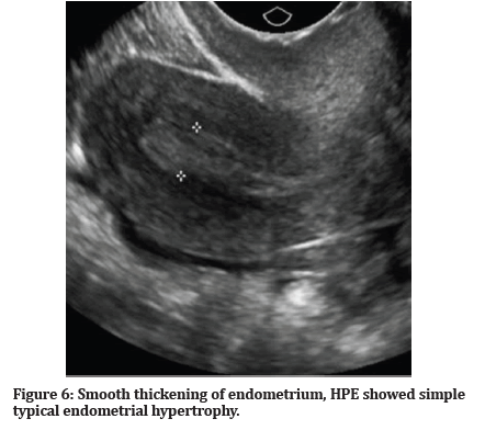
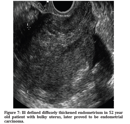
Among the 31 cases of endometrial hyperplasia the TAS diagnosed thickened endometrium in 8 cases and TVS in 11 cases.23 patients had simple typical endometrial hyperplasia.4 subjects showed complex atypical hyperplasia. Endometrial carcinoma was diagnosed in 4 cases on HPE. Endometrial carcinoma was diagnosed in 4 cases on HPE. Inflammatory cervix was seen in 24 (40%) of cases. There was 1 case which was diagnosed with carcinoma cervix (Tables 1-4) and (Figures 1-14).
| Age group | Number | % |
|---|---|---|
| 40-49 | 38 | 63.30% |
| 50-59 | 22 | 36.67% |
Table 1: Distribution of age.
| Complaints | <12 Months | >12 Months | Total % |
|---|---|---|---|
| Menorrhagia | 16 | 11 | 27(45%) |
| Polymenorrhagia | 5 | 2 | 7(11.7%) |
| Metrorrhagia | 4 | 3 | 7(11.7%) |
| Menometrorrhagia | 1 | 5 | 6 (10%) |
| Postmenopausal Bleeding | 5 | 8 | 13 (21.7%) |
| Total | 31 | 29 | 60 (100%) |
Table 2: Distribution of menstrual complaints.
| Diagnosis | TAS | TVS |
|---|---|---|
| Uterine Fibroid | 31 | 30 |
| Bulky Uterus | 15 | 15 |
| Bulky Cervix | 17 | 17 |
| Adenomyosis | 10 | 13 |
| Thickened Endometrium | 8 | 11 |
Table 3: Ultrasound findings.
| Types of hyperplasia | No |
|---|---|
| Simple typical | 23 |
| Complex typical | 3 |
| Simple atypical | 1 |
| Complex atypical | 4 |
Table 4: Endometrial hyperplasia on histopathological examination.
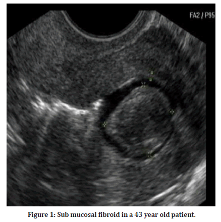
Figure 1: Sub mucosal fibroid in a 43 year old patient.
Figure 2: Large intramural fibroid in a 41 year old patient with menorrhagia.
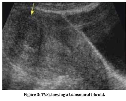
Figure 3: TVS showing a transmural fibroid.
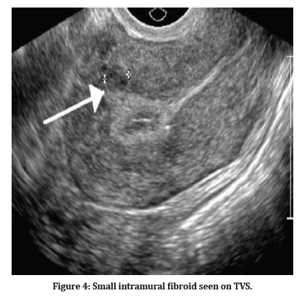
Figure 4: Small intramural fibroid seen on TVS.
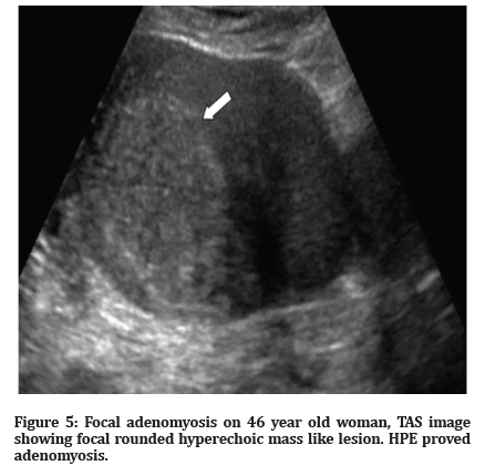
Figure 5:Focal adenomyosis on 46 year old woman, TAS image showing focal rounded hyperechoic mass like lesion. HPE proved adenomyosis.
Figure 6:Smooth thickening of endometrium, HPE showed simple typical endometrial hypertrophy.
Figure 7:Ill defined diffusely thickened endometrium in 52 year old patient with bulky uterus, later proved to be endometrial carcinoma.
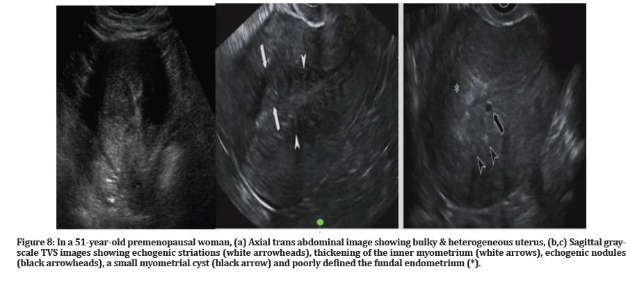
Figure 8:In a 51-year-old premenopausal woman, (a) Axial trans abdominal image showing bulky & heterogeneous uterus, (b,c) Sagittal grayscale TVS images showing echogenic striations (white arrowheads), thickening of the inner myometrium (white arrows), echogenic nodules (black arrowheads), a small myometrial cyst (black arrow) and poorly defined the fundal endometrium (*).
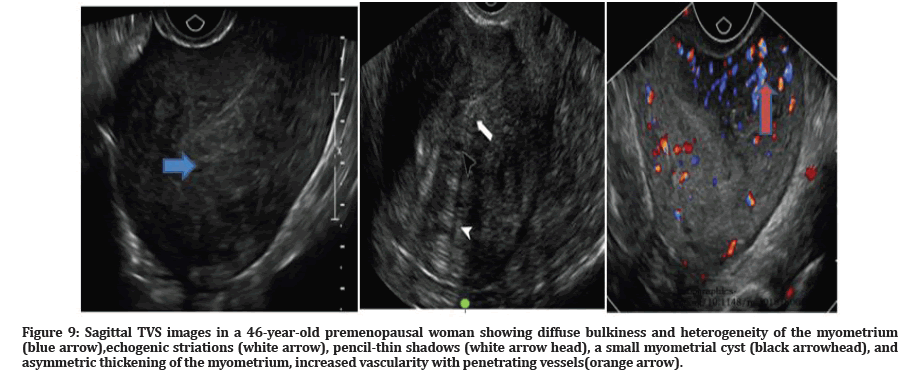
Figure 9:Sagittal TVS images in a 46-year-old premenopausal woman showing diffuse bulkiness and heterogeneity of the myometrium (blue arrow),echogenic striations (white arrow), pencil-thin shadows (white arrow head), a small myometrial cyst (black arrowhead), and asymmetric thickening of the myometrium, increased vascularity with penetrating vessels(orange arrow).
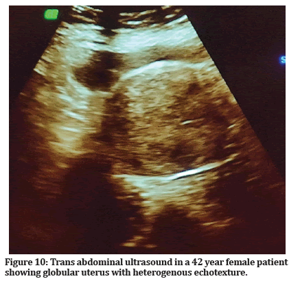
Figure 10:Trans abdominal ultrasound in a 42 year female patient showing globular uterus with heterogenous echotexture.
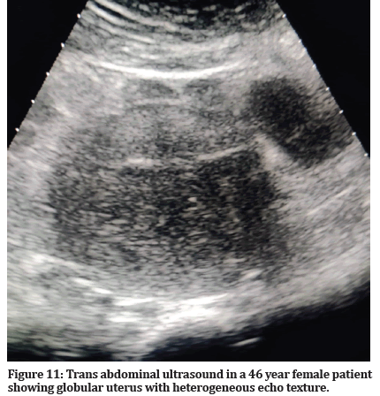
Figure 11:Trans abdominal ultrasound in a 46 year female patient showing globular uterus with heterogeneous echo texture.
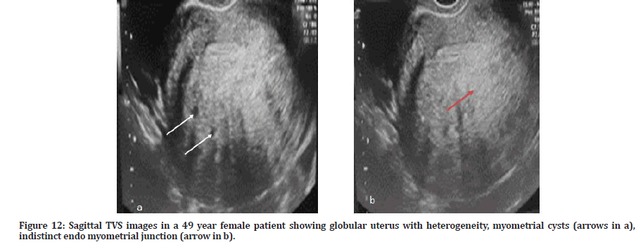
Figure 12:Sagittal TVS images in a 49 year female patient showing globular uterus with heterogeneity, myometrial cysts (arrows in a), indistinct endo myometrial junction (arrow in b).
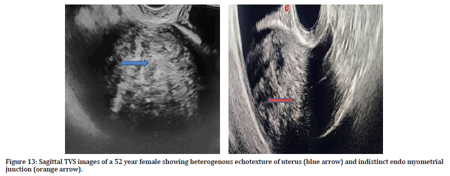
Figure 13:Sagittal TVS images of a 52 year female showing heterogenous echotexture of uterus (blue arrow) and indistinct endo myometrial junction (orange arrow).
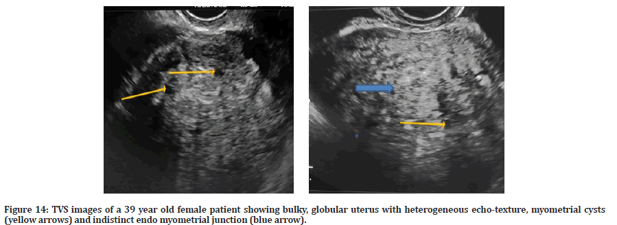
Figure 14:TVS images of a 39 year old female patient showing bulky, globular uterus with heterogeneous echo-texture, myometrial cysts (yellow arrows) and indistinct endo myometrial junction (blue arrow).
Discussion
AUB is one of the main gynecological cause of hysterectomy and accounts for two‑thirds of all hysterectomies [6]. USG forms the baseline imaging modality, & helps exclude organic pathology for AUB and is well accepted for various diseases whose pathology is accurately detected by histopathological examination (HPE) [7] In this study, 60 numbers of perimenopausal hysterectomized patients were analyzed. Most number of patients (63.3%) was between 40 and 49 years age group. Thus the demographic findings of our study were consistent with other studies. In a study by Modak R et al in which the majority of the patients were in the age group of 40-44 yrs [8] In another study by J. Meena, the maximum number of patients were in the age group of 41-50 yrs [9]. There can be various manifestations of abnormal uterine bleeding like bleeding between menses known as metrorrhagia, irregular length of cycles with or without a variable duration and amount of bleeding, called as menometrorrhagia or an increased number named as polymenorrhea or decreased number of bleeding episodes termed as oligomenorrhea may be present. In addition, overall increased volume labelled as menorrhagia or, conversely, a reduction in blood loss per cycle known as hypomenorrhea are described [10]. The common menstrual problem in our study was menorrhagia (45%).This finding was consistent with the study of Jetley et al [11]. and Shobha [12] in which clinical presentation as menorrhagia in AUB evaluation revealed 46.4% and 46.6%, respectively. A similar result was shown by study of Pillai [12]. In another study by Talukdar et al. [13] also menorrhagia was the dominant presentation. In our study, ultrasonographic diagnosis of Uterine fibroid was seen in 31 (51.7%) cases seen on TAS and 30 (50%) cases on TVS which was consistent with the findings of a study by Talukdar et al. [13]. In a another study by Meena et al. [9] also, the most prevalent histopathological finding, amongst the cases diagnosed on USG. In a study conducted by N Bhavani et al., among the causes of abnormal uterine bleeding, myoma seen in 38.5% was the commonest cause of abnormal uterine bleeding [14]. Leiomyoma is a proliferation of smooth muscle surrounded by pseudocapsule, unopposed estrogens may accelerate its growth. Leiomyomas are characterized by the layer they occupy, whether submucosal (or subendometrial), intramural within the myometrium, or subserosal. Intramural lesions do not involve the endometrial cavity, whereas submucosal lesions are intracavitary and are best detected with saline hysterosonography. Hypoechoic areas within a leiomyoma may represent cystic degeneration [15]. In our study Adenomyosis was found in 10 (16.7%) cases on TAS and 13 (21.7%) cases on TVS. Adenomyosis is a condition in which there is functional endometrial tissue within the myometrium. The US appearance of adenomyosis includes myometrial cysts, ill-defined areas of myometrial echotexture, heterogeneous and distorted myometrial echotexture, and a globular or enlarged uterus with asymmetry [16,17]. In this study, HPE confirmed endometrial hyperplasia in 31 (51.7%) of cases of which simple typical type was the predominant. In a study by Talukdar and Mahela endometrial hyperplasia was seen in (56.31%) of cases of which simple typical type was the leading type in 46.60% cases. In the studies done by Khare endometrial hyperplasia was seen in 23%, 22.6%, and 36.2%, respectively which were lower than our study [18-20].
Conclusion
Based on the above study we conclude that Ultrasonography is an easy and relatively inexpensive method of first choice to find out various etiologies of AUB. Histopathology plays a major role in the definitive diagnosis and for accurate evaluation of uterine endometrium. Uterine fibroid was the leading cause of AUB. Radiological and pathological evaluation correlated well to diagnose fibroids. TVS had higher sensitivity in diagnosis of uterine fibroids in comparison to TAS. Other major cause of AUB was adenomyosis, where TVS had high sensitivity and specificity. TAS was less sensitive to diagnose adenomyosis (50% sensitivity). Both TAS and TVS were not very sensitive for diagnosing endometrial pathologies, not correlating well on HPE.
References
- Soules MR, Sherman S, Parrott E. Stages of reproductive aging workshop (STRAW). J Womens Health Gender Based Med 2001; 10:843-8.
- Mahajan N, Aggarwal M, Bagga A. Health issues of menopausal women in North India. J Midlife Health 2012; 3:847.
- Kumar P, Malhotra N. Clinical types of abnormal uterine bleeding. In: Kumar P. Jeffcoate’s Principle of Gynecology. 7th Ed. New Delhi: Jaypee Brothers Medical Publishers (P) Ltd 2008; 599.
- Munro MG, Critchley HO, Border MS, et al. FIGO classification system–PALM-COIEN for causes of AUB in non-gravid women of reproductive age. Int J Gynaecol Obstetr 2011; 113:3-13.
- Dueholm M, Lundorf E, Hansen ES, et al. Evaluation of the uterine cavity with magnetic resonance imaging, transvaginal sonography, hysterosonographic examination, and diagnostic hysteroscopy. Fertil Steril 2001; 76:350-357.
- Telner DE, Jakubovicz D. Approach to diagnosis and management of abnormal uterine bleeding. Can Fam Physician 2007; 53:58‑64.
- Fraser IS, Critchley HO, Munro MG, et al. A process designed to lead to international agreement on terminologies and definitions used to describe abnormalities of menstrual bleeding. Fertil Steril 2007; 7:466-476.
- Modak R, Pal A, Pal A, et al. Abnormal uterine bleeding in perimenopausal women: Sonographic and histopathological correlation and evaluation of uterine endometrium. Int J Repr Contraception Obstetr Gynecol 2020; 9:1959-1965.
- Meena Jain, Prehit Chate. TAS/TVS verus histopathological diagnosis in abnormal uterine bleeding: A comparative study. IntJ Clin Obtet Gynaecol 2020; 4:282-286.
- Shagam JY. Ultrasound assessment of uterine disorders. Radiol Technol 2000; 72:11–25.
- Jetley S, Rana S, Jairajpuri ZS. Morphological spectrum of endometrial pathology in middleaged women with atypical uterine bleeding: A study of 219 cases. J Midlife Health 2013; 4:216-220.
- Shobha PS. Sonographic and histopathological correlation and evaluation of endometrium in perimenopausal women with abnormal uterine bleeding. Int J Reprod Contracept Obstet Gynaecol 2014; 3:113‑117.
- Talukdar B, Mahela S. Abnormal uterine bleeding in perimenopausal women: Correlation with sonographic findings and histopathological examination of hysterectomy specimens. J Mid-life Health 2016; 7:73-77.
- Al-Bajalan TH, Khalid SI. Thyroid dysfunction and abnormal uterine bleeding. Indian J Public Health Res Develop 2019; 10.
- Tranquart F, Brunereau L, Cottier JP, et al. Prospective sonographic assessment of uterine artery embolization for the treatment of fibroids. Ultrasound Obstet Gynecol 2002; 19:81–87
- Bazot M, Cartez A, Darai E, et al. Ultrasonography compared with magnetic resonance imaging for the diagnosis of adenomyosis: Correlation with histopathology. Hum Reprod 2001; 16:2427–2433.
- Fedele L, Bianchi S, Dorta M, et al. Transvaginal ultrasonography in the diagnosis of diffuse adenomyosis. Fertil Steril 1992; 58:94–97.
- Dangal G. A study of endometrium of patients with abnormal uterine bleeding at Chitwan valley. Kathmandu Univ Med J 2003; 1:110‑112.
- Sloboda L, Molnar E, Popovic Z, et al. Analysis of pathohistological results from the uterine mucosa 1965‑1998 at the gynecology department in Senta. Med Pregl 1999; 52:263‑265. l>
- Khare A, Bansal R, Sarma S, et al. Morphological spectrum of endometrium in patients presenting with dysfunctional uterine bleeding. People J Sci Res 2012; 5:13‑16.
Indexed at, Google Scholar, Cross Ref
Indexed at, Google Scholar, Cross Ref
Indexed at, Google Scholar, Cross Ref
Indexed at, Google Scholar, Cross Ref
Indexed at, Google Scholar, Cross Ref
Indexed at, Google Scholar, Cross Ref
Indexed at, Google Scholar, Cross Ref
Indexed at, Google Scholar, Cross Ref
Indexed at, Google Scholar, Cross Ref
Indexed at, Google Scholar, Cross Ref
Author Info
Bhanupriya Singh1*, Tanvi Khanna1, Devyani Mishra2 and Gaurav Raj1
1Department of Radiodiagnosis, Dr. Ram Manohar Lohiya Institute Of Medical Sciences, Vibhuti Khand, Gomti Nagar, Lucknow, UP, India2Department of Obstetrics and Gynecology, Dr. Ram Manohar Lohiya Institute Of Medical Sciences, Vibhuti Khand, Gomti Nagar, Lucknow, UP, India
Citation: Bhanupriya Singh, Tanvi Khanna, Devyani Mishra, Gaurav Raj, Transabdominal and Transvaginal Ultrasound Evaluation of Abnormal Uterine Bleeding in Post-Menopausal Women, J Res Med Dent Sci, 2022, 10 (6): 79-85.
Received: 09-May-2022, Manuscript No. JRMDS-22-62370; , Pre QC No. JRMDS-22-62370 (PQ); Editor assigned: 11-May-2022, Pre QC No. JRMDS-22-62370 (PQ); Reviewed: 26-May-2022, QC No. JRMDS-22-62370; Revised: 31-May-2022, Manuscript No. JRMDS-22-62370 (R); Published: 07-Jun-2022

