Case Report - (2022) Volume 10, Issue 1
Tibia Hemimelia-A Rare Case Report
Dharun Kumar M* and Venkat kiran
*Correspondence: Dharun Kumar M, Department of Orthopaedics, Sree Balaji Medical College and Hospital, Chromepet, Chennai, Tamilnadu, India, Email:
Abstract
Tibial hemimelia (TH) is a rare congenital condition characterised by limb length discrepancy often associated with other congenital anomalies and deficiencies. There are limited reconstruction options, and management of choice remains amputation with prosthesis. The fibula is usually normally present, or duplicate or dysplastic. The quadriceps muscle is formed normally or absent or maybe associated with weakness, and the patella maybe formed normally, abnormally developed, or absent. Based on new classification scheme for jones type 4 tibia hemimelia, a new technique for reconstruction of limb is discussed in this report.
Keywords
Tibial hemimelia, Fibular osteotomy, Limb reconstructive system, Jones type 4 is equivalent to paley type 3A
Introduction
Hemimelia of tibia can have wide spectrum of pathological presentations, ranging from tibial hypoplasia to absent tibia, much more than that of the congenital fibular hemimelia. Tibial hemimelia can be present unilaterally or bilaterally. Bilateral presentation accounts for about 30% of cases [1]. Hemimelia of Tibia can be related to other congenital anomalies like those affecting the same side limb like syndactyly, dysplastic radius, polydactyly [2]. It also has many syndromic associations.
Jones classification [3]
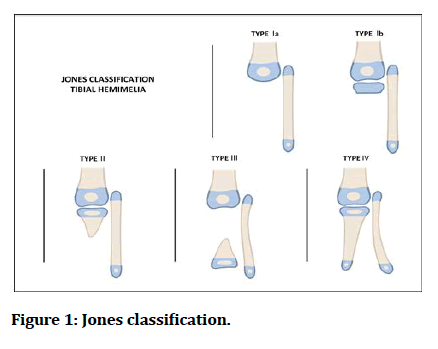
This classification is based on plain x-ray findings, divided into 4 types, ranging from the most deficient to the least deficient.
• Jones type I -Tibia is absent.
• Ia–Femoral distal epiphysis hypoplasia.
• Ib-Tibia is not seen and with normal femoral lower epiphysis.
• Jones type II-Distal tibia is absent.
• Jones type III-Proximal tibia is not seen.
• Jones type IV-Presence of diastasis (Figure 1).
Figure 1: Jones classification.
Paley proposed a new modified classification
• Paley type 1: Hypoplasia of tibia: Genu valgum with excessive growth of fibula proximal part with normal plafond.
• Paley type 2: Tibial proximal and distal epiphysis present with dysplasia of ankle from proximal aspect of tibial; dysplastic tibial plafond; associated proximal fibula over growth.
• Paley type 3: knee joint and proximal tibia, medial malleolus present, no tibial distal plafond, with diastasis of tibiofibular.
• Paley type 4: Distal tibial aplasia.
• Paley type 5: Complete aplasia of tibial.
In tibial Jones type I b or II, there are some reports having good outcomes with tibiofibular synostosis [4-7]. The use of an external fixator prior to reconstruction, contractures can be reduced6. Schoe-necker [8] recommendation for Jones types Ib and II patients was TF synostosis in combination with a Syme’s amputation. These patients were functional as B-K amputees.
Spiegel et al. [9] described about side effect of amputation, in patients with Jones type II managed with amputation (Chopart or Syme) due to irritation of prosthesis because of the overgrowth and fibular head prominence.
Jones type IV treatment of choice is ankle stabilization, arthrodesis or amputation [10,11].
Case Report
History
A 10-year-old patient came with c/o deformity and shortened left leg since birth. History of difficulty in walking. No history of developmental disorder. No syndromic features.
Clinical features
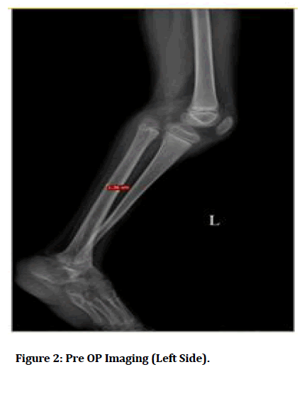
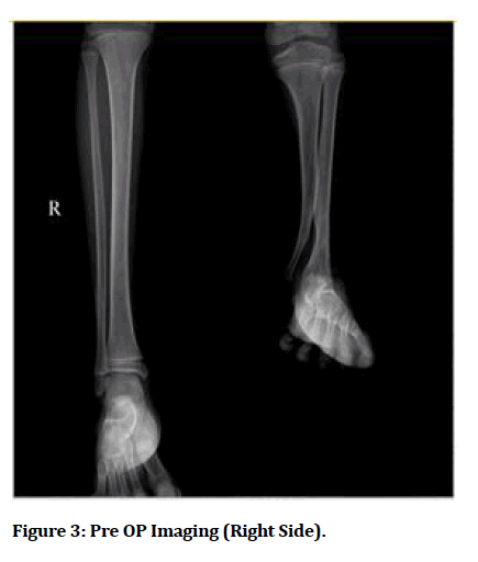
Limb length discrepancy – left leg shorter than right leg by 12cm.Distal tibia could not be palpated. Tarsal and metatarsal appears to be normal. Foot in varus deformity (Figure 2 and 3).
Figure 2: Pre OP Imaging (Left Side).
Figure 3: Pre OP Imaging (Right Side).
Surgical treatment
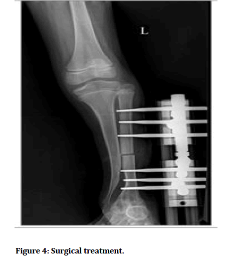
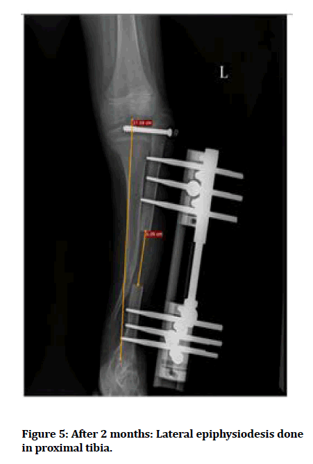
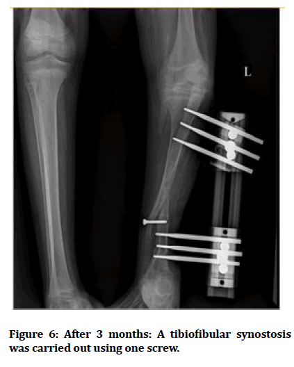
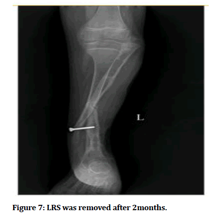
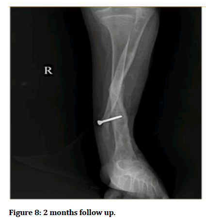
According to type 4 Jones, we decided to treat the patient with fibular osteotomy under limb reconstructive system (Figures 4-8).
Figure 4:Surgical treatment.
Figure 5:After 2 months: Lateral epiphysiodesis done in proximal tibia.
Figure 6:After 3 months: A tibiofibular synostosis was carried out using one screw.
Figure 7:LRS was removed after 2months.
Figure 8:2 months follow up.
Discussion
Based on the Jones classification, our case corresponds to type IV. Jones classified Tibial hemimelia into 4 types, ranging from the most to the least deficient. Type 4 is tibia shortening with diastasis. They are various surgical treatments based on the type of tibial hemimelia, such as amputation, reconstruction and deformity correction, arthrodesis of ankle, tendons repair. Jones type 4 is equivalent to Paley type 3A. Paley did ankle arthrodesis, external fixator for type 3A, whereas in our study patient underwent above mentioned procedure. The outcome proved to be effective as paleys. Hence our treatment modality can be considered as an alternative option.
Conclusion
Jones type 4 is equivalent to Paley type 3A. Paley did ankle arthrodesis, external fixator for type 3A, whereas in our study patient underwent above mentioned procedure. The outcome proved to be effective as Paleys. Hence our treatment modality can be considered as an alternative option.
References
- Aitkin GT, Bose K, Brown FW et al. Tibial hemimelia. In: Canale ST (Edn) Campbell’s operative orthopaedics. Mosby-Year Book, St Louis 1998; 93-1003.
- Chinnakkannan S, Das RR, Rughmini K, et al. A case of bilateral tibial hemimelia type VIIa. Indian J Human Genetics 2013; 19:108.
- Jones DA, Barnes JO, Lloyd-Roberts GC. Congenital aplasia and dysplasia of the tibia with intact fibula. Classification and management. J Bone Joint Surg 1978; 60:31-9.
- https://www.us.elsevierhealth.com/pediatric-orthopaedic-secrets-9781416029571.html
- https://link.springer.com/book/10.1007/978-3-319-17097-8
- Kalamchi A, Dawe RV. Congenital deficiency of the tibia. J Bone Joint Surg Br 1985; 67:581–584.
- Weber M. New classification and score for tibial hemi- melia. J Child Orthop 2008; 2:169–175.
- Schoenecker PL, Capelli AM, Millar EA, et al. Congenital longitudinal deficiency of the tibia. J Bone Joint Surg Am 1989; 71:278–287.
- Spiegel DA, Loder RT, Crandall RC. Congenital longitudinal deficiency of the tibia. Int Orthop 2003; 27:338–342.
- Fujii H, Doi K, Baliarsing AS. Transtibial amputation with plantar flap for congenital deficiency of the tibia. Clin Orthop Relat Res 2002; 403:186–190.
- Tokmakova K, Riddle EC, Kumar SJ. Type IV congenital deficiency of the tibia. J Pediatr Orthop 2003; 23.
Indexed at, Google Scholar, Cross Ref
Indexed at, Google Scholar, Cross Ref
Indexed at, Google Scholar, Cross Ref
Indexed at, Google Scholar, Cross Ref
Indexed at, Google Scholar, Cross Ref
Indexed at, Google Scholar, Cross Ref
Author Info
Dharun Kumar M* and Venkat kiran
Department of Orthopaedics, Sree Balaji Medical College and Hospital, Chromepet, Chennai, Tamilnadu, IndiaCitation: Dharun Kumar M, Venkat kiran, Tibia Hemimelia-A Rare Case Report, J Res Med Dent Sci, 2022, 10(1): 346-349
Received: 06-Dec-2021, Manuscript No. Jrmds-22-43928; , Pre QC No. Jrmds-22-43928 (PQ); Editor assigned: 08-Dec-2021, Pre QC No. Jrmds-22-43928 (PQ); Reviewed: 22-Dec-2021, QC No. Jrmds-22-43928; Revised: 27-Dec-2021, Manuscript No. Jrmds-22-43928 (R); Published: 03-Jan-2022

