Research - (2022) Volume 10, Issue 9
The Role of Selenium , Vitamin C and Glutathione Reductase Enzyme and Disease activity in Rheu-matoid Arthritis Patient of Medical city In Baghdad
Muthanna Abd Al-Rubaie*, Hallah Ghazi Mahmood and Mohammed Hadi Munshed Alosami
*Correspondence: Muthanna Abd Al-Rubaie, Department of Clinical Biochemistry, College of Medicine, University of Baghdad, Iraq, Email:
Abstract
Background: The "rheumatoid arthritis (RA)" is a multi-factorial chronic autoimmune disorders that influences a few organs and joints transcendently the synovial joints, Early diagnosis is important to ideal helpful achievement treatment; RA is a condition that is related with oxidative stress. "Oxidative stress" is defined the imbalance of oxidant/antioxidant forces in favor of the oxidant, During normal cellular metabolic processes free radicals and reactive metabolites are continuously generated, These oxidant products can injure and destroy the structures of cells or tissues. Objective: We aimed on this study to evaluate serum Glutathione Reductase (GR) Enzyme concentration, and see if there a correlation with disease activity in patient with RA, and compare it with healthy control. Subject and Method: The study had included on (130) subjects, (100) patient with RA only and (30) age, sex matched healthy control. This study and sample collection performed during the period from November 2021 to January 2022. In Medical City Baghdad Hospital. History was taken and Disease activity had measured by (DAS28-ESR) Disease activity score 28 – ESR from each patient and Ten, milliliters of blood were withdrawing from each control and patient, used for measurements of the GR Result : In studying of Glutathione reductase (GR) enzyme there are high enzyme concentration in human serum patient with Rheumatoid arthritis than the healthy control subject serum ( µ=10.622 ± 5.213, µ=6.522 ± 1.867 ng/ml) respectively with highly statically significant (T-Value 6.58, P- Value<0.05 ) the 95% CI for Difference is (-5.333;-2.867), I had found there are a positive correlation between the increase serum glutathione serum level and high disease activity when I study the correlation between Disease activity (DAS28-ESR) and GR, with high statistically significant ( P-Value<0.05) (Pearson correlation 0.265). Conclusion: Glutathione Reductase Enzyme gives a Picture for damaged cells due to Oxidative stress during chronic inflammation in Patient with RA. Patient with RA is more prone to Oxidative Stress related disease than other patient with autoimmune disease that hasn’t high oxidative stress.
Keywords
Glutathione, Rheumatoid arthritis, Selenium, Vitamin C
Introduction
Rheumatoid Arthritis (RA) is an immune system disorders that influences a few organs and joints, transcendently the synovial joints. The pathogenesis of this sickness isn't totally perceived, which possibly engaged with the genomic varieties, gene expression, protein translation and post translational changes [1]. Early diagnosis is important to ideal helpful achievement treatment, especially in patients with all around described danger factors for helpless results like high disease activity [2]. Rheumatoid Arthritis (RA) is a condition that is related with oxidative stress. Blood trace elements Such as Selenium (Se) and their transporter proteins, e.g., albumin and ceruloplasmin, and some water soluble vitamins such as Vitamin C has a significant part of cell protection against free radicals.
As anti-Oxidant components [3]. Rheumatoid arthritis has a world-wide distribution and affects all ethnic groups with prevalence of RA for most of the world is between 0.5 and 1 percent [4]. The disease can occur at any age thus no age group is exempted but its prevalence increases with age, The disease starts most commonly between the third and fifth decades but the age of onset follows a normal distribution curve [5-7]. In the absence tests to establish the diagnosis of RA, incidence and prevalence studies are based on a set of criteria developed for classification purposes. The most widely used criteria are the American College of Rheumatology 1987 revised criteria for the classification of RA [8].
Rheumatoid arthritis is a disease of unknown cause, though inherited and non-inherited factors may play a role in the etiology of RA [9]. The major sources of ROS include complexes I, III and the dehydrogenases that use ubiquinone as an acceptor, and succinate dehydrogenase In mitochondria. When electrons from other mitochondrial appropriate forum in substrate oxidation and oxidative phosphorylation circulate to oxygen, superoxide or hydrogen peroxide are formed [10]. "Glutathione (GSH) is a simple sulfur compound composed of three amino acids and the major non-protein thiol in many organisms", GSH is a major endogenous antioxidant which plays a key role in various cellular processes such as redox-homeostatic buffering, signaling, Detoxification and cell death & proliferation [11–13]. "GSH is a tripeptide, γ-L-glutamyl-L-cysteinylglycine", that is present in all mammalian tissues at 1-10 mM concentrations (highest concentration in liver) as being the most abundant non-protein thiol that protects against oxidative stress. GSH is also a key determinant of redox signaling, essential in xenobiotic detoxification, and modulates cell proliferation, apoptosis [14], Figure 1 shows the structure and synthesis pathway of glutathione synthesis by a two-step ATP-dependent enzymatic process. Glutamate-cysteine ligase (GCL) catalyzes the first step, which is made up of catalytic and modifier subunits (GCLC and GCLM).

Figure 1: The structure and synthesis pathway of glutathione synthesis.
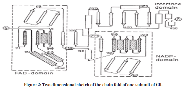
This process combines cysteine and glutamate to formglutamylcysteine. GSH synthase catalyzes the second step, which adds glycine to-glutamylcysteine to generateglutamylcysteinylglycine or GSH. GSH inhibits GCL via negative feedback inhibition [14]. Glutathione reductase (GR) is a flavoprotein catalyzing the NADPH-dependent reduction of glutathione disulfide (GSSG) to glutathione (GSH) The reaction is essential for the maintenance of glutathione levels. Glutathione has a major role as a reductants in oxidation-reduction processes, and also serves in detoxication and several other cellular functions [15]. GR is a member from the family of disulfide reductase. This family also includes lipoamide dehydrogenase and thioredoxin reductase, both are FAD-containing anti-oxidant enzymes that interact with disulfide substrates, Lipoamide dehydrogenase especially is very similar to glutathione reductase in structural and mechanism of effect, Also the bacterial enzyme mercuric reductase (disulfide reductases) very similar to GR [16]. The high cellular GSH/GSSG ratio is maintained by flavoproteins GRs, which are with high affinity for both GSSG and NADPH, Classical fractionation studies described high GR activities in chloroplasts but GR has also been detected in the cytosol, mitochondria, and peroxisomes [13,17]. "Glutathione reductase" from human erythrocytes a dimer of two identical subunits Each subunit contains 478 residues and one FAD molecule with a total molecular weight of each subunit is 52,400 (52,4 kD) [18,19]. Two dimensional sketch of the chain fold of one subunit of GR showen Strands of β-sheets are given as arrows and helices as rectangles [20] as showen in Figure 2.

Figure 2: Two dimensional sketch of the chain fold of one subunit of GR.
Vitamin C (Ascorbic Acid) Is one of water-soluble vitamins and one of non-enzymatic anti oxidation family, Vitamin C (Vit. C) can't be synthesized by humans, and has to be obtained from diet. Vitamin C is an electron donor and acts as a cofactor for many mammalian enzymes, two sodium-dependent transporters are specific for it, and its oxidation product dehydroascorbic acid (DHA) is transported by glucose transporters. Ascorbic acid is accumulated by most tissues and body fluids. Plasma and tissue vitamin C concentrations are dependent on amount consumed & bioavailability and renal excretion [21,22]. Vitamin C circulates basically as unbound vitamin C and is available in reducing form in blood. Vitamin C oxidation forms the transient ascorbyl radical, monodehydro- L-ascorbic acid, which is either quickly recycled to vitamin C,(DHA) is rapidly transported into cells (eg, erythrocytes, leukocytes, and many insulin-sensitive tissues) and rapidly recycled to vitamin C, as important source of intracellular vitamin C. Because these transporters are also responsible for glucose absorption, glucose is a competitive inhibitor of dehydroascorbic acid transport [22]. "Selenium is an atomic number 34 chemical element with the symbol (Se)". It is a nonmetal (rarely a metalloid) with features transitional between the elements above and below in the periodic table, and it also has properties comparable to arsenic. It is rarely found in the Earth's crust in its elemental form or as pure ore complexes [23], Although trace amounts of selenium are required for cellular activity in many animals, especially humans, selenium and (particularly) selenium salts are hazardous in even low concentrations, resulting in selenosis, Selenium is a component of the protective enzymes glutathione peroxidase as well as thioredoxin reductase (which effectively reduce various oxidized compounds in mammals and some plants), as well as three deiodinase enzymes found in the thyroid gland [24,25]. Selenium (Se) is a critical trace element for the ROS antioxidant process and overall health 3. Brazil nuts, wheat, whole grains, and seafood are among the sources. Se has anti-proliferative and antioxidant characteristics, as well as the ability to control anti-inflammatory and immunological responses [26,27]. Se found in the inorganic forms selenite and selenate, as well as the organic forms selenocysteine (Sec) and selenomethionine (SeMet). The organic forms when Se interacts with the sulfur in specific amino acids to integrate and produce (Sec) and (SeMet), Sec is mostly found in animal proteins, while SeMet is mostly found in plant proteins. Although the methods of dietary selenium absorption are unknown, SeMet and Sec have been shown to be transported through intestinal amino acid transporters, specifically the sodium-dependent basic amino acid transporter and the neutral and basic amino acid transporter protein [26]. The liver can capture them from the bloodstream. SeMet is delivered to the liver in the type of Se-albumin, while Sec and the inorganic types are delivered intact or by an unknown method. Some proteins, such as albumin, can be incorporated by hepatocytes with SeMet in place of methionine, while the remaining SeMet is metabolized to Sec in the liver and other tissues, Furthermore, selenite can be transformed to selenide (HSe-). Selenodiglutathione is formed when selenite combines with glutathione. Then, glutathione reductase reduces selenodiglutathione to glutathioselenol as a substrate. Finally, glutathioselenol combines with glutathione to produce glutathione (HSe- ). (HSe-) will be useful in the formation of Sec tRNA, which is essential for the manufacture of selenoproteins by the body [27]. Selenium can appear in both inorganic and organic chemical forms in biomolecules and foods, but the organic form (SeMet) is more bioavailable. Many countries' National Research Councils have established dietary requirements to ensure sufficient selenium intake, such as in New Zealand and Australia, where the suggested daily consumption of selenium is 60 μg/day for women and 70 μg/day for adult men [28], in 1987 the American College of Rheumatology(ACR) proposed classification criteria for rheumatoid arthritis to demonstrate between rheumatoid arthritis from other rheumatic diseases The now requirements proposed by the (American College of Rheumatology/European League Against Rheumatism) (ACR/EULAR 2010) allow for an earlier rheumatoid arthritis classification this prevent of bone degradation and progression through the use of disease modifying drugs [29]. EULAR guidelines for the use of radiological imaging in RA states that where evaluate and diagnosis by Magnetic resonance imaging (MRI) or ultrasound (U/S) to increase the accuracy of RA diagnosis. U/S or MRI can be used to predict development from undifferentiated inflammation to clinical rheumatoid arthritis. Since Magnetic resonance imaging and ultrasound more superiority clinical testing for the identification of joint inflammation, should be used for more accurate inflammation assessment 29. It is noteworthy while feet were not included in some of the activity index (DAS- 28 and CDAI), feet were included in the EULAR / ACR 2010 classification because of their clinical significance [29]. The disease activity score (DAS- 28) is a tool used to monitor disease activity in patient with RA, is recommended by the American College of Rheumatology (ACR). (DAS-28) is a score system that is commonly used in daily clinical practice to evaluate the disease activity of patients with RA. DAS-28 scores range from 0 to 9.4 and are calculating by tender joints, swollen joints, general health, and a laboratory measure of acute inflammation. DAS28 can be characterized using (ESR) or (CRP). Despite the routine use of disease activity scores in guiding treatment [30,31]. In clinical cases, there are another disease activity score defining by Clinical disease activity index (CDAI) is evaluate by painful joint, swelling joint and general health and patient evaluating score clinical examinations and laboratory tests assess disease activity by regularly assessing acute phase reagents such as erythrocyte sedimentation rate and C-reactive proteins that are elevated in most rheumatoid arthritis The DAS-28 score is important for the conversion of patients Drugs and treatment modifying [30-32].
Materials and Methods
The study had included on (130) subjects, (100) patient with RA only and (30) age, sex matched healthy control. This study and sample collection performed during the period from November 2021 to January 2022. these participants were selected from Medical City Baghdad Hospital Rheumatology and Rehabilitation Consultation Unit attending the outpatient clinic. Where the anthropometric tests were, performed. The additional tests were done in same hospital lab and in the Department of Clinical Biochemistry college of Medicine. Two groups of interest which included:
The First group: individuals with rheumatoid arthritis only.
The second group: consisted of healthy controls who had no history of RA or other chronic diseases and showed no clinical symptoms either. 100 already diagnosed as RA and 30 healthy controls subjects group, were enrolled in this study. Both groups members were attended Baghdad Hospital/Medical City Rheumatology and Rehabilitation Consultation Unit. according to ACR/EULAR 2010 All RA patients had diagnosed. Ten, milliliters of blood were withdrawing from each control and patient, divided into two parts, the first one (8 ml) transferred into gel tube, allows 15 minutes to clot, and centrifuge after that at 3000 rpm, the serum was then isolated for 10 minutes' centrifugation used for measurements of the GR, Se and vitamin C. While, the second part (2 ml) was transferred into tri sodium citrate tube to be used for hematological calculation of erythrocyte sedimentation rate (ESR). Each patient disease activity had calculated by disease activity score for 28 joints (DAS-28-ESR). the number of sore or painful and swollen joints (28-joint count), patient self-assessment of disease activity (VAS) and ESR were used to calculate DAS28-ESR by formula:
"DAS-28=0.56 x √TJC + 0.28 x √SJC + 0.7 x ln (ESR) + 0.014 x VAS x10"
Were SJC: Swollen Joint Count, TJC : Tender Joint Count, VAS: Visual Analog Scale
It is noteworthy A score of DAS-28 ≤ 2.6 considered as Remission and 2.7 ≤ DAS-28 ≤ 3.2 is considered as mild disease activity and 3.3 ≤ DAS-28 ≤ 5.1 is moderate, when DAS-28>5.1 that mean is patient with high disease activity [30-33].
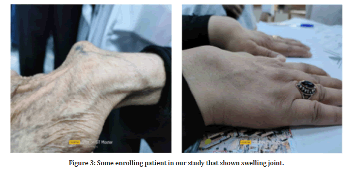
Figure 3 showing picture for some enrolling patient in our study that shown swelling joint.

Figure 3:Some enrolling patient in our study that shown swelling joint.
Results
A total of 100 RA patients and 30 healthy controls were enrolled in this study, the mean age for the patients and controls were (46.3 ± 11.3) and (46.4 ± 11.6) respectively with (P>0.05) with no deference between tow mean, statistical showed the patients 74 (74.0 %) female and 26 (26.0 %) were males and for the control group 22 (73.3%) were females and 8 (26.7%) were males, In our study viewed there are high enzyme concertation in human serum patient with Rheumatoid arthritis than the healthy control subject serum there are deference between patient mean and healthy control mean with highly statically significant (T-Value-6.58, P- Value<0.05) the 95% CI for Difference is (-5.333,-2.867). we had found there are a positive correlation between the increase serum glutathione serum level and high disease activity when we had study the correlation between parameters, with high statistically significant (P-Value<0.05) (Pearson correlation 0.265). In this study we had found there are a Significant decreased in serum vitamin C level in patient with RA than the control subjects, the deference between the mean of patient and control subject the statistical infertial shown that are highly statistically significant (P<0.05) with (T-Value=-9.628), The 95% Confidence Interval of the Difference (- 1.081,- 0.704), There are a significant moderate negative correlation between Vitamin C Serum level with the disease activity (DAS-28-ESR) (Pearson correlation-0.684, P<0.05).The statistical result had obtund from study showing there are low serum selenium level in patients with RA than the healthy control subjects with highly statistically significant (P<0.05),. the 95% CI (-52.31,-42.37) and the (T=-19.46) with highly statistically significant deference between tow means. Serum selenium level in RA patients had showing a negative correlation with disease activity by studying the correlation between Se level and DAS28- ESR(Pearson correlation-0.846, P<0.05), the correlation had statistically significant. The study descriptive statistics for parameter shown in Table 1 and Inferential statistics had mentioned above shown brifely in Table 2.
| Sample State | N | Mean | Std. Deviation | Std. Error Mean | |
|---|---|---|---|---|---|
| Glutathione reductase Enzyme (ng/ml) | Patient | 100 | 10.6225 | 5.213129 | 0.521313 |
| Healthy Control | 30 | 6.5227 | 1.867729 | 0.340999 | |
| Serum selenium level (µg/l) | Patient | 100 | 69.2323 | 1.78252 | 0.17825 |
| Healthy Control | 30 | 116.5797 | 13.28568 | 2.42562 | |
| Serum Vitamin C (mg/dl) | Patient | 100 | 0.43916 | 0.249317 | 0.024932 |
| Healthy Control | 30 | 1.33173 | 0.489043 | 0.089287 |
Table 1: Descriptive statistics for parameter through the enrolling subject.
| t-test for Equality of Means | |||||||
|---|---|---|---|---|---|---|---|
| t | df | Sig. (2-tailed) | Mean Difference | Std. Error Difference | 95% Confidence Interval of the Difference | ||
| Lower | Upper | ||||||
| Glutathione | 4.217 | 128 | 0 | 4.0998 | 0.972159 | 2.176218 | 6.023382 |
| reductase Enzyme (ng/ml) | |||||||
| Serum selenium level (µg/l) | -34.911- | 128 | 0 | -47.34737- | 1.35625 | -50.03094- | -44.66380- |
| Serum Vitamien C (mg/dl) | -13.408- | 128 | 0 | -.892562- | 0.066568 | -1.024279- | -.760846- |
Table 2: Inferential statistics (Independent sample T-test) for our study.
Discussion
This increase in GR serum level from damaged cell due to Oxidative Stress during chronic inflammation in RA joint [17,34,35]. Decreased in serum vitamin C level due to consumed the vitamin during Non- Enzymatic anti-oxidant action due to highly reactive oxygen species generated due to chronic inflammation in patient with RA [36] the high disease activity means there is high inflammation responded in patient, this high respond lead to formation more ROS [37], it has been hypothesized that the variation in Se levels in RA patients is due to a redistribution of this mineral from the circulation to the tissues as a defensive mechanism against Inflammation 38, nevertheless, could result in selenoprotein turnover and Se depletion Similar to this, it has been proposed that the liver's production of antiinflammatory protein may inhibit the growth of SeP, the protein responsible for transporting selenium, leading to a reduction in the circulation of selenium. It was important to note that nutrition has a significant impact on Se serum level [37,38].
Conclusions
Glutathione Reductase Enzyme gives a Picture for damaged cells due to Oxidative stress during chronic inflammation in Patient with RA and Vitamin C can be considering as approved parameter for oxidative stress due to inflammation respond that will consume by Non- Enzymatic Anti-Oxidant effect. the Evaluation Selenium level to show the activity of Glutathione peroxidase enzyme that show significant decrease, that will be consider low enzyme activity. Patient with RA is more prone to Oxidative Stress related disease than other patient with Autoimmune disease that haven’t high oxidative stress.
References
- Song X, Lin Q. Genomics, transcriptomics and proteomics to elucidate the pathogenesis of rheumatoid arthritis. Rheumatol Int 2017; 37:1257–1265.
- Firestein GS. Evolving concepts of rheumatoid arthritis. Nature 2003; 423:356–361.
- Sahebari M, Ayati R, Mirzaei H, et al. Serum trace element concentrations in rheumatoid arthritis. Biol Trace Elem Res 2016; 171:237–245.
- Lawrence RC, Helmick CG, Arnett FC, et al. Estimates of the prevalence of arthritis and selected musculoskeletal disorders in the United States. Arthritis Rheum 1998; 41:778–799.
- Turesson C, Jacobsson L, Bergstrӧm U. Extra-articular rheumatoid arthritis: Prevalence and mortality. Rheumatology 1999; 38:668–674.
- Gabriel SE, Crowson CS, O’Fallon M. The epidemiology of rheumatoid arthritis in Rochester, Minnesota, 1955-1985. Arthritis 1999; 42:415–420.
- Albers J, Kuper H, Van Riel P, et al. Socio-economic consequences of rheumatoid arthritis in the first years of the disease. Rheumatol 1999; 38:423–430.
- Arnett FC, Edworthy SM, Bloch DA, et al. The American Rheumatism Association 1987 revised criteria for the classification of rheumatoid arthritis. Arthritis 1988; 31:315–324.
- Yamanishi Y, Firestein GS. Pathogenesis of rheumatoid arthritis: The role of synoviocytes. Rheum Dis Clin North Am 2001; 27:355–371.
- Brand MD. Mitochondrial generation of superoxide and hydrogen peroxide as the source of mitochondrial redox signaling. Free Radical Biology Med Elsevier 2016; 100:14–31.
- Aydemir D, Hashemkhani M, Durmusoglu EG, et al. A new substrate for glutathione reductase: Glutathione coated Ag2S quantum dots. Talanta Elsevier 2019; 194:501–506.
- Meister A, Anderson ME. Glutathione. Annu Rev Biochem 1983; 52:711–760.
- Noctor G, Mhamdi A, Chaouch S, et al. Glutathione in plants: An integrated overview. Plant Cell Environ 2012; 35:454-484.
- Lu SC. Glutathione synthesis. Biochem 2013; 1830:3143–353.
- Gutteridge JM, Halliwell B. Free radicals and antioxidants in the year 2000: A historical look to the future. Ann N Y Acad Sci 2000; 899:136–147.
- Pai EF, Schulz GE. The catalytic mechanism of glutathione reductase as derived from x-ray diffraction analyses of reaction intermediates. J Biol Chem 1983; 258:1752–1757.
- Romero-Puertas MC, Corpas FJ, Sandalio LM, et al. Glutathione reductase from pea leaves: response to abiotic stress and characterization of the peroxisomal isozyme. New Phytol 2006; 170:43–52.
- Karplus PA, Schulz GE. Refined structure of glutathione reductase at 1.54 Å resolution. J Mol Bi 1987; 195:701–729.
- Lorestani S, Hashemy SI, Mojarad M, et al. Increased glutathione reductase expression and activity in colorectal cancer tissue samples: An investigational study in Mashhad, Iran. Middle East J Cancer 2018; 9:99–104.
- Schulz G, Schirmer RH, Sachsenheimer W, et al. The structure of the flavoenzyme glutathione reductase. Nature 1978; 273:120–124.
- Guaiquil VH, Vera JC, Golde DW. Mechanism of vitamin C inhibition of cell death induced by oxidative stress in glutathione-depleted HL-60 cells. J Biol Chem 2001; 276:40955–40961.
- Padayatty SJ, Levine M. Vitamin C. The known and the unknown and Goldilocks. Oral Dis 2016; 22:463.
- Frankenberger WT, Engberg RA. Environmental chemistry of selenium. CRC Press; 1998.
- Bodnar M, Konieczka P, Namiesnik J. The properties, functions, and use of selenium compounds in living organisms. J Environ Sci Health 2012; 30:225–252.
- Ursini F, Bindoli A. The role of selenium peroxidases in the protection against oxidative damage of membranes. Chem Phys Lipids 1987; 44:255–276.
- Turrubiates-Hernández FJ, Márquez-Sandoval YF, González-Estevez G, et al. The relevance of selenium status in rheumatoid arthritis. Nutrients 2020; 12:3007.
- Qamar N, John P, Bhatti A. Emerging role of selenium in treatment of rheumatoid arthritis: An insight on its antioxidant properties. J Trace Elem Med Biol 2021; 66:126737.
- Oropeza-Moe M, Wisløff H, Bernhoft A. Selenium deficiency associated porcine and human cardiomyopathies. J Trace Elem Med Biol 2015; 31:148–156.
- Kourilovitch M, Galarza-Maldonado C, Ortiz-Prado E. Diagnosis and classification of rheumatoid arthritis. J Autoimmun 2014; 48:26–30.
- Felson DT, Smolen JS, Wells G, et al. American College of Rheumatology/European league against rheumatism provisional definition of remission in rheumatoid arthritis for clinical trials. Arthritis Rheum 2011; 63:573-586.
- Greenmyer JR, Stacy JM, Sahmoun AE, et al. DAS28-CRP cutoffs for high disease activity and remission are lower than DAS28-ESR in rheumatoid arthritis. ACR Open Rheumatol 2020; 2:507–511.
- Orr CK, Najm A, Young F, et al. The utility and limitations of CRP, ESR and DAS28-CRP in appraising disease activity in rheumatoid arthritis. Front Med 2018; 5:185.
- Fransen J, van Riel PLCM. The disease activity score and the EULAR response criteria. Rheum Dis Clin North Am 2009; 35:745–757.
- Mulherin DM, Thurnham DI, Situnayake RD. Glutathione reductase activity, riboflavin status, and disease activity in rheumatoid arthritis. Ann Rheum Dis 1996; 55:837–840.
- Hassan M, Hadi R, Al-Rawi Z, et al. The glutathione defense system in the pathogenesis of rheumatoid arthritis. J Appl Toxicol 2001; 21:69–73.
- Mateen S, Moin S, Khan AQ, et al. Increased reactive oxygen species formation and oxidative stress in rheumatoid arthritis. PLoS One 2016; 11:e0152925.
- Das DC, Jahan I, Uddin M, et al. Serum CRP, MDA, vitamin C, and trace elements in Bangladeshi patients with rheumatoid arthritis. Biol Trace Elem Res 2021; 199:76–84.
- Afridi HI, Talpur FN, Kazi TG, et al. Estimation of toxic elements in the samples of different cigarettes and their effect on the essential elemental status in the biological samples of Irish smoker rheumatoid arthritis consumers. Environ Monit Assess 2015; 187:1–16.
Indexed at, Google Scholar, Cross Ref
Indexed at, Google Scholar, Cross Ref
Indexed at, Google Scholar, Cross Ref
Indexed at, Google Scholar, Cross Ref
Indexed at, Google Scholar, Cross Ref
Indexed at, Google Scholar, Cross Ref
Indexed at, Google Scholar, Cross Ref
Indexed at, Google Scholar, Cross Ref
Indexed at, Google Scholar, Cross Ref
Indexed at, Google Scholar, Cross Ref
Indexed at, Google Scholar, Cross Ref
Indexed at, Google Scholar, Cross Ref
Indexed at, Google Scholar, Cross Ref
Indexed at, Google Scholar, Cross Ref
Indexed at, Google Scholar, Cross Ref
Indexed at, Google Scholar, Cross Ref
Indexed at, Google Scholar, Cross Ref
Indexed at, Google Scholar, Cross Ref
Indexed at, Google Scholar, Cross Ref
Indexed at, Google Scholar, Cross Ref
Indexed at, Google Scholar, Cross Ref
Indexed at, Google Scholar, Cross Ref
Indexed at, Google Scholar, Cross Ref
Indexed at, Google Scholar, Cross Ref
Indexed at, Google Scholar, Cross Ref
Indexed at, Google Scholar, Cross Ref
Indexed at, Google Scholar, Cross Ref
Indexed at, Google Scholar, Cross Ref
Indexed at, Google Scholar, Cross Ref
Indexed at, Google Scholar, Cross Ref
Indexed at, Google Scholar, Cross Ref
Indexed at, Google Scholar, Cross Ref
Indexed at, Google Scholar, Cross Ref
Indexed at, Google Scholar, Cross Ref
Author Info
Muthanna Abd Al-Rubaie*, Hallah Ghazi Mahmood and Mohammed Hadi Munshed Alosami
Department of Clinical Biochemistry, College of Medicine, University of Baghdad, IraqReceived: 01-Sep-2022, Manuscript No. jrmds-22-74955; , Pre QC No. jrmds-22-74955(PQ); Editor assigned: 03-Sep-2022, Pre QC No. jrmds-22-74955(PQ); Reviewed: 19-Sep-2022, QC No. jrmds-22-74955(Q); Revised: 23-Sep-2022, Manuscript No. jrmds-22-74955(R); Published: 30-Sep-2022
