Research - (2021) Volume 9, Issue 9
The Relationship between the Maxillary Sinus and Dental Root Apices Using Cone-Beam Computed Tomography (CBCT)
Amani Mirdad1*, Razan Alaqeely1, Sumayah Ajlan1, Nahed Ashri1 and Mazen Aldosimani2
*Correspondence: Amani Mirdad, Faculty in Periodontics, College of Dentistry, King saud university, Saudi Arabia, Email:
Abstract
The maxillary sinus is an essential anatomical structure in close relation to maxillary teeth roots. The aim of the present study is to evaluate the anatomic proximity of the maxillary posterior roots’ apices to the maxillary sinus floor using cone-beam CT in a population attending King Saud University. Materials and methods: CBCT images for patients attending the dental school of king Saud university were screened, and images containing maxillary sinuses were evaluated. The relation between teeth roots and sinus floor was divided into Root tips penetrating the sinus (In the sinus, Type 1), Root tips in contact with the sinus floor (On the sinus, Type 2), and Root tips below the sinus floor (Type 3). Results: around 288 scans were included. The average age was 40.63 ±6.53. Root tips contacting the sinus floor (root on the sinus) formulated the largest category (45.1%). A strong correlation was found between all age groups and root relation to sinus (p<0.000), with most roots penetrating the sinus belongs to younger patients. Around (41.7%) of molar roots were in direct contact with the sinus floor (on the sinus), while 50% of premolar roots had no relation to the sinus floor. Conclusion: Molar roots appear closer to the sinus floor than premolars, with age appearing to influence the root relation to the sinus.
Keywords
Maxillary sinus, Maxillary molars, Maxillary premolars, Roots contacting the sinus, Roots into the sinus, Cone-beam computed tomography (CBCT).
Introduction
The maxillary sinus is a major anatomical structure positioned in the midfacial region next to the nasal cavity and close to posterior maxillary teeth root apices [1]. It is pyramidal in shape, and its floor is formed by the alveolar process of the maxilla. The sinus walls are lined by the Schneiderian membrane, which is changeable during relevant disease development [2].
Maxillary sinuses are the first Para nasal sinuses to develop. A common sinus phenomenon is sinus pneumatisation, which indicates an increase in maxillary sinus volume. Studies reported that the maxillary sinus volume is dynamic and controlled through metabolic processes leading to size increase throughout the person's growth. Development is considered complete around the age of 20 years after the complete eruption of maxillary third molars [3,4]. Further pneumatisation may still occur and is often associated with pathological processes or teeth extraction [5,6].
The adult sinus extension varies between people [7]. The maxillary sinus floor might extend between adjacent teeth or roots, creating what are commonly known as ‘hillocks’ [8]. Hillocks are defined as elevations in the antral surface. Roots of maxillary molars, premolars, and often maxillary canines might project into the maxillary sinus [9]. This close anatomical proximity of the root apices to maxillary sinus floor (MSF) can result in the spread of periapical or periodontal infection beyond the confines of the supporting dental tissue into the maxillary sinus itself, causing sinusitis [7,10,11]. In addition, root canal therapy or extraction of these teeth can result in sinus penetration [12], oroantral fistulae, or root displacement into the sinus cavity [13]. Orthodontic tooth movement against cortical walls of the sinus might also be challenging and require careful planning [14]. Further, the location geometry and volume of the maxillary sinus should be carefully evaluated before surgical placement of dental implants in the maxillary arch [1].
Accordingly, a proper understanding of the relationship between the root apices of maxillary teeth and the maxillary sinus floor (MSF) is of great importance for preventing complications and selecting the best treatment options. Several authors have attempted to evaluate this relationship. Some studies have reported that around 10% to 36.7% of maxillary molar roots protrude into the sinus [15,16]. Other studies, however, have attempted to measure the distance between root tips and sinus floor, and correlate it with age, gender, type of teeth, and history of edentulism with different results [17-19].
Maxillary sinus anatomy can be evaluated through cadaveric dissection, clinical intra-operative evaluation, and the use of different radiographic modalities [20]. Radiographic imaging can range from simple two-dimensional images (periapical and panoramic radiographs) to more advanced three-dimensional modalities, including cone-beam computed tomography (CBCT). Cone-beam computed tomography (CBCT) provides high-quality cross-sectional images of oral and maxillofacial regions. It can thus more precisely evaluate the relationship between the maxillary root apices and the maxillary sinus [21-23].
Therefore, the current study aimed to evaluate the anatomic proximity of the maxillary posterior roots apices to the maxillary sinus floor using cone-beam computed tomography (CBCT) in a population attending King Saud University and evaluate the impact of age and gender on this relation.
Materials and Methods
This retrospective study was approved by the Institutional Review Board (IRB) of King Saud University Medical City (KSUMC) E-20-4695.
For case selection, high-quality CBCT scans of patients obtained between January 2018 to September 2020 were screened from the radiology department of Dental University Hospital, King Saud University. The inclusion criteria were:
• Clear radiographic images that were covering a complete bilateral maxillary sinus and maxillary molars area.
• Adult patients (>18 years).
• Complete root development of maxillary second premolars and molars, excluding the third molar.
• Images showing posterior maxillary teeth, excluding images with loss of both molars and premolars.
• Absence of pathological changes, e.g., maxillary sinus mucosal thickening, maxillary sinus inflammation, cyst or tumours, malformations, and maxillary fractures.
Scanning and analysis
Computed tomography scans were obtained using CBCT scanner (ProMax 3D Mid, Planmeca, USA) with 110kVp, 3.6–4.8 mA, a voxel size of 0.2 mm, and a field of view of 12×8 cm or 15 X 15 cm settings. Scans were viewed and analyzed using Romexis software (Planmeca Romexis, Planmeca, USA).
Evaluations were done by two calibrated examiners (A.M, RA). Calibration was done by interpreting 10 CBCT images for intra and inter-examiner reliability.
The relationship between maxillary teeth roots and maxillary sinus was evaluated, where the relationship of the first maxillary molar root tips (#16 or 26) was selected to represent the molar teeth, and if the 1st molar was missing, it was replaced by the second molar. For the premolars, the comparison depended on the second maxillary premolar (#15, 25), with no compensation done if that tooth was missing. The relationship between maxillary teeth roots and maxillary sinus was determined in sagittal and coronal planes. The brightness and contrast of the images were adjusted to ensure optimal visualization as needed.
The relation between teeth roots and sinus floor was divided as follows:
• Type 1: Root tips penetrating the sinus (In the sinus).
• Type 2: Root tips in contact with the sinus floor (On the sinus).
• Type 3: Root tips below the sinus floor (No relation with sinus floor).
Statistical analysis
Statistical analysis was performed using SPSS Statistics, version 22.0 (IBM Corp., Armonk, NY, USA). Age and gender were analysed to determine association with radiographic findings using nonparametric tests. A pvalue of <0.05 was considered statistically significant.
Results
A total of (288) patient scans met the inclusion criteria and were analysed. Most of the cases were for females (n= 206, 71.5%), while male patients presented only 28.5% of the sample.
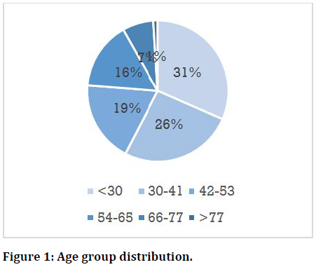
The population age ranged between 18 to 84 years old, with an average of 40.63 ±6.53. Most patients were younger than 30 (Figure 1).
Figure 1. Age group distribution.
The relationship between Sinus and root tips
Root tips contacting the sinus floor (Type 2) formulated the most significant category (45.1%). This was followed by root tips away from the sinus floor (Type 3), which was the case for around one-third of the examined population (33.7%). The root tips appeared penetrating the sinus (Type 1) in only 21% of the population. Table 1 explains the distribution of this relationship.
| Root relation to sinus | Number of cases (%) |
|---|---|
| Type 1: Roots in sinus | 61 (21.2%) |
| Type 2: Roots on sinus | 130 (45.1%) |
| Type 3: No relation between roots and sinus | 97 (33.7%) |
| Total | 288 (100%) |
Table 1: Distribution of the relationship between roots and sinus.
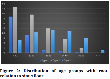
A strong correlation was found between all age groups and root relation to sinus (p<0.000). Type 2 relation was highest in younger age groups and statistically significant for the same age group compared to the other types (P<0.00) (Figure 2). Type 1 relation was statistically significantly lower in groups older than 30 years (P<0.00). The gender was not significant with different root relations to sinus (P<0.14).
Figure 2. Distribution of age groups with root relation to sinus floor.
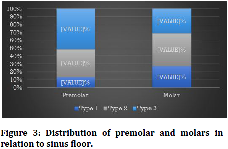
For the 288 patients, a total of 576 molar teeth were evaluated. However, there were only 567 premolar teeth present for the evaluation. Figure 3 shows the distribution of teeth in relation to the sinus.
Figure 3. Distribution of premolar and molars in relation to sinus floor.
Around (41.7%) of molar roots were in direct contact with the sinus floor (Type 2); however, no significant difference was found regarding the different categories of molar root tip relation to the sinus (p<0.1).
On the other hand, around 50% of premolar roots did not contact the sinus floor (type 3) compared to other types (p<0.03). Comparing molars to premolars, molars had more of type 1 and 2 compared to premolars with no significant difference except for type 3 where difference was significant (p<0.01).
Discussion
The maxillary sinus represents a crucial anatomical structure commonly present in the field of operation of the dentist. Proper knowledge of the location of maxillary teeth and the relation of their root tips are of great importance for proper case management and treatment planning [24]. The present study aimed at the evaluation of this relationship among the Saudi population.
In this study, the maxillary molars had more root tips at the sinus floor or penetrating the sinus than premolar teeth. Several studies found maxillary molar root tips closer to the sinus than premolars [15,24,25]. However, they varied in the determination of the exact tooth (1st Vs. 2nd molar) and in the identification of the exact root that is closest to the sinus floor (mesiobuccal, distobuccal, palatal) [15,24,26,27].
Regarding the premolars, several studies have reported that the first premolar rarely contacted the sinus [18,28]. Additionally, von Arx, et al. [18] found that age, gender, side of the mouth, and the presence or absence of premolars did not significantly influence the mean distances between premolar roots and the maxillary sinus. In fact, the only possible effect was associated with the absence of maxillary first molars [19].
Our findings indicated a correlation between age groups and contact with the sinus, where most roots penetrating the sinus floor belonged to the younger age group. Similar findings were reported by multiple other authors [17,19,29]. The gender did not affect the relation of the roots as well, and this was the case in Gu, et al. [19] study where no difference was found in the distance between the maxillary molars and maxillary sinus floor, according to both sex and side [19].
The relation of maxillary roots to the sinus floor was measured differently in previous research [25]. Despite the diversity, it is of great importance to carefully assess the relation of the roots to the sinus in cases of endodontic surgical and non-surgical treatment, dental implant placement, extraction complications, and sinus pathology [27].
Conclusion
Root relation to MSF is of paramount importance. The largest group of cases had root tips associated with the sinus floor (on the sinus). Molar roots appear closer to the sinus floor than premolars, with age influencing the root relation to the sinus. Careful evaluation of tooth relation to the sinus is highly recommended during all dental procedures.
References
- Misch C. Contemporary implant dentistry. 2nd Edn. 1999.
- Lu Y, Liu Z, Zhang L, et al. Associations between maxillary sinus mucosal thickening and apical periodontitis using cone-beam computed tomography scanning: A retrospective study. J Endod 2012; 38:1069–74.
- Asaumi R, Sato I, Miwa Y, et al. Understanding the formation of maxillary sinus in Japanese human foetuses using cone beam CT. Surg Radiol Anat 2010; 32:745–51.
- Alfaraje L. Surgical and radiologic anatomy for oral implantology. US: Quintessence. 2013.
- Sharan A, Madjar D. Maxillary sinus pneumatization following extractions: a radiographic study. Int J Oral Maxillofac Implant 2008; 23:48–56.
- Alqahtani S, Alsheraimi A, Alshareef A, et al. Maxillary Sinus Pneumatization following extractions in Riyadh, Saudi Arabia: A cross-sectional study. Cureus 2020; 12:e6611.
- Hauman C, Chandler N, Tong D. Endodontic implications of the maxillary sinus: A review. Int Endod J 2002; 35:127-41.
- Waite D. Maxillary sinus. Dent Clin North Am 1971; 15:349-368.
- Tank P. Grant’s Dissector. 13 Edn. Philadelphia:: Lippincott Williams & Wilkins 2005.
- Engstrom H, Chamberlain D, Kiger R, et al. Radiographic evaluation of the effect of initial periodontal therapy on thickness of the maxillary sinus mucosa. J Periodontol 1988; 59:604-698.
- Monkhouse S. Cranial Nerves functional anatomy. 2nd Edn. New York: Cambridge University Press 2006.
- Williams P, Bannister L, Berry M. Gray's anatomy. 38th Edn. New York: Churchill-Livingstone 1995.
- Harrison D. Oro-antral fistula. Br J Clin Pract 1961; 15:169-174.
- Sun W, Xia K, Huang X, et al. Knowledge of orthodontic tooth movement through the maxillary sinus: a systematic review. BMC Oral Health 2018; 18:91.
- Kilic C, Kamburoglu K, Yuksel S, et al. An assessment of the relationship between the maxillary sinus floor and the maxillary posterior teeth root tips using dental cone-beam computerized tomography. Eur J Dent 2010; 4:462–467.
- Tian X, Qian L, Xin X, et al. An analysis of the proximity of maxillary posterior teeth to the maxillary sinus using cone- beam computed tomography. J Endod 2016; 42:371–377.
- Ok E, GuÌ?ngoÌ?r E, Colak M, et al. Evaluation of the relationship between the maxillary posterior teeth and the sinus floor using cone-beam computed tomography. Surg Radiol Anat 2014; 36:907–914.
- von Arx T, Fodich I, Bornstein M. Proximity of premolar roots to maxillary sinus: A radiographic survey using cone-beam computed tomography. J Endod 2014; 40:1541–1548.
- Gu Y, Sun C, Wu D, et al. Evaluation of the relationship between maxillary posterior teeth and the maxillary sinus floor using cone-beam computed tomography. BMC Oral Health 2018; 18:164.
- Neugebauer J, Ritter L, Mischkowski RA, et al. Evaluation of maxillary sinus anatomy by cone-beam CT prior to sinus floor elevation. Int J Oral Maxillofac Implant 2010; 25:258-65.
- Newman MG, Takei HH, Klokkevold PR, et al. Newman and carranza’s clinical periodontology. 13th Edn. St. Louis, MO: Saunders Elsevier 2012.
- Bornstein MM, Horner K. Use of cone-beam computed tomography in implant dentistry: current concepts, indications, and limitations for clinical practice and research. Periodontol 2017; 73:51-72.
- Baheerati MM, Don KR. Applications of cone-beam computerized tomography in dental practice: A brief review. Drug Invention Today 2019; 11:214-218.
- Shaul Hameed K, Abd Elaleem E, Alasmari D. Radiographic evaluation of the anatomical relationship of maxillary sinus floor with maxillary posterior teeth apices in the population of Al-Qassim, Saudi Arabia, using cone beam computed tomography. Saudi Dent J 2020.
- Kwak HH, Park HD, Yoon HR, et al. Topographic anatomy of the inferior wall of the maxillary sinus in Koreans. Int J Oral Maxillofac Surg 2004; 33:382-388.
- Jung Y, Cho B. Assessment of the relationship between the maxillary molars and adjacent structures using cone beam computed tomography. Imaging Sci Dent 2012; 42:219–224.
- Lupoi D, Dragomir M, Coada G, et al. CT scan evaluation of the distance between maxillary sinus floor and maxillary teeth apices. Romanian J Rhinol 2021; 11:18-23.
- Lavasani SA, Tyler C, Roach SH, et al. Cone-beam computed tomography: Anatomic analysis of maxillary posterior teeth-impact on endodontic microsurgery. J Endod 2016; 42:890-895.
- Qin Y, Shu G. Evaluation of the relationship between maxillary sinus wall and maxillary canines and posterior teeth using cone-beam computed tomography. Med Sci Monit 2020; 26.
Author Info
Amani Mirdad1*, Razan Alaqeely1, Sumayah Ajlan1, Nahed Ashri1 and Mazen Aldosimani2
1Faculty in Periodontics, College of Dentistry, King saud university, Saudi Arabia2Faculty in Radiology, College of Dentistry, King saud university, Saudi Arabia
Citation: Amani Mirdad, Razan Alaqeely, Sumayah Ajlan, Nahed Ashri, Mazen Aldosimani,The Relationship between the Maxillary Sinus and Dental Root Apices Using Cone-Beam Computed Tomography (CBCT), J Res Med Dent Sci, 2021, 9(9): 5-9
Received: 07-Aug-2021 Accepted: 31-Aug-2021


