Research - (2020) Volume 8, Issue 6
The Occlusion Time Evaluation in Iraqi Patients with TMJ Internal Derangement Utilizing T-Scan (NOVUS) System
Zena Kamel Kadhem* and Fawaz Aswad
*Correspondence: Zena Kamel Kadhem, Department of Oral Medicine, College of Dentistry, Mustansiriyah University, Baghdad, Iraq, Email:
Abstract
The aim of the study is to evaluate the occlusion time is prolonged in patients with TMJ internal derangement (TMJ ID) as compared to healthy control subjects. Materials and Methods: Subjects with full dentition and angle class I relation. DC/TMD criteria was used to diagnose the patients, the occlusion time OT registered in the maximum intercuspation digitally by using T-Scan NOVUS device. Results: Non-significant differences between the age groups, disc displacement with reduction is the more prevalent disorder. Highly significant differences between the participant’s genders and the female demonstrated the higher percentage of the TMJ ID patients. Highly significant difference in the occlusion time is reported between the TMJ ID patients and the healthy control. Conclusion: In this study, the Disc displacement with reduction is the more prevalent and females represent the higher percentage in having TMJ internal derangement. Prolonged occlusion time was reported in both healthy control and the patients with signs and symptoms of TMJ internal derangement. But it was higher in the TMJ ID patients with a significant difference. Regarding the different conditions of disc displacement, there were a non-significant difference in OT between them.
Keywords
Occlusion time, T-scan, Internal derangement, Disc displacement, TMD, OT
Introduction
The masticatory system is made up of the teeth, periodontal tissues, masticatory muscles, and temporomandibular joint (TMJ). A balanced dental occlusion plays an important role in the healthy functioning of the masticatory system [1]. Normal occlusal and the articular relations between the jaws ensure balanced distribution of the generated forces in them during mastication [2]. TMJ disc displacement represented abnormal relationship between the disc and the head of the condyle. Four common stages of internal derangements of the TMJ are: 1. Vibrating loose capsules that have nondisplacing disks. 2. Partial disk displacements with reduction. 3. Complete disk displacements with reduction and 4. Nonreducing permanent disk displacements [3].
The T-Scan system provided a result that can be easily reproducible and documented the occlusal contacts, occlusal forces, and occlusal times quantitatively and dynamically, even during a continuous movement of the mandible [4,5]. The time reported from the first occlusal contact until reaching the maximum intercuspation is known as Occlusion Time (OT) [6]. The length of Occlusion Time is clear to be correlated to the existence of occlusal instability, premature occlusal contacts, and occlusal interferences. Additionally, the computerized system can display the relative occlusal force variance from the first point of contact to MIP, in real time [7].
Materials and Methods
The participants were recruited from the attendants to the teaching clinic of oral medicine in the teaching hospital of College of Dentistry/ University of Baghdad during the period from April 2019 to January 2020. All participants were subjected to questionnaire about name, age, past dental treatment, medical history, and medication used. The diagnosis of the patients was established according to the diagnostic criteria for temporomandibular joint disorders, clinical protocol, and assessment instrument [8]. The study protocol was approved by the ethical committee of the College of Dentistry/ University of Baghdad. An informed consent was obtained from the patients.
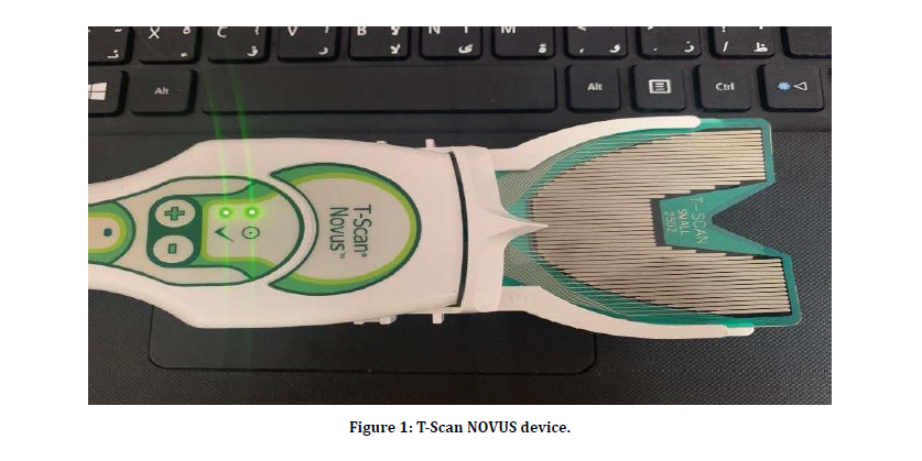
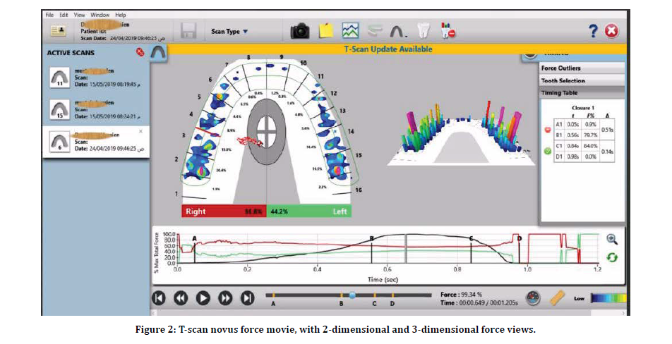
Digital evaluation of occlusion time in the maximum intercuspation of all the included subjects was performed using the T-Scan NOVUS system (T-Scan, Tekscan, Inc., S. Boston, MA, USA) system, shown in Figure 1. These clinical examinations were done by one examiner and supervised by special expert. The inclusion criteria were: Subjects of both groups (patients and controls) should have full dentition with Angle class I relation. Good general health with no history of any systemic diseases. Patients should not have ongoing treatment (medication or occlusal splint) or recently treated from TMJ disorders. Figure 2 show the T-scan Novus timed and force movie in the maximum intercuspation position of patients with TMJ internal derangement.

Figure 1: T-Scan NOVUS device.

Figure 2: T-scan novus force movie, with 2-dimensional and 3-dimensional force views.
Statistical data analysis approaches were demonstrated by the application of the statistical package (SPSS) ver. (22.0). Descriptive data analysis presented in Mean value and Standard Deviation. ANOVA test, T-test, and Least Significant Difference-LSD test with Games Howell-GH test were used for data analysis. Significant at P<0.05.
Results
One hundred and nine (109) subjects were participated in this study with age range (19-45 years old) and divided into two main groups: study group (show signs and symptoms of internal derangement ID) with (84) patients (64:20 females: male number) and (25) healthy controlled free from signs and symptoms of TMD with (14:11 female: male number). Table 1 illustrated the demographical characteristics variables, as well as comparisons significant of TMJ ID group with control group. Nonsignificant difference at P>0.05 in age between patients and control while highly significant difference at P<0.01 demonstrated in gender between the two-study group. In addition to that, gender distribution concerning TMJ ID group showed that highly significant difference was accounted at P<0.01, and TMD is higher in females (76.2%) than males (23.8%)., whereas the control group revealed a non- significant differences. Regarding the TMJ ID group, the group is subdivided into four subgroups, included:
| Characteristics | TMJ ID No=84 | Control No=25 | ||||
| No | % | No | % | 0.779 | ||
| Age (years) | <20years | 12 | 14.3 | 6 | 24 | |
| 20---29 | 26 | 31 | 7 | 28 | ||
| 30---39 | 33 | 39.3 | 9 | 36 | ||
| =>40years | 13 | 15.5 | 3 | 12 | ||
| Mean±SD | 29.82 ± 7.90 (19-45) | 29.30 ± 7.80 (19-45) | ||||
| Gender | Male | 20 | 23.8 | 11 | 44 | 0.002* |
| Female | 64 | 76.2 | 14 | 56 | ||
| Total | 84 | 100 | 25 | 100 | ||
| P-value | 0.000* | 0.69 | ||||
(*) TMJ ID (Temporomandibular joint Internal derangement); HS: Highly Significant at P<0.01; S: Significant at P<0.05; NS: Non Significant at P>0.05; Testing based on ANOVA test, T-test.
Table 1: Distribution of the demographical characteristic’s variables for the tmd and control groups with comparison's significant.
Regarding the ID patients, the group is subdivided into four subgroups, included:
Group1: includes 26 (30.95%) patients with a disc displacement with reduction, 4(15.4%) were males and 22 (84.6%) were females.
Group 2: includes 22 (26.19%) patients with disc displacement with reduction, with intermittent locking, 5 (22.7%) were males and 17 (77.3%) were females.
Group 3: includes 21 (25%) patients with disc displacement without reduction, without limited opening; 5 (23.8%) were males and 16 (76.2%) were females.
Group 4: includes 15 (17.86%) patients with disc displacement without reduction, with a limited opening, showed 6(40%) were males and 9 (60%) were females.
Figure 2 represents a summary statistic for (occlusion time OT) parameter concerning studied groups in the (Maximum Intercuspation MIC). Data were presented as Mean ± SD. Table 2 represents a data statistic of (Occlusion time test) parameter, with respect to (MIC location), result showed a highly significant difference accounted at P<0.01 and according to the obvious results, (Least Significant Difference- LSD test, and Games Howell-GH test) illustrated in the Table 3 the multiple comparisons of (Occlusion time test) between the studied four TMD subgroups and with the control group at the studied location.
| Locations | Groups | Mean ± SD | t-test | P-value (*) |
|---|---|---|---|---|
| MIC | ID | 0.59±0.41 | 3.547 | 0.001 |
| Control | 0.35±0.25 |
(*) Highly Significant at P< 0.01; Significant at P< 0.05; Non-Significant at P>0.05; t-test for testing equality of means of two independent groups.
Table 2: Descriptive Statistics of (Occlusion time) parameter in the studied groups distributed for different locations.
| Studied Locations | MIC | ||
|---|---|---|---|
| Sig. | C.S. (*) | ||
| Group 1 | Group 2 | 0.519 | NS |
| Group 3 | 0.954 | NS | |
| Group 4 | 0.889 | NS | |
| Control | 0.438 | NS | |
| Group 2 | Group 3 | 0.909 | NS |
| Group 4 | 0.97 | NS | |
| Control | 0.04 | S | |
| Group 3 | Group 4 | 0.999 | NS |
| Control | 0.196 | NS | |
| Group 4 | Control | 0.164 | NS |
(*) S: Sig. at P>0.05; NS: Non-Sig. at P>0.05; Testing based on LSD, and GH tests.
Table 3: All probable pair's comparisons by using (LSD, and GH) tests among studied groups for studied locations.
Results showed that (MIC) location accounted no significant difference compared among multiple comparisons at P>0.05. except between group 2 and controlled since significant difference was accounted at P<0.05.
Discussion
Disc displacement with reduction was the most prevalent group which diagnosed in the present study. The present result is like previous studies which mentioned that the disc displacement with reduction is more common than other groups of internal derangement disorders [9].
This study presented the internal derangement clicking in female more than male. These results agree with similar results reported by [10-15]. The pattern of onset of TMD after puberty and lowered prevalence rates in the postmenopausal years of female suggest that female reproductive hormones may play an etiologic role in temporomandibular disorders [16]. This is also supported by the longitudinal data reported by Magnusson [17]. The prevalence of disc displacement with reduction is higher in female patients which had been also reported by previous study [18]. This fact may derive from the influence of some female-specific characteristics such as greater joint laxity, [19] and greater intra-articular pressure [20].
The present study showed that no differences in age groups. The average age of patients with internal derangement clicking in the present study was close to studies done by [15,21-23]. This result concluded that younger individuals run a greater risk of having precipitating TMJ noises.
As a part of inspection in the TMJ disorders it is difficult to correlate signs and symptoms of TMD and dental occlusion because all the published papers in this field are not well defined and showed a disparity of results, and is likely resulted from the low reproducibility of variables assessment methods of the dental occlusion.
The T-Scan system provided a result that can be easily reproducible and documented the occlusal contacts, occlusal forces, and occlusal times quantitatively and dynamically, even during a continuous movement of the mandible [4,5,24]. Factors influences the length of Occlusion Time showed correlated to the existence of occlusal instability, premature occlusal contacts, and occlusal interferences. In this study, patients with TMJ internal derangement in comparable with control group (free from signs and symptoms of TMD) showed significant differences. In fact, the patients complained from signs and symptoms of Intraarticular disorders had Occlusion Times about longer than the control. Occlusion Time is causally related with patients’ occlusal contact pattern [5] and has been considered as a capable description of occlusion [25,26]. According to the manufacturer, Occlusion Time is recommended as less than 0.2 seconds [6].
Generally, Occlusion Time (OT) in patients with TMJ internal derangement were consistently longer than control. These results were in agreement with many previous studies [5,26-30]. More precisely, [26,30] found significantly high occlusion time in participants with certain signs and symptoms of TMD and specifically intra-articular joint disorders as compared to the control group. The reported results suggest that there is a deterioration in occlusal stability in subjects with TMD.
Haralur et al. [27] reported a significant difference between TMD patients and control regarding OT in MIC position. The average OT in normal dentate subjects with healthy TMJs in Haralur study where longer than registered in this study. These discrepancies may be probably explained by individual difference. The findings of this study were partly in disagreement with Dzingute et al. [29] when they also reported that TMD patients showed higher OT but with no significant differences from controls. The results of OT in this study agreed with study done by Baldini et al. [28] who reported the occlusion time longer in patients with TMDs and the results were statistically significant. Baldini et al. [28] and Cheng et al. [25] reported OT in the MIC position like OT in the healthy control of this study. Ciavarella et al. [31] also reported the asymmetry in the occlusal force in TMD (intracapsular joint) disorders.
The disparity of results may be attributed inhomogeneity in population samples and diversity among data collection procedures. Larger size of the sample could have influences higher average values, but it still can be noted that a longer occlusion time was recorded in patients with temporomandibular joint disorders.
Conclusion
In this study, Prolonged occlusion time was reported in both healthy control and the patients with signs and symptoms of TMJ internal derangement. But it was higher in the ID patients with a significant difference. Regarding the different conditions of disc displacement, there were a non-significant difference in OT between them.
References
- Agbaje JO, Casteele EV de, Salem AS. Assessment of occlusion with the T-Scan system in patients undergoing orthognathic surgery. Sci Rep 2017; 7:53-56.
- Bozhkova TP. The T-SCAN system in evaluating occlusal contacts. Folia Medica 2016; 58:122-130.
- Kuwahara T, Bessette RW, Maruyama T. Characteristic chewing parameters for specific types of temporomandibular joint internal derangements. Cranio 1996; 14:12-22.
- Kerstein R, Chapman R, Klein MA. A comparison of ICAGD to mock ICAGD for symptom reductions in chronic myofascial pain dysfunction patients. Cranio J Craniomandibular Practice. 1997; 15:21-37.
- Wang C, Yin X. Occlusal risk factors associated with temporomandibular disorders in young adults with normal occlusions. Oral surgery, oral medicine, oral pathology and oral radiology. 2012; 114:419-423.
- Kerstein R, Grundset K. Obtaining measurable bilateral simultaneous occlusal contacts with computer-analyzed and guided occlusal adjustments. Quintessence Int 2001; 32:7-18.
- Qadeer S, Kerstein R, Kim RJ, et al. Relationship between articulation paper mark size and percentage of force measured with computerized occlusal analysis. J Adv Prosthodont 2012; 4:7–12.
- Schiffman E, Ohrbach R, Truelove E, et al. Diagnostic criteria for temporomandibular disorders (DC/ TMD) for clinical and research applications: Recommendations of the international RDC/TMD consortium network and orofacial pain special interest group. J Oral Facial Pain Headache 2014; 28: 6–27.
- Manfredini D, Chiappe G, Bosco M. Research diagnostic criteria for temporomandibular disorders (RDC/TMD) axis I diagnoses in an Italian patient population. J Oral Rehabil 2006; 33:551-558.
- Farsi NM. Symptoms and signs of temporomandibular disorders and oral parafunctions among Saudi children. J Oral Rehabil 2003; 30:1200-1208.
- Feteih RM. Signs and symptoms of temporomandibular disorders and oral parafunctions in urban Saudi Arabian adolescents: A research report. Head Face Med 2006; 2:25.
- Poveda Roda R, Bagan JV, Díaz Fernández JM, et al. Review of temporomandibular joint pathology. Part I: Classification, epidemiology and risk factors: Review. Med Oral Patol Oral Cir Bucal 2007; 12:292-298.
- Troeltzsch M, Cronin RJ, Brodine AH, et al. Prevalence and association of headaches, temporomandibular joint disorders, and occlusal interferences. J Prosthet Dent 2011; 105:410–417.
- Bagis B, Ayaz EA, Turgut S, et al. Gender difference in prevalence of signs and symptoms of temporomandibular joint disorders: A retrospective study on 243 consecutive patients. Int J Med Sci 2012; 9:539-544.
- Leissner O, Yanez MM, Bella WM, et al. Assessment of mandibular kinematics values and its relevance for the diagnosis of temporomandibular joint disorders. J Dent Sci 2020; 15.
- Nekora-Azak A. Temporomandibular disorders in relation to female reproductive hormones a literature review. J Prosthet Dent 2004; 91:491-493.
- Magnusson T, Egermark I, Carlsson GE. A prospective investigation over two decades on signs and symptoms of temporomandibular disorders and associated variables. A final summary. Acta Odontol Scand 2005; 63:99-109.
- Lazarin RO, Previdelli IT, Silva RS, et al. Correlation of gender and age with magnetic resonance imaging findings in patients with arthrogenic temporomandibular disorders: a cross-sectional study. Int J Oral Maxillofac Surg 2016; 45:1222-1228.
- McCarroll RS, Hesse JR, Naeije M, et al. Mandibular border positions and their relationships with peripheral joint mobility. J Oral Rehabil 1987; 14:125-131.
- Nitzan DW. Intraarticular pressure in the functioning human temporomandibular joint and its alteration by uniform elevation of the occlusal plane. J Oral Maxillofac Surg 1994; 52:671-679.
- Kamisaka M, Yatani H, Kuboki T, et al. Four-year longitudinal course of TMD symptoms in an adult population and the estimation of risk factors in relation to symptoms. J Orofac Pain 2000; 14:224–232
- Köhler AA, Hugoson A, Magnusson T. Prevalence of symptoms indicative of temporomandibular disorders in adults: Cross-sectional epidemiological investigations covering two decades. Acta Odontologica Scandinavica 2012; 70:213-223.
- Maixner W, Diatchenko L, Dubner R. Orofacial pain prospective evaluation and risk assessment study—The oppera study. J Pain 2011; 12:T4-T11.
- Sutter B, Girouard P, Radke J, et al. A review of comparison between conventional and computerized methods in the assessment of an occlusal scheme. Adv Den Tech 2020; 2:84-89.
- Cheng HJ, Geng Y, Zhang FQ. The evaluation of intercuspal occlusion of healthy people with T-Scan II system. Shanghai J Stomatology 2012; 21:62–65.
- Baldini A, Nota A, Cozza P. The association between occlusion time and temporomandibular disorders. J Electromyogr Kinesiol 2015; 25:15-154.
- Haralur SB. Digital Evaluation of functional occlusion parameters and their association with temporomandibular disorders. J Clin Diagnostic Res 2013; 7:1772–1775.
- Baldini A, Nota A, Coza P. The association between occlusion time and temporomandibular disorders. J Electromyogr Kinesiol 2014; 25:151-154.
- Dzingute A, Pileicikiene G, Baltrusaityte A. Digital occlusal evaluation in patients with temporomandibular joint disorders. Mol Med Ther 2017; 1:8-13.
- Jivnani HM, Tripathi S, Shanker R, et al. A study to determine the prevalence of temporomandibular disorders in a young adult population and its association with psychological and functional occlusal parameters. J Prosthodont 2019; 28:e445-e449.
- Ciavarella D, Parziale V, Mastrovincenzo M, et al. Condylar position indicator and T-scan system II in clinical evaluation of temporomandibular intracapsular disease. J Craniomaxillofac Surg 2012; 40:449-455.
Author Info
Zena Kamel Kadhem* and Fawaz Aswad
Department of Oral Medicine, College of Dentistry, Mustansiriyah University, Baghdad, IraqCitation: Meer Zena Kamel Kadhem, Fawaz Aswad, The Occlusion Time Evaluation of in Iraqi Patients with TMJ Internal Derangement Utilizing T-Scan (NOVUS) System, J Res Med Dent Sci, 2020, 8 (6): 77-82.
Received: 19-Aug-2020 Accepted: 17-Sep-2020
