Research Article - (2022) Volume 10, Issue 8
The Effect of Three Different Obturation Techniques on Push Out Bond Strength of Bioceramic (Total Fill) and AH Plus Sealers
Batool Basim Mounes* and Raghad A Al-Hashimi
*Correspondence: Batool Basim Mounes, Department of Endodontics, University of Baghdad, Baghdad, Iraq, Email:
Abstract
Background: The adhesion strength of the sealer and the appropriate technique used with it is important to achieve single adhesion unite (mono block) and prevent any leakage.
The aim of the study to evaluate and compare the efficacy of different obturation techniques Cold Lateral Compaction (CLC), Single Cone (SC), carrier based obturation techniques (Gutta Core) (GC) on push out bond strength of bio ceramic (total fill) and AH plus sealers at different root levels.
Materials and methods: Sixty maxillary first molars with a straight round palatal root canal, after sectioning of the palatal roots, the canal were instrumented with Edge Endo X7 Rotary system files up to(40/04)Then the samples divided into two groups according to sealer used A group "(bioceramic sealer)" B group "AH PLUS sealer" each group subdivided into 3 subgroup according to obturation techniques each group (n=10): group 1: (CLC), group 2: (SC), group 3: (GC). The universal testing machine used for a push-out test to evaluate the bond strength, mode of failure evaluate by digital microscope. The data were statistically analysed at (p<0.05) significance level.
Result: There were GC in total fill showed high bond strength in different root canal regions, while GC in AH plus showed lowest bond strength among all groups, and CLC in AH plus highest bond strength among all groups, cohesive mode of failure most predominant in all groups.
Conclusion: ClC, SC in AH plus GC in total fill BC. The use of these techniques with these sealers may improve the success rate with better prognosis for endodontic treatment outcomes.
Keywords: Obturation techniques, Total fill bc sealer, Single cone technique, Guttacore obturation technique, Ah plus, Push out bond strength
Introduction
The goal of a root canal filling is three dimensional obturation with the complete seal of the root canal system. Controlling pulp space infection was essential for effective root canal therapy. Many techniques and materials have been developed to improve root canal obturation [1]. New generation of obturation systems from manufacturers has increased the use of adhesive dentistry in endodontic, generating a "mono block" where the core material, sealing agent, and root canal dentin all function together as a single unit [2]. BC total fill (FKG Dentaire SA, LA-Chaux-de-founds, Switzerland) were premixed, injectable, hydrophilic, and bioactive root-filling materials, they immediately drew the attention of the dental community [3-4]. Because the sealer exhibits minimal shrinkage and some degree of expansion, it recommended to use with a single cone hydraulic condensation technique however, single cone will not be sufficient to close the wider canal space if the root canal is oval [5]. The significance of the research is to determine the best technique to use with these sealers in order to achieve high bond strength and maximum adaptation of filling materials to the different anatomy and levels of the root canal space [6]. The most commonly used technique was cold lateral compaction obturation; however, it results in an inhomogeneous poorly adapted gutta-percha mass with a high percentage of sealer, particularly in the apical portion and it was a difficult and time-consuming technique [7]. Thermafill was the most commonly used carrier-based obturation technique. Its carrier is made of plastic, and the guttacore obturator is made of a proprietary cross-linked gutta percha. In this study guttacore obturater was used However, studies on its bond strength and adhesion are limited [8].
Push-out bond strength testing was chosen because it is simple to perform and has been cited as one of the best adhesion tests available for predicting the bond of the root canal sealer and core material to dentine [9].
Till now limited evidence is available regarding the effect of obturation techniques on bioceramic sealers so the objective of this study was to evaluate and compare the effect of CLC which is most commonly used, SC which is recommended by total fill manufacture and GC obturation technique on bond strength of two type sealer total fill BC sealer and AH plus resin sealer at different regions of palatal roots of maxillary molars. The null hypothesis is there are no significant differences regarding efficacy of different obturation techniques on push out bond strength of total fill bio ceramic and AH PLUS sealers.
Materials and Methods
Sixty molars with a straight and round palatal root canal and a mature centrally placed apical foramen were chosen. And the initial size equal to 200023 K-file (Dentsply Maillefer, Ballaigues, Switzerland). By using a diamond disc bur with straight hand piece. At the furcation area, the palatal roots of teeth were sectioned perpendicular to the long axis of the root at 11 mm length. The Edge Endo X7 rotary system was used to instrument the teeth (Edge Endo, EDGEFILE®, U.S.A.) in sequence (20/04–40/04) at the working length of 11 mm before instrumentation, the canals were irrigated with 1 ml of NaOCl during canal preparation, a 30 gauge needle with a working length of 2 mm was utilized to remove debris with 1 ml of 2.5% NaOCl irrigation. After instrumentation, the canals were irrigated with 2 ml of 2.5% NaOCl and finally, 1 ml of EDTA were used for 1 min using passive sonic agitation by endoactivator, then final rinsing with 5 ml saline solution and the canals will be dried with paper points. They were divided into two equal groups of 30 teeth each group obturated (total fill+total fill coated guttapercha, AH plus+gutta-percha). A total of three subgroups (n=10) according to obturation technique (cold lateral compaction, single cone, and carrier-based guttacor obturation technique).
Group 1: Cold lateral compaction
We used a Total Fill BC premixed sealer and GP master cones coated with bio ceramics (40/04)to seal the canals (Total Fill, FKG, Dentaire, Switzerland). The intra canal sealer tip was inserted into the coronal one-third of the canal after it had been cleaned (FKG, Dentaire, Switzerland). In AH plus sealer was mixed according to manufacture instruction and inserted to the canal by master cone (40/04) after that Cold lateral-compaction was performed with a stainless-steel finger spreader size 25 (Dentsply Tulsa) and fine accessory gutta-percha cones (Diadent, North Fraser Way, Burnaby, BC, Canada) to fill the canal space after the master cone was coated with a thin layer of sealer and slowly inserted into the working length. The master cone was coated with a thin layer of sealer and slowly inserted into the working length. In the end, the orifice level was trimmed off the cones, and the plugged was used to lightly pack the cones in a vertical position.
Group 2: Single Cone
A bio ceramic-coated Total Fill GP cone and Total Fill BC sealer and AH plus and gutta-percha were used to obturate the canals in accordance with the manufacturer's instructions. With an intra canal tip to the BC sealer, a small amount of sealer was injected into the canal's upper portion. Sealers were applied to the master cone, and it was slowly inserted to its working length. A plugged was used to lightly pack the cone vertically at the orifice level.
Group 3: Gutta core obturator
The intra canal tip of the BC sealer was inserted into the coronal one-third of the canal, where it was sealed. In AH plus sealer was applied by using paper point (40/04). Thermaprep 2 ovens (Tulsa Dental Dentsply, Tulsa OK, USA) was used to thermos plasticize the obturator. The obturator began to beep once the oven's obturator size was selected and the holder was pushed down, indicating that it was ready for use. After removing it from the oven, the obturator was inserted into the canal using a downward pressing motion. The handle of the obturator was bent right and left to remove excess material extruded from the orifice, and the excess material was removed from the orifice.
After obturation, the canal entry was sealed with a quick-setting temporary filling temporary filling (Dent-a-cav, WP Dental, Germany) placed in an incubator at 37°C and 100 percent humidity for two weeks to give the sealer time to complete set. After the incubation period root inserted vertically in acrylic resin For convenience of placement during the push out test, To obtain three 2 mm thick sections, the roots were sectioned at three levels: apical, middle, and coronal at (2,4.5,7) each slice was marked from the apical surface for easy placement during push out test.
Push out bond strength test
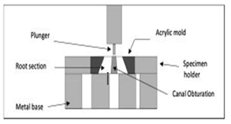
Universal testing machine (Z050, Zwick Roell, Xlorce HP, and Germany) used to measure push out bond strength. Sections on metal bases were held in place by a specimen holder (mold), which was designed to hold the section in place. Stainless-steel plungers of three different sizes (0.4,0.5,0.7) (corresponding to the coronal, middle, and apical sections) that cover 90% of canal diameter without touching canal dentine vertical force applied on the obturation materials. Using a universal testing machine, the load was applied in the apico-coronal direction with a crosshead speed of 0.5 mm/minutes across the filling materials in each area (Figure 1). "The maximum force in Newton (F-max) at which the filling materials were dislodged was recorded, and the push-out bond strength in Mega Pascal (MPa) was calculated for each specimen using the equation:
Push-out bond strength (MPa)=F-max/adhesion surface area (mm²)"
"F-max: Maximum force.
Adhesion surface area was calculated according to the following formula: Adhesion surface area=(D1+D2/2) × μ × h"
"Where D1=apical diameter, D2=coronal diameter calculated by image j program.
μ=3.14, and h=the section thickness confirmed by digital calibre".
Figure 1: Illustration of push out test.
Mode of failure evaluated by digital microscope at 60X magnification and adhesive (dentine without sealer) or adhesive between sealer and gutta-percha, cohesive in (sealer or gutta-percha), and mixed combination of (adhesive and cohesive).
Statistical analyses of the results were done by the use of the (SPSS Statistics for Windows, Version 25.0; IBM Corp., Armonk, NY, USA). The data were analysed by tested for normality using Shapiro-Wilk test One-way Analysis of Variance (ANOVA) and Tukey HSD were used to determine if there is statistically significant difference between each group and independent t test for comparison between groups. If the p-value (p ≤ 0.05) will be considered statistically significant.
Results
180 slices are subjected to push out test the finding of test are summarized in independent t test in Table 1 which showed At the apical level, the push out strength's mean values were significantly higher in total fill sealer for the CLC and GC sealers, while there was non-significant difference in SC group.
At the middle level, the push out strength's mean values were significantly higher in total fill sealer for the CLC while there was non-significant difference in SC and GC group.
At the coronal level, the push out strength’s means values was significantly higher in AH plus sealer for the CLC, SC and GC sealers. As comparison between AH plus and total fill BC for all obturation technique at all root levels each sealer group alone by One way Analysis of variance (ANOVA) test was performed and show that there were very high significant differences at all level except CLC, SC significant Tables 2 and 3.
| Levels | Obturation techniques | Descriptive Statistics | Group | ||||
|---|---|---|---|---|---|---|---|
| AH plus | Total Fill | difference | |||||
| Mean | S.D. | Mean | S.D. | t-test | p-value | ||
| Apical | CLC | 3.588 | 0.848 | 4.453 | 0.841 | -2.291 | 0.034 S |
| SC | 3.94 | 0.892 | 3.617 | 0.548 | 0.976 | 0.342 NS | |
| GC | 3.585 | 0.835 | 9.482 | 1.239 | -12.484 | 0.000 HS | |
| Middle | CLC | 2.443 | 0.876 | 4.181 | 1.41 | -3.311 | 0.004 HS |
| SC | 3.258 | 0.853 | 3.242 | 0.726 | 0.045 | 0.964 NS | |
| GC | 1.96 | 0.84 | 2.191 | 0.8 | -0.63 | 0.537 NS | |
| Coronal | CLC | 13.363 | 1.966 | 3.003 | 0.921 | 15.093 | 0.000 HS |
| SC | 5.94 | 0.87 | 4.205 | 0.796 | 4.651 | 0.000 HS | |
| GC | 1.182 | 0.311 | 4.916 | 1.386 | -8.314 | 0.000 HS. |
Table 1: Descriptive statistics and sealers difference".
| Obturation techniques | Levels | Descriptive statistics | Levels difference | ||||||||
|---|---|---|---|---|---|---|---|---|---|---|---|
| N | Mean | S.D. | S.E. | Confidence Interval | Min. | Max. | |||||
| Lower | Upper | F-test | p-value | ||||||||
| Bound | Bound | ||||||||||
| CLC | Apical | 10 | 3.588 | 0.848 | 0.268 | 2.982 | 4.194 | 2.11 | 4.84 | 202.01 | 0.000 HS |
| Middle | 10 | 2.443 | 0.876 | 0.277 | 1.816 | 3.07 | 1.25 | 3.71 | |||
| Coronal | 10 | 13.363 | 1.966 | 0.622 | 11.957 | 14.769 | 11.63 | 16.6 | |||
| SC | Apical | 10 | 3.94 | 0.892 | 0.282 | 3.302 | 4.578 | 2.49 | 5.1 | 25.559 | 0.000 HS |
| Middle | 10 | 3.258 | 0.853 | 0.27 | 2.648 | 3.868 | 2.17 | 4.35 | |||
| Coronal | 10 | 5.94 | 0.87 | 0.275 | 5.317 | 6.563 | 4.86 | 7.37 | |||
| GC | Apical | 10 | 3.585 | 0.835 | 0.264 | 2.988 | 4.182 | 2.35 | 4.57 | 30.103 | 0.000 HS |
| Middle | 10 | 1.96 | 0.84 | 0.265 | 1.359 | 2.561 | 1.13 | 3.22 | |||
| Coronal | 10 | 1.182 | 0.311 | 0.098 | 0.96 | 1.404 | 0.69 | 1.7 |
Table 2: Descriptive statistics and root level difference of the push out bond strength of the AH+ obturation techniques".
| techniques | Levels | Descriptive statistics | Levels difference | ||||||||
|---|---|---|---|---|---|---|---|---|---|---|---|
| N | Mean | S.D. | S.E. | Confidence Interval | Min. | Max. | |||||
| Lower | Upper | F-test | p-value | ||||||||
| Bound | Bound | ||||||||||
| CLC | Apical | 10 | 4.453 | 0.841 | 0.266 | 3.852 | 5.054 | 3.31 | 5.47 | 5.029 | 0.014 S |
| Middle | 10 | 4.181 | 1.41 | 0.446 | 3.172 | 5.19 | 2.14 | 5.9 | |||
| Coronal | 10 | 3.003 | 0.921 | 0.291 | 2.344 | 3.662 | 1.75 | 4.4 | |||
| SC | Apical | 10 | 3.617 | 0.548 | 0.173 | 3.225 | 4.009 | 2.67 | 4.35 | 4.834 | 0.016 S |
| Middle | 10 | 3.242 | 0.726 | 0.23 | 2.723 | 3.761 | 2.18 | 4.26 | |||
| Coronal | 10 | 4.205 | 0.796 | 0.252 | 3.635 | 4.775 | 2.59 | 5.29 | |||
| GC | Apical | 10 | 9.482 | 1.239 | 0.392 | 8.596 | 10.368 | 8.25 | 11.6 | 99.426 | 0.000 HS |
| Middle | 10 | 2.191 | 0.8 | 0.253 | 1.618 | 2.764 | 1.02 | 3.43 | |||
| Coronal | 10 | 4.916 | 1.386 | 0.438 | 3.925 | 5.907 | 3.01 | 6.56 |
Table 3: Descriptive statistics and root level difference of the push out bond strength of the total fill obturation techniques".
Further comparison using Tukey's HSD test after ANOVA in AH plus revealed that in CLC and SC groups, there were high significant difference between the coronal part with the apical and middle parts while there was non-significant difference between the apical and middle parts and just the reverse was true for the other two sealers.
Further comparison using Tukey's HSD test after ANOVA in total fill (BC) revealed that in CLC, there were high significant difference between apical and coronal and there was non-significant difference between apical and middle and coronal SC groups, there were high significant difference between the coronal and middle, while there was non-significant difference between the apical and middle parts, apical and coronal and (GC) there were high significant difference for all parts in this technique.
Figure 2: Showing types of failure mode, A-Adhesive (D\S), B-adhesive (S\G) C-mixed, D-cohesive in Guttapercha, and E-cohesive in Sealer. S sealer, D dentine, G gutta-percha.
Analysis of Failure Mode
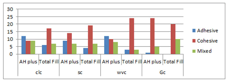
In AH plus sealer, the highest failure mode was recorded as followed:
CLC (adhesive), SC (cohesive), and GC (cohesive)
In the total fill technique, the highest failure mode was recorded as followed:
CLC (cohesive), SC (cohesive), and GC (cohesive) (Figure 3).
Figure 3: Bar chart of failure mode distribution.
Discussion
A variety of root canal system filling materials and techniques has been developed as a result, in order to eliminate any voids or spaces within the root canal system and prevent reinfection [10]. A root canal filling's ability to adhere to the dentinal wall is critical under both static and dynamic conditions. Static situations call for the elimination of any space between the filling and the wall that could allow fluids to percolate through the filling. Preventing filling dislodgement during subsequent manipulation is essential in highly dynamic situations [11]. Cold lateral compaction (coronal, middle thirds) had the highest significant effect of obturation techniques and root third bond strength groups in this study in AH plus. These findings could be attributed to the pressure exerted by the spreaders over the master and accessory cones, which can generate forces in the lateral and apical directions and favour sealer interlocking with dentin irregularities and/or tubules [12]. These findings are consistent with [13]. Which reported similar results when comparing CLC to WVC techniques using the AH Plus sealer. The explanation for higher bond strength in the coronal third compared to the apical third may be related to less patent apical tubules than coronal dentin, resulting in better infiltration in the coronal dentin compared to the sclerotic apical counterpart, when compared to the apical third [14]. They have a higher density of dentinal tubules with larger diameter. The bond strength of SC is highly significant difference in (coronal, middle) thirds in comparison to GC in this study, the AH plus penetrate better in to micro irregularities because of their creep capacity and long polymerization period, thus the bond strength was improved by mechanical locking between the canal dentine w and the canal dentine [15]. While with AH plus the lower bond strength associated with GC in our results, which could be related to the influence of heat on epoxy resin sealer composition and setting time [17]. It was observed that when AH plus was exposed to heat; its chemical structure was altered [16]. When heat is applied to AH plus, the amine groups are lost, these amine groups operate as setting initiators, which are required for the polymerization reaction to take place. The film thickness was raised while the setting time of AH Plus was lowered significantly [18]. However, the clinical implications of these alterations in the AH Plus sealer are still unknown, necessitating more research to determine their clinical significance. When Regardless of the obturation materials utilized, there was no statistical significance difference between the tested levels, which is consistent with the findings of [19]. Who found that differences in tubular density along the canal are inadequate to affect sealer adherence. This study used BC sealer entire fill+BC coated gutta-percha (total fill) to increase bonding by forming a real gap-free seal single cohesive unity (mono block) as claimed [20]. But result demonstrates reduction in bond strength in single cone and clc in comparison to GC. Total Fill BC sealer's increased bond strength in our investigation could be attributable to its bioactivity, as bio ceramic sealers attach to root dentine through a process known as alkaline etching [21]. The minerals in the bio ceramic sealer permeate the dentine, allowing ions to exchange and create a mineral infiltration zone at the dentine-sealer interface, which could lead to fewer gaps [22-23]. Bio ceramic sealer's better flow, along with smaller particle size and a low contact angle, result in deep penetration into dentinal tubules for mechanical interlocking and hence superior adhesion [24]. In terms of the obturation technique's impact on bond strength. The higher bond strength in the apical and middle third of the root as There were less voids and a low density of dentinal tubules as a result of anatomical heterogeneity in root canal thirds sealer thickness and very limited canal widening in the apical section, making push-out tests impossible without values that have a frictional component with the canal walls, and by Babb [25]. Several investigations have shown that the softening gutta-percha used in thermos plasticized procedures has the flexibility to flow into deep depressions, lateral canals, auxiliary canals, and defects not filled by sealing cement. The most predominant mode of failure is cohesive in these two sealers that come in agreement with [26].
Conclusion
Total fill BC bond strength was not considerably higher than that of Ah plus sealer. AH plus sealer is not preferable to be used with GC while total fill is used with GC showing good bond strength.
References
- Deshpande PM, Naik RR. Comprehensive review on recent root canal filling materials and techniques-An update. Int J Appl Dent Sci 2015; 1:30-34.
- Fisher MA, Berzins DW, Bahcall JKJ, et al. An in vitro comparison of bond strength of various obturation materials to root canal dentin using a push-out test design. J Endod 2007; 33:856-858.
- Chang S-W, Lee S-Y, Kang S-K, et al. In vitro biocompatibility, inflammatory response, and osteogenic potential of 4 root canal sealers: Sealapex, Sankin apatite root sealer, MTA Fillapex, and iRoot SP root canal sealer. J Endod 2014; 40:1642-1648.
- Chen I, Salhab I, Setzer FC, et al. A new calcium silicate-based bioceramic material promotes human osteo-and odontogenic stem cell proliferation and survival via the extracellular signal-regulated kinase signaling pathway. J Endod 2016; 42:480-486.
- Celikten B, Uzuntas CF, Orhan AI, et al. Evaluation of root canal sealer filling quality using a single-cone technique in oval shaped canals: An In vitro Micro-CT study. Scanning 2016; 38:133-140.
- Whitworth J. Methods of filling root canals: principles and practices. J Endod Topics 2005; 12:2-24.
- Nagas E, Altundasar E, Serper AJOS, et al. The effect of master point taper on bond strength and apical sealing ability of different root canal sealers. Oral Surg Oral Med Oral Pathol Oral Radiol Endod 2009; 107:e61-e4.
- Schroeder A, Ford N, Coil J, et al. Micro-computed tomography analysis of post space preparation in root canals filled with carrier-based thermoplasticized gutta-percha. Int Endod J 2017; 50:293-302.
- Dem K, Wu Y, Kaminga AC, et al. The push out bond strength of polydimethylsiloxane endodontic sealers to dentin. J B Oral Health 2019; 19:1-6.
- Schilder H. Filling root canals in three dimensions. J Endod 2006; 32:281-290.
- Ungor M, Onay E, Orucoglu HJ, et al. Push-out bond strengths: The Epiphany-Resilon endodontic obturation system compared with different pairings of Epiphany, Resilon, AH Plus and gutta-percha. Int Endod J 2006; 39:643-7.
- Rached-Junior FJA, Souza AM, Macedo LMD, et al. Effect of root canal filling techniques on the bond strength of epoxy resin-based sealers. Barz Oral Res 2016; 30.
- Carneiro S, Sousa-Neto M, Rached-Junior F, et al. Push-out strength of root fillings with or without thermomechanical compaction. Int Endod J 2012; 45:821-828.
- Hammad M, Qualtrough A, Silikas NJJoe, et al. Evaluation of root canal obturation: A three-dimensional in vitro study. J Endod 2009; 35:541-544.
- Sousa-Neto M, Silva Coelho F, Marchesan M, et al. Ex vivo study of the adhesion of an epoxy-based sealer to human dentine submitted to irradiation with Er: YAG and Nd: YAG lasers. J Endod 2005; 38:866-870.
- Viapiana R, Guerreiro-Tanomaru JM, Tanomaru-Filho M, et al. Investigation of the effect of sealer use on the heat generated at the external root surface during root canal obturation using warm vertical compaction technique with System B heat source. J Endod 2014; 40(4):555-561.
- Sakr O, Abdelhafeez M, AlKhalifah S, et al. Evaluation of microleakage in endodontically treated teeth with two different types of sealers: Bioceramic based sealer and AH26; an in vitro Int J Curr Microbiol Appl Sci 2017; 6:3556-3564.
- Camilleri J. Sealers and warm gutta-percha obturation techniques. J Endod 2015; 41:72-78.
- Costa J, Rached-Junior F, Souza-Gabriel A, et al. Push-out strength of methacrylate resin-based sealers to root canal walls. Int Endod J 2010; 43:698-706.
- Trope M, Bunes A, Debelian GJET, et al. Root filling materials and techniques: bioceramics a new hope. Endod Topics 2015; 32:86-96.
- Kakoura F, Pantelidou OJO-IJoDS. Retreatment efficacy of endodontic bioceramic sealers: a review of the literature. Odo Int J Dent Sci 2018; 20:39-50.
- Eltair M, Pitchika V, Hickel R, et al. Evaluation of the interface between gutta-percha and two types of sealers using scanning electron microscopy (SEM). Clinic Oral Investig 2018; 22:1631-1639.
- Abarca AM, Bustos A, Navia MJ, et al. A comparison of apical sealing and extrusion between Thermafil and lateral condensation techniques. J Endod 2001; 27:670-672.
- Babb BR, Loushine RJ, Bryan TE, et al. Bonding of self-adhesive (self-etching) root canal sealers to radicular dentin. J Endod 2009; 35:578-582.
- Natera M, Pileggi R, Nair UJOS, et al. A comparison of two gutta-percha obturation techniques to replicate canal irregularities in a split-tooth model. Oral Surg Oral Med Oral Pathol Oral Radiol Endod 2011; 112:29-34.
- Shokouhinejad N, Gorjestani H, Nasseh AA, et al. Push-out bond strength of gutta-percha with a new bioceramic sealer in the presence or absence of smear layer. Austr Endod J 2013; 39:102-106.
Author Info
Batool Basim Mounes* and Raghad A Al-Hashimi
Department of Endodontics, University of Baghdad, Baghdad, IraqCitation: Batool Basim Mounes, Raghad A Al-Hashimi, The Effect of Three Different Obturation Techniques on Push Out Bond Strength of Bio Ceramic (Total Fill) and Ah Plus Sealers, J Res Med Dent Sci, 2022, 10 (7): 000-000.
Received: 24-May-2022, Manuscript No. JRMDS-22-48335; , Pre QC No. JRMDS-22-48335; Editor assigned: 27-May-2022, Pre QC No. JRMDS-22-48335; Reviewed: 10-Jun-2022, QC No. JRMDS-22-48335; Revised: 25-Jul-2022, Manuscript No. JRMDS-22-48335; Published: 03-Aug-2022


