Research Article - (2022) Volume 10, Issue 10
The Effect of Nano Barium Titanate on Candida Albicans Adherence and Other Properties of Heat-Cured Soft Acrylic Denture Lining Material
Fatima Basil Sadiq* and Hikmat Jameel Abdulbaqi
*Correspondence: Fatima Basil Sadiq, Department of Prosthodontics, College of Dentistry, University of Baghdad, Baghdad, Iraq, Email:
Abstract
Introduction: Permanent denture soft liners are widely applied in prosthodontics. The unsolved problem of Candida albicans colonization of soft denture liners is especially troubling. Recently, many research tried to enhance soft liner properties by incorporating nano fillers into the polymeric matrix. In this study BaTiO3 NPs powder was incorporated in acrylic soft liner to assess its effect on adherence of candida albicans, radiopacity and surface hardness.
Materials and Methods: Based on the findings of the pilot study the percentage (1% and 1.5% by wt. of BaTiO3 NPs) chosen to be added to soft liner. Main: one hundred specimens prepared in accordance with the tests to be conducted. Firstly, BaTiO3 NPs powder was dispersed into the monomer of the soft lining material by using probe sonicator and then, measured amount of the soft liner's powder blended into this suspension. C. albicans adherence test was performed to assess the antifungal efficacy of BaTiO3 NPs/soft liner composite. Also, radiopacity test and Shore a hardness test were performed.
Results and Discussion: there was a highly significant drop in Candida albicans adherence to soft lining material after the addition of BaTiO3 NPs. Also, the radiopacity significantly increased, but the surface hardness not significantly increased after the addition of BaTiO3 NPs.
Conclusion: the addition of BaTiO3 NPs can improve the anti-fungal properties and the radiopacity of soft denture liner, while surface hardness Non-significantly changed with the addition of BaTiO3 NPs.
Keywords
Barium titanate, Soft liner, Candida albicans, Nano particles
Introduction
Soft denture liners are class of resilient material that have been popularly used in dentistry to redefine prosthetic surfaces that come into contact with soft tissues in the oral cavity to achieve an optimum denture fit, conditioning the traumatized tissues underneath the denture base, and trying to improve prosthetic retention [1].
There are two types of soft denture liners; silicon or acrylic based. Each type can be auto or heat cured [2]. Acrylic polymers and copolymers are used to make plasticized acrylic resin, as well as a liquid including an acrylic monomer and plasticizers that protects the material's flexibility [3].
Ideally, soft denture liners should be non-toxic, comfortable and durable, not allow the adherence and growth of microorganisms and have good mechanical properties [4]. But there are several problems associated with the use of soft denture liners; one of the major problems is Candida albicans colonization which causes denture stomatitis infection [5].
Another problem associated with soft liners is they are radiolucent that cannot be detected by radiograph if the patient accidently swallowed a fractured piece which can threaten the patient’s life [6]. Also, soft denture liners lose their resiliency after a period of use as a result of plasticizer leaking [7].
Several studies have attempted in order to improve the qualities of soft denture liners by adding different type of fillers. Recently nano fillers have been widely used in dentistry like zirconia, glass fiber and silver nanoparticles [8-10].
Barium titanate nano particles is known for its antimicrobial and antibiofilm activity [11,12]. It has been applied widely in medical field as in tissue engineering and drug delivery [13-15]. Also, it has been added to PMMA denture base to enhance its radiopacity and mechanical properties [16].
Hence, in this study BaTiO3 NPs incorporated in acrylic based heat cured soft denture liner to evaluate its effect on Candida albicans adherence and other properties.
Materials and Methods
Polyethylmethacrylate (PEMA) Heat cured soft denture liner (moon star) was used in this study. According to pilot study findings 1% by wt. and 1.5% by wt. of BaTiO3 NPs were the most appropriate percentage to be added as they shown good effect in Candid albicans adherence test.
Sampling
A total of 100 the specimens were processed and categorized into three groups based on the test to be conducted. Then every group was further sorted into three subgroups that reflect the percentage of BaTiO3 NPs incorporated. (Control 0%, 1% by wt. BaTiO3 NPs and 1.5% by wt. BaTiO3 NPs), except for Candida albicans adherence test which had a fourth control positive group (1.5% of Nystatin), 10 specimens for each subgroup [17].
Acrylic soft liner preparation
According to manufacturer instruction soft liner mixed in dry glass container in powder liquid ratio (1.2 g/1 ml.) till reaching the dough stage then packed in stone mold and pressed in hydraulic press at 100 Kg/cm2 pressure to make sure that the soft liner is distributed evenly throughout the mold. Then transferred to water bath for curing that was heated up to 70°C for 90 minutes. And then, the temperature was raised to 100°C for 30 minutes. Later the entire specimens were finished and before testing, the samples were kept in purified distilled water at 37°C for 48 hours.
Nano filler incorporation
For experimental groups the BaTiO3 nano filler weighted in clean dry glass container using electronic balance of 0.0001 accuracy then the liquid monomer of soft liner was added to the powder and mixed using sonication prob at 120 W, 60 KHz (Soniprep-150, England) for 3 minutes to break them down into individual nanoparticles. The powder was then added, mixed, packed and cured according with manufacturer guidelines.
FTIR analysis was carried out to determine whether or not there was a chemical reaction of PEMA soft liner and BaTiO3 NPs. Also, Scanning Electron Microscopy (SEM) was made for control group and experimental groups (1% by wt. and 1.5% by wt. BaTiO3 NPs) to evaluate the degree of dispersion of Nano particles inside PEMA soft liner matrix. In addition Energy Dispersive X-ray Spectroscopy (EDS) was carried out in order to tell the composite filler percentages in terms of both weight and atoms.
Candida albicans adherence test
Forty specimens of soft liner in disc shape with the dimension 10 mm. in diameter and 2 mm. in thickness were prepared for Candida albicans adherence test [18].
Isolation and identification of Candida albicans
Candida albicans isolated from patients with "dentureinduced stomatitis" symptoms using a sterile cotton swab. Then the swab was inoculated immediately in Sabouraud Dextrose Agar (SDA) and incubated for 48 h at 37°C [19].
Candida albicans identification was done by morphological examination as it has a pearl shape with smooth, creamy and pasty appearance [20]. Microscopical examination also conducted using light microscope (Olympus, Japan) using a gram stain procedure. Then biochemical examination was done using VITEK 2 fully automated system with fluorescence based technology for microorganisms’ identification [21].
Candida albicans adherence test procedure
Sabouraud dextrose broth was prepared according to manufacturer instructions and small proportion of candida albicans added to the media to make a suspension equal to 0.5 McFarland standards [22]. In sterile tubes, the soft lining samples that were sterilized by autoclave were placed in the prepared broth and incubated at room temperature for 24 hours. Next, the specimens were taken out of the tubes and for 1 minute, samples were rinsed with phosphate buffered saline. Using a gentle rocking to eliminate any yeast cells that didn’t adhere then dried using a filter paper. Candida albicans cells attached to the surface of soft liner specimens were fixed with methanol, stained for 1 min with crystal violet, rinsed with PBS solution, left to dry on filter paper, and inverted light microscope used to examine them [23,24]. Inverted light microscope used to examine the soft liner specimens, three fields taken for each specimen and the adhered Candida albicans cell counted for each field then, for each specimen, the mean of three fields is taken.
Radiopacity test
Thirty specimens of soft liner in rectangular shape of 30 mm. in length and 10 mm. in width and 2.5 mm in thickness were constructed to undergo radiopacity test [25].
Using aluminum plate an aluminum step wedge was made with ten steps starting from a step of 1 mm thickness of aluminum and progressing to 10 mm of aluminum thickness step with each step increasing by 1 mm [26]. Thirty specimens (10 for each percentage of concentration) were positioned on a 10 mm in thickness wax plate to approximate soft tissue media in absorption and reflection. The aluminum step wedge was placed next to soft liner specimens in order to standardize the radiographic film density [27].
The soft liner specimens, along with the aluminum step wedge, and wax plate were laid on top of a cassette (kodak medical x-ray film) and with 50 Kv irradiation, 200 mA, and exposure time of 0.1 sec at a distance of 1 m from the x-ray source. As an x-ray source, a Computed Radiographic System (CR) was used (Chest X-ray machine, Germany).
In accordance with the manufacturer's instructions, the processing was carried out using an automatic x-ray film processor (JP-33, Korea). The optical density was measure for of every aluminium step and soft liner specimens using a light transmission densitometer. Three measurements were made for each specimen, and the average value of these readings was calculated.
Surface hardness test
30 specimens were prepared of soft liner in disk shape of 30 mm. in diameter and 3 mm. in thickness for Shore a surface hardness test [28,29].
The soft liner samples' hardness was measured using a shore A durometer (Time group-TH200, China) [30]. Five durometer measurements were made for every sample, with 5 sec. contact time with every penetration, with the average taken into account as the test value.
Statistical analysis
For analysing the study's results, SPSS (Statistical Package for Social Science) computer software (version 21) was used, as well as descriptive and inferential statistics. A one-way ANOVA (analysis of variance) test was used. A "P" value of > 0.05 was considered statistically non-significant, a "P" value of 0.05 was considered significant, and a "P" value of 0.01 was considered highly significant.
Results
FTIR analysis: FTIR analysis revealed that there has been only physical interaction between the acrylic soft liner and the BaTiO3 NPs (Figure 1).
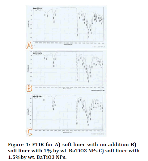
Figure 1: FTIR for A) soft liner with no addition B) soft liner with 1% by wt. BaTiO3 NPs C) soft liner with 1.5%by wt. BaTiO3 NPs.
Scanning electron microscopy: The morphology of crosssectional samples and mapping of samples with soft liners before and after the addition of BaTiO3 NPs are studied (Figures 2-6).
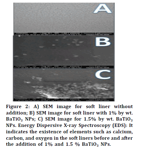
Figure 2: A) SEM image for soft liner without addition; B) SEM image for soft liner with 1% by wt. BaTiO3 NPs; C) SEM image for 1.5% by wt. BaTiO3 NPs. Energy Dispersive X-ray Spectroscopy (EDS): It indicates the existence of elements such as calcium, carbon, and oxygen in the soft liners before and after the addition of 1% and 1.5 % BaTiO3 NPs.
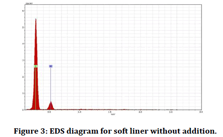
Figure 3: EDS diagram for soft liner without addition.
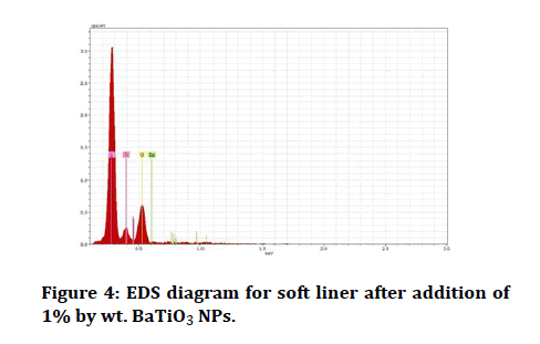
Figure 4: EDS diagram for soft liner after addition of 1% by wt. BaTiO3 NPs.
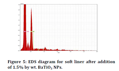
Figure 5: EDS diagram for soft liner after addition of 1.5% by wt. BaTiO3 NPs.
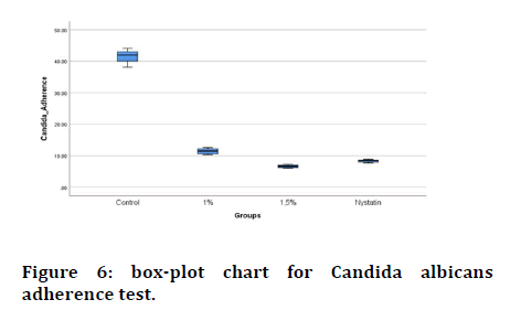
Figure 6: box-plot chart for Candida albicans adherence test.
Candida albicans adherence test results: under an inverted light microscope stained specimens for each group were inspected, the control group has a higher mean value (41.49), while the experimental group (1.5% by wt. of BaTiO3 NPs) has the lowest mean value (6.65) (Figure 6).
Comparison of means of candida albicans adherence test results using one way ANOVA table test showed a highly significant decrease in Candida albicans adherence to the surface of soft denture liner as shown in Table 1.
| Group | N | Mean | SD | SE | Minimum | Maximum | F |
|---|---|---|---|---|---|---|---|
| Control | 10 | 41.492 | 1.89672 | 0.59979 | 38.1 | 44.1 | 2293.417 |
| 1% BaTiO3 nps | 10 | 11.418 | 0.85008 | 0.26882 | 10.3 | 12.6 | |
| 1.5% BaTiO3 nps | 10 | 6.6567 | 0.4622 | 0.14616 | 6 | 7.3 | |
| Nystatin | 10 | 8.325 | 0.44074 | 0.13937 | 7.68 | 8.91 |
Table 1: Mean of Candida albicans adherence test and one-way ANOVA table for all studied groups.
According to the results of Levene's test for data homogeneity, which show highly significant differences between groups, the Games-Howell post-hoc test was chosen for candida adherence multiple comparisons (Table 2).
| Tested groups | Mean difference | Sig | |
|---|---|---|---|
| Control | 1% | 30.07* | 0.000HS |
| 1.50% | 34.83* | 0.000HS | |
| Nystatin | 33.16* | 0.000HS | |
| 1% | 1.50% | 4.76* | 0.000HS |
| Nystatin | 3.09* | 0.000HS | |
| 1.50% | Nystatin | -1.66* | 0.000HS |
| *The mean difference is significant at the 0.05 level. | |||
Table 2: Games-Howell post hoc test for Candida albicans adherence.
Radiopacity test results: The control group had the highest mean score of optical density (1.3112), while the experimental group had the lowest mean score (1.5% incorporation of BaTiO3 NP) (1.0353) (Figure 7).
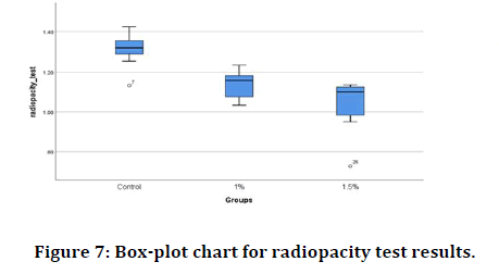
Figure 7: Box-plot chart for radiopacity test results.
A comparison of the means of all groups' radiopacity test results using one-way ANOVA table test showed a highly significant increase in radiopacity in experimental groups when compared to the control group as shown in Table 3.
| Groups | N | Mean | SD | SE | Minimum | Maximum | F | P value |
|---|---|---|---|---|---|---|---|---|
| Control | 10 | 1.3112 | 0.08158 | 0.0258 | 1.13 | 1.42 | 20.736 | 0.000HS |
| 1% BaTiO3 nps | 10 | 1.1377 | 0.0688 | 0.02176 | 1.03 | 1.24 | ||
| 1.5% BaTiO3 nps | 10 | 1.0353 | 0.12943 | 0.04093 | 0.73 | 1.14 |
Table 3: Mean for radiopacity test and one-way ANOVA table for all tested groups.
Bonferroni post-hoc test was used to compare mean value between each two groups (Table 4), there was high significant difference between (control and 1% groups) and (control and 1.5% groups), while there was nonsignificant difference between 1% and 1.5% groups.
| Tested groups | Mean difference | Sig. | |
|---|---|---|---|
| Control | 1% | .17350* | 0.001 HS |
| 1.50% | .27590* | 0.000 HS | |
| 1% | 1.50% | 0.1024 | 0.077 NS |
Table 4: Bonferroni post hoc test for radiopacity test.
Surface hardness test results: Highest mean value of surface hardness test was for the 1.5% incorporation of BaTiO3 NP group (50.1600) while the lowest mean value was for control group (48.5200) as shown in Figure 8.
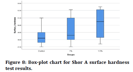
Figure 8: Box-plot chart for Shor A surface hardness test results.
Comparison of means of surface hardness test results of all groups using one way ANOVA table test revealed a non-significant increase in surface hardness of soft lining material after addition of 1% and 1.5% by wt. barium titanate nano particles as shown in Table 5.
| Group | N | Mean | SD | SE | Minimum | Maximum | F | P value |
|---|---|---|---|---|---|---|---|---|
| Control | 10 | 48.52 | 1.30792 | 0.4136 | 47 | 51 | 2.283 | 0.121NS |
| 1% BaTiO3 nps | 10 | 49.28 | 1.90193 | 0.60144 | 47 | 52.1 | ||
| 1.5% BaTiO3 nps | 10 | 50.16 | 1.87688 | 0.59352 | 47.4 | 52.5 |
Table 5: Mean for surface hardness test and one-way ANOVA table for all tested groups.
Discussion
Liners are commonly used in dentistry to alter prosthetic surfaces that come into contact with soft tissues in the oral cavity. Soft liners are a type of flexible material that is used to reline denture base surfaces that come into contact with the occlusal stress bearing oral mucosa [31].
Several studies have tried to combat fungi colonization by incorporating antifungal agents into the soft denture liner material, for example; when compared to conventional organic antimicrobial agents, metal oxide nanoparticles such as TiO2, MgO, and ZnO are thought to be superior antimicrobial agents in terms of safety, durability, and heat resistance [32,33].
The current study tried to enhance soft lining material properties against C. albicans adherence by incorporating BaTiO3 NPs into soft liner.
In Candida albicans adherence test incorporation of BaTiO3 NP powder in two different precents of weight (1%, 1.5%) caused a highly significant decrease in Candida albicans adherence to acrylic based heat cured soft liner compared to control group and in comparison, with Nystatin control positive group the 1.5% group showed significant difference in decreasing Candida albicans adherence.
BaTiO3's antifungal mode of action include two proposed method: Due to the increase in positive surface potential, which may have resulted in bacteria being neutralized on their surfaces, resulting in the creation of ROS (Reactive Oxygen Species), that can cause membrane and DNA damage [34].
When Candida albicans is exposed to nano BaTiO3, the nanoparticles can covalently connect to the membrane components. This results in a disturbance of the cell wall architecture, which ultimately results in the death of Candida albicans via suppression of ergosterol production.
In radiopacity test a highly significant increase in radiopacity observed after addition of barium titanate nanoparticles to soft denture liner compared to control group. This finding can be explained that barium and titanium have high atomic number (56 for barium and 22 for titanium) compared to the elements of polymer which produce more radiopacity when added to the polymer.
Furthermore, the addition of barium titanate nanoparticles to polymer should result in an increase in density, as NBT has a higher density than polymer. (6.08 g/cm3, 0.906 g/cm3 respectively) [35], this increase in material density result in increased radiopacity of the material as the radiopacity determined by the size, density, chemical composition of the filler molecules, and their concentration within the polymer matrix [36].
In shore a surface hardness test the results revealed that there is no significant increase in surface hardness between control group and experimental groups, thus they were within the ISO specification limit for long-term soft lining materials. Because only trace amounts of BaTiO3 NPs were introduced to the soft denture liner, they had no discernible effect on the material's surface hardness due to low network density and small particle size of BaTiO3 NPs.
Conclusion
The results of this study indicate that integration of BaTiO3 nanoparticles into soft denture liner resulted in a significantly substantial increase in antifungal activity as it reduced the adherence of Candida albicans to soft denture liner. Also, a highly significant increase in radiopacity of denture liner resulted from BaTiO3 NPs incorporation, while there was non-significant increase in surface hardness.
References
- Sanchez-Aliaga A, Pellissari CVG, Arrais CAG, et al. Peel bond strength of soft lining materials with antifungal to a denture base acrylic resin. Dent Mater J 2016; 35:194–203.
- Khaledi AAR, Bahrani M, Shirzadi S. Effect of Food Simulating Agents on the Hardness and Bond Strength of a Silicone Soft Liner to a Denture Base Acrylic Resin. Open Dent J 2015; 9:402–408.
- Hashem MI. Advances in Soft Denture Liners: An Update. J Contemp Dent Pract 2015; 16:314–318.
- Iwasaki N, Yamaki C, Takahashi H, et al. Effect of long-time immersion of soft denture liners in water on viscoelastic properties. Dent Mater J 2017; 36:584–589.
- Chladek G, Mertas A, Barszczewska-Rybarek I, et al. Antifungal activity of denture soft lining material modified by silver nanoparticles-a pilot study. Int J Mol Sci 2011; 12:4735–4744.
- Goshima T, Gettleman L, Goshima Y, et al. Evaluation of radiopaque denture liner. Oral Surgery Oral Med Oral Pathol 1992; 74:379–382.
- Maeda T, Hong G, Sadamori S,et al. Durability of peel bond of resilient denture liners to acrylic denture base resin. J Prosthodont Res 2012; 56:136–141.
- Habibzadeh S, Omidvaran A, Eskandarion S, et al. Effect of Incorporation of Silver Nanoparticles on the Tensile Bond Strength of a Long term Soft Denture Liner. Eur J Dent 2020; 14:268–273.
- Hatamleh MM, Maryan CJ, Silikas N, et al. Effect of net fibre reinforcement surface treatment on soft denture liner retention and longevity. J Prosthodont 2010; 19:258–262.
- Yasser, Fatah A. The Effect of Addition of Zirconium Nano Particles on Antifungal Activity and Some Properties of Soft Denture Lining Material. Bagh Coll Dent 2017; 29:27–32.
- Shah AA, Khan A, Dwivedi S, et al. Antibacterial and Antibiofilm Activity of Barium Titanate Nanoparticles. Mater Lett 2018; 229:130–133.
- Sasikumar M, Ganeshkumar A, Chandraprabha MN, et al. Investigation of Antimicrobial activity of CTAB assisted hydrothermally derived Nano BaTiO3. Mater Res Express 2019; 6:0–31.
- Schult M, Buckow E, Seitz H. Experimental studies on 3D printing of barium titanate ceramics for medical applications. Curr Dir Biomed Eng 2016; 2:95–99.
- Genchi GG, Marino A, Rocca A, et al. Barium titanate nanoparticles: Promising multitasking vectors in nanomedicine. Nanotechnology 2016; 27:1–19.
- Bai Y, Dai X, Yin Y, et al. Biomimetic piezoelectric nanocomposite membranes synergistically enhance osteogenesis of deproteinized bovine bone grafts. Int J Nanomedicine 2019; 14:3015–3026.
- Elshereksi NW, Mohamed SH, Arifin A, et al. Evaluation of the mechanical and radiopacity properties of poly (methyl methacrylate)/barium Titanate-denture base composites. Polym Polym Compos 2016; 24:365–374.
- Urban VM, de Souza RF, Galvao Arrais CA, et al. Effect of the association of nystatin with a tissue conditioner on its ultimate tensile strength. J Prosthodont 2006; 15:295–299.
- Karakis D, Akay C, Oncul B, et al. Effectiveness of disinfectants on the adherence of Candida albicans to denture base resins with different surface textures. J Oral Sci 2016; 58:431–437.
- Manikandan C, Amsath A. Isolation and Rapid Identification of Candida Species from the Oral Cavity. Int J Pure Appl Zool 2013; 1:172–177.
- ElFeky DS, Gohar NM, El-Seidi EA, et al. Species identification and antifungal susceptibility pattern of Candida isolates in cases of vulvovaginal candidiasis. Alexandria J Med 2016; 52:269–277.
- Sundaram M, Navaneethakrishnan MR. Evaluation of Vitek 2 System for Clinical Identification of Candida Species and Their Antifungal Susceptibility Test. J Evol Med Dent Sci 2016; 5:2948–2951.
- de Sousa LVNF, Santos VL, de Souza Monteiro A, et al. Isolation and identification of Candida species in patients with orogastric cancer: Susceptibility to antifungal drugs attributes of virulence in vitro and immune response phenotype. BMC Infect Dis 2016; 16:1–12.
- Gedik H, Ozkan YK. In vitro evaluation of Candida albicans adherence to soft denture-lining materials. J Prosthet Dent 2009; 68:804–808.
- Govindswamy, Rodrigues S, Shenoy VK, et al. The Influence of Surface Roughness on The Retention of Candida Albicans to Denture Base Acrylic Resins–an in vitro study. J Nepal Dent Assoc 2014; 14:1–9.
- Mikael J, Al-Samaraie S, Ikram F. Evaluation of Some Properties of Acrylic Resin Denture Base Reinforced with Calcium Carbonate Nano-Particles. Erbil Dent J 2018; 1:41–47.
- Salem SA. Reinforced denture base material. Ph. D. Thesis. University of Manchester, 1979.
- Abdulmajeed SK, Abdulbaqi HJ. Evaluation of some properties of heat cured soft denture liner reinforced with calcium carbonate nano-particles. Pakistan J Med Heal Sci 2021; 14:1728–1733.
- Abraham AQ, Abdul-Fattah N. The Influence of Chlorhexidine Diacetate Salt Incorporation into Soft Denture Lining Material on Its Antifungal and Some Mechanical Properties. J Baghdad Coll Dent 2017; 29:9–15.
- Mancuso DN, Goiato MC, Zuccolotti BCR, et al. Effect of thermocycling on hardness, absorption, solubility and colour change of soft liners. Gerodontol 2012; 29:1–5.
- Basima MA, Hussein BDS MsP. Effect of some disinfectant solutions on the hardness property of selected soft denture liners after certain immersion periods. J Fac Med 2009; 51:259–264.
[Crossref]
- Mainieri VC, Beck J, Oshima HM, et al. Surface changes in denture soft liners with and without sealer coating following abrasion with mechanical brushing. Gerodontol 2011; 28:146–151.
- Daistan S, Aktas AE, Caglayan F, et al. Differential diagnosis of denture-induced stomatitis, Candida, and their variations in patients using complete denture: A clinical and mycological study. Mycoses 2009; 52:266–271.
- Sawai J, Yoshikawa T. Quantitative evaluation of antifungal activity of metallic oxide powders (MgO, CaO and ZnO) by an indirect conductimetric assay. J Appl Microbiol 2004; 96:803–809.
- Marin E, Boschetto F, Sunthar TPM, et al. Antibacterial effects of barium titanate reinforced polyvinyl-siloxane scaffolds. Int J Polym Mater Polym Biomater 2020; 1–12.
- Nidal W Elshereksi, Mariyam J Ghazali, et al. Investigation on the Physical Properties of Denture Base Resin Filled with Nano-Barium Titanate. Aust J Basic Appl Sci 2016; 249–257.
- Pekkan G. Radiopacity of Dental Materials: An Overview. Avicenna J Dent Res 2016; 8:8–8.
[Crossref]
Author Info
Fatima Basil Sadiq* and Hikmat Jameel Abdulbaqi
Department of Prosthodontics, College of Dentistry, University of Baghdad, Baghdad, IraqCitation: Fatima Basil Sadiq, Hikmat Jameel Abdulbaqi, The Effect of Nano Barium Titanate on Candida Albicans Adherence and Other Properties of Heat-Cured Soft Acrylic Denture Lining Material, J Res Med Dent Sci, 2022, 10 (10): 109-116.
Received: 02-Aug-2022, Manuscript No. JRMDS-22-53722; , Pre QC No. JRMDS-22-53722(PQ); Editor assigned: 04-Aug-2022, Pre QC No. JRMDS-22-53722(PQ); Reviewed: 18-Aug-2022, QC No. JRMDS-22-53722; Revised: 03-Oct-2022, Manuscript No. JRMDS-22-53722(R); Published: 13-Oct-2022
