Research Article - (2022) Volume 10, Issue 8
The Effect of Different Placement Strategies of Bulk Fill Composite on Marginal Leakage of Classii Restoration (Comparative in Vitro Study)
Sura Y Alani and Abdulla MW Al-Shamma*
*Correspondence: Abdulla MW Al-Shamma, Department of Restorative and Esthetic Dentistry, College of Dentistry, Baghdad University, Iraq, Email:
Abstract
Introduction: In spite of huge revolution in composite materials and techniques, some drawback still exist which may hindered the success of composite especially in class II restorations such as volumetric shrinkage, micro-leakage and secondary caries.
Aims: The purpose of this in vitro study was to evaluate the effects of different placement strategies of bulk fill composites on the gingival micro leakage of class II restorations.
Material and methods: forty human extracted premolars teeth had received 2 proximal box preparation with the following criteria (Mesiodistal width: 2 mm, buccolingual width: 3 mm and the gingival margin located 1 mm above CEJ on mesial cavity while for the distal cavity located 1 mm below CEJ. All prepared cavities had received etch and rinse adhesive strategy and had been divided randomly to four groups. Group 1: Restored with bulk fill composite alone. Group 2: Restored with 1 mm flow able composite followed by bulk fill composite cured separately. Group 3: (Snow plow technique) which restored by application of 1 mm flow able and bulk fill composite and curing them simultaneously. Group 4: (Injection molding technique) Teeth were restored with adhedive+1 mm flowable+bulk fill composite and co-curing the three layers concurrently.
After one week storage in water, all specimens were thermo cycled for 500 cycle between 5°C and 55°C for dwell time of 30 sec. Using model wax, the root apices of the teeth were sealed. All specimens had received double coats of nail varnish except 1 mm around the restoration, and then all specimens had been immersed with 2% methylene blue dye for 24 hrs. After rinsing thoroughly with water, all specimens were embedded in self-curing epoxy resin and allowed to set for 24 h before sectioning.
Then one section had been made from mesial to distal margins and then examined under digital microscope 40X and the depth of dye penetration had been analysed according to 0-3 scoring system. All data were subjected to kruskal Wallis followed by Mann Whitney tests at p>0.05.
Results: Statistical significant differences between the tested groups was observed in enamel margin (mesial) with lower micro leakage were revealed in snow plow and injection molding techniques, while no significant difference was noticed in dentin (distal) margin. For intragroup comparison, all tested groups showed higher micro leakage score in dentin than enamel margin.
Conclusion: According to the results the present study, co-curing the flow able with overlaying bulk fill composite reduce gingival micro leakage. For dentinal margin, higher micro leakage score were presented compared to enamel
Keywords: Bulk fills, Snow plow and Injection molding technique
Introduction
Tooth colored posterior restoration has emerged as an essential and vital component of the restorative procedure instead of dental amalgam due to aesthetic and conservative demand. So composite material received large attention and development in its properties such as wear resistance, adhesion, mechanical properties, shade selection, colour stability, polish ability and handling properties [1,2].
Even though many researches documented high failure rate in class II posterior composite restorations as a result of some restriction. One of them is polymerizations shrinkage with its detriments effect such as micro leakage, marginal gap, marginal staining, recurrent caries and post-operative sensitivity [3].
Polymerization shrinkage may result from: Volumetric shrinkage, viscoelastic behaviour, effect of Configuration factor (C-factor) and water sorption [3].
Many techniques were suggested to reduce the adverse effects of shrinkage stress. Among these suggestions are: Altered light curing cycles (Intensity and time curing), polymerization-induced phase separation, ring-opening polymerization, hybrid polymerization reactions, thiol-ene photo polymerization, preheating, vibration, stress absorbing layers with low elastic modulus liners and diverse composite layering techniques [4].
Different placement techniques of posterior composite were adopted to reduce polymerization shrinkage. Among these techniques, a widely used method named Incremental Technique which relies on applying composite in 2 mm increment in different suggested ways such as: Horizontal technique, oblique successive cusp build-up technique, centripetal incremental technique, split horizontal technique and three-site technique [5].
Although polymerization shrinkage has been reduced by incremental technique, researches documented some drawback such as voids, cuspal deflection and time consuming. Bulk fill composite which allow 4-5 mm bulk placement, has been developed to overcome some of these shortenings [6].
Some authors advocated the use of flow able composite underneath the bulk fill to improve the adaptation of composite and help in increase relaxation stress [7].
In addition to traditional technique in the application of composite, two advanced methods have been suggested. First technique known as snow plow technique which involves the placement of a layer of flow able composite on the pulpal floor and the gingival margin of the proximal box of a posterior composite resin restoration. However, the layer of flow able composite is not cured followed by application of packable composite and curing the 2 layers simultaneously [8].
While another technique were suggested by David Clark named injection molding technique, who proposed using one layer of adhesive followed by flow able composite then finally injecting the paste composite. The resin flow able paste mass is polymerized together in a single [9].
So the aim of this study is to evaluate the effects of different placement strategies of bulk fill composites on the gingival micro leakage of class II restorations the null hypothesis stated that neither the placement techniques of bulk fill composite nor the location of margin can affect the marginal leakage of class II restoration.
Materials and Methods
Specimens
Human maxillary first premolar teeth free from caries, restorations, defect and cracks were collected from specialized orthodontic centre in Baghdad University. 40 teeth with comparable size and morphology are selected (average crown length 6.9: mesiodistal: 6.5 and buccolingual dimension: 9.2) To preserve homogeneity of tooth measurements between groups, a one-way ANOVA test was done for each measurement among the four groups, which revealed no statistically significant difference.
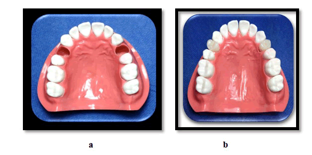
Any extrinsic stains, soft tissue or calculus deposits were removed by ultrasonic scalar then all teeth stored in (0.1%) thymol solution for 2 days in order to prevent bacterial and fungal growth and then kept in deionized distilled water at room temperature to avoid dehydration of extracted teeth. the teeth were kept no more than 1 month The sample teeth was positioned by dental surveyor in dental manikin (maxilla)and fixed by aid of light body polyvinyl siloxane impression material in the socket of maxillary first premolar. All teeth attached to manikin to stimulate clinical condition (Figure 1).
Figure 1: a) Dental manikin with adjustment of place of first premolar; b) Dental manikin after attachment of examined teeth.
Cavity preparation: Each teeth had received 2 proximal box preparations with following criteria (mesiodistal width: 2 mm, buccolingual width: 3 mm and gingival margin located 1 mm above Cementoenamel Junction (CEJ) on mesial cavity and for distal cavity the gingival margin located 1 mm below CEJ
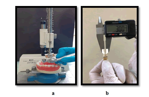
The cavity preparation had done by parallel sided; flat ended carbide fissure burs with high speed water cooled hand piece which was fixed to modified dental survey or each bur was discarded after four cavity preparation to ensure maximum cutting efficiency Figure 2.
Figure 2: a) Cavity preparation by dental surveyor; b) Checking the dimension of cavity preparation by dental Vernier.
The dimension was checked by dental Vernier and Williams graduated periodontal probe the teeth were rinsed and dried. The teeth randomly divided into 4 groups based on restoration placement techniques.
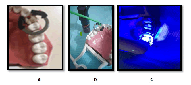
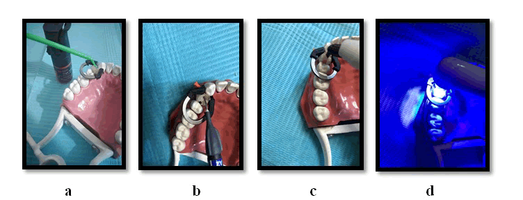
Bioclear matrice was placed adjacent to each preparation and secured by ring and plastic wedge Figure 3a. All teeth received etch and rinse strategies by etching with phosphoric acid 37% for 15 second and washed for another 15 second then dried with cotton pellets one layer of adhesive Scotchbond Universal bonding adhesive (3 M, USA) were applied and rub it for 20 seconds. After that, a gentle stream of air was directed over the adhesive for about 10 seconds until it stopped moving after that, an LED light was used to cure the adhesive (x lif Cure-L LED curing light, wavelength (430-480 nm) light intensity, (1300 mw/cm2 cured for 10 seconds Figures 3b and c.
Figure 3: a) Adjustment of bio clear matrices, plastic wedge and ring; b) Adhesive application; c) Curing for 10 seconds.
Restorative procedure
Group 1: Bulk fill composite (Filtek, 3 m, USA) composite was applied to fill the deepest 4 mm of the cavity and cured for 40 seconds from occlusal direction then another bulk increment was applied to fill the full cavity and cured as for the first increment, to ensure optimum polymerization. Additional curing from buccal and palatal directions for 10 seconds had been done after removal of the ring (Figure 4).
Figure 4: a) Bulk fills application in 4 mm increment; b) Curing of composite for 40 seconds.
Group 2: the cavity restored by 1 mm layer of flow able composite (3 M, USA) cured for 20 seconds (the amount of flow able composite required to fill 1 mm in the cavity was predetermined in a pilot study) after that 4 mm of bulk fill composite was applied and cured for 40 second as in group 1 (Figure 5).
Figure 5: a) 1 mm flow able application; b) Curing of flow able for 10 seconds; c) Application of 4 mm bulk fill composite d) Curing of bulk fill for 40 seconds.
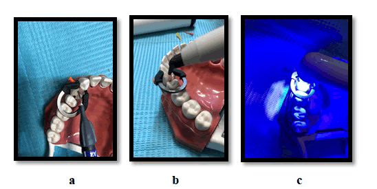
Group 3 (Snow plow technique): Similar to group 2, 1 mm flow able composite was injected in the gingival floor however; this layer was left uncured. Followed by application of predetermine amount of bulk fill composite (that fill 4 mm depth of the cavity). A condenser was used to pack the two layers ending with final thickness of 4 mm, all the excess of composite was removed thoroughly and then the 2 layers was curing simultaneously for 40 seconds. The rest of the cavity restored as group 1 Figure 6.
Figure 6: a) Application of 1 mm flow able composite 6; b) Application of 4 mm bulk fill composite; c) Curing of the two layers for 40 seconds.
Group 4 (Injection molding technique): similar to group 3 except the application of adhesive coating without curing before flow able and bulk fill composite. And the three layers were occurred as a one layer for 40 seconds. The full of the cavity restored as group Figure 7.
Figure 7: a) Adhesive coating; b) Application of 1 mm flow able composite; c) Application of 4 mm bulk fill composite; d) Curing of the three layers simultaneously.
All the restorations were finished and polished by enhancing finishing system The teeth were placed in deionized distilled water and subjected to 500 thermo cycling ISO/TR 11405:1994. After completing the thermo cycling, the teeth were dried and the apex of the roots sealed by model wax and the teeth were painted with double coating of nail varnish al around except 1 mm around the restoration. Then all examined teeth were submerged in 2% methylene blue for 24 hours at 37áµ?C.
After 24 h the teeth removed from the dye and rinsed thoroughly by running water and dried for 5 seconds. The teeth were embedded in self-curing epoxy resin and allowed for complete setting before sectioning.
Then the teeth were sectioned from mesial to distal side with water cooled slow speed diamond sectioning saw to obtain to similar fragment. The sectioned restoration were examined under stereomicroscope 40X to evaluate the extent of gingival marginal micro leakage.
The degree of gingival micro leakage were analysed according to scoring system suggested by Araujo as following [10].
- Score 0-no dye penetration
- Score 1-dye penetration less than ½ gingival floors
- Score 2-dye penetration more than ½ gingival floors
- Score 3-dye penetration involving the axial wall.
All data analysed by SPSS (BM Corp. Released 2013) shapero Wilk normality test was used and showed that the data were not normally distributed. The statistical analysis for micro leakage had been performed using the Kruskal Wallis test followed by the Mann Whitney U-tests with a significance level of p<0.05.
Results
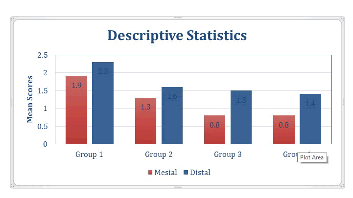
The scoring data showed variable range from 0-3 score in four groups as show in Figure 8 (Table 1).
Figure 8: Scoring data showed variable range from 0-3 score in four groups.
| margin | Subgroups | N | Minimum | Maximum | Mean | Medium | SD | Mean rank | P value | SIGNIFICANCE |
|---|---|---|---|---|---|---|---|---|---|---|
| Mesial | Groups 1 | 10 | 1 | 3 | 1.9 | 2 | 0.876 | 28.85 | 0.008 | S |
| Groups 2 | 10 | 1 | 3 | 1.3 | 1 | 0.823 | 22.85 | |||
| Groups 3 | 10 | 0 | 2 | 0.8 | 1 | 0.675 | 15.6 | |||
| Groups 4 | 10 | 0 | 3 | 0.8 | 1 | 0.966 | 14.7 | |||
| Distal | Groups 1 | 10 | 0 | 1 | 2.3 | 2.5 | 0.422 | 27.85 | 0.96 | NS |
| Groups 2 | 10 | 0 | 3 | 1.6 | 1.5 | 0.85 | 19.4 | |||
| Groups 3 | 10 | 0 | 3 | 1.5 | 1.5 | 0.853 | 18.35 | |||
| Groups 4 | 10 | 1 | 3 | 1.4 | 1 | 0.883 | 16.4 |
Table 1: kruskal wallis test.
The kruskal wallis test revealed significant difference among tested groups in mesial margin (enamel) with the highest marginal leakage score in group 1 (bulk fill) while the lowest score reported in both group 3 and group 4. In distal margin (dentin), non-significant differences had been resulted.
Figure 9: Bar chart descriptive the mean values for each group and subgroups.
Mann-Whitney U Test
Mann-Whitney U test results show in Table 2 and 3.
| Groups | Mean Rank | Mann-Whitney U | p-value | Significance |
|---|---|---|---|---|
| Groups 1 | 12.3 | 32 | 0.143 | NS |
| Groups 2 | 8.7 | |||
| Groups 1 | 13.9 | 16 | 0.004 | S |
| Groups 3 | 7.1 | |||
| Groups 1 | 13.65 | 18.5 | 0.012 | S |
| Groups 4 | 7.35 | |||
| Groups 2 | 12.6 | 29 | 0.061 | NS |
| Groups 3 | 8.4 | |||
| Groups 2 | 12.55 | 29.5 | 0.093 | NS |
| Groups 4 | 8.45 | |||
| Groups 3 | 11.1 | 44 | 0.588 | NS |
| Groups 4 | 9.9 |
Table 2: Mann-Whitney U test results.
| Subgroup | Mean rank | Mann–Whitney U | p-value | Significance | |
|---|---|---|---|---|---|
| Control | enamel | 7.65 | 21.5 | 0.021 | S |
| dentin | 13.35 | ||||
| Flow able | enamel | 7.95 | 24.5 | 0.038 | S |
| dentin | 13.05 | ||||
| Snowplow | enamel | 8 | 25 | 0.032 | S |
| dentin | 13 | ||||
| Injection | enamel | 8.2 | 27 | 0.048 | S |
| dentin | 12.8 |
Table 3: Mesial and Distal margins Mann-Whitney U test results.
Discussion
Micro leakage is one of these detrimental drawback in in class II composite restorations, which define as clinically dynamic undetectable passage of chemical substances bacteria, fluids, molecules and ions between the cavity walls and the restoration material applied [11].
Micro leakage is regarded as indication of marginal gap which result in Postoperative sensitivity, Marginal percolation and secondary caries.
The aim of present study was to evaluate the effect of bulk fill composite with different placement technique and the effect of margin location on marginal leakage of class II composite restoration.
To our knowledge this is the first study that evaluates the marginal leakage of snow plow and injection molding techniques using of bulk fill composite.
The result of this study revealed the different placement techniques significantly affect the magnitude of marginal leakage and thus the null hypotheses had been rejected.
Group 1 and group 2 showed the highest marginal leakage with no significant difference among them. The highest mean marginal leakage in bulk fill composite may attributed to high viscosity of bulk fill which result in less adaptation to cavity walls hence increased magnitude of micro leakage [12]. Non-significant difference between group 1 and 2 was result. This had agreement with other study which advocate that the use of intermediate layer underneath the bulk fill composite did not reduce the micro leakage. This may result from the higher resin content and less filler loading of the flow able composite resins causes more polymerization shrinkage and eventually more micro leakage [11,13].
Although non-significant difference between group 1 and group 2 had been reported in this study, slight reduction in marginal leakage result from the use of flow able underneath the bulk fill composite which attributed to the high adaptation of flow able composite at the gingival margin and it is act as a flexible intermediate layer which helps to relieve stresses during polymerization shrinkage. Also, the flow ability and inject ability of fluid composites make them very attractive when placing in difficult areas, such as the proximal boxes [14] and this agree with results obtained from two other studies in which mentioned that the addition of flow able composite in first layer before bulk fill can greatly reduce the micro leakage and the least micro leakage had reported in both snow plow and injection molding techniques with no significant different between them. While both of these two technique reported significant difference with group 1 [7,15].
This result has agreement with many studies that mentioned that the use of uncured flow able composite underneath the packable composite result in marked reduction in micro leakage in class II restoration [16,17].
The use of these techniques in which most of the flow able composite and therefore its potential disadvantages are removed from the cavity. Instead only a small amount of flow able composite remains in the areas of the cavity in which the higher viscosity resin composite does not completely adapt to the preparation. Therefore this technique may reduce void formation and increase marginal adaptation. In addition the use uncured resin (adhesive and flow able) increase the wettability so can penetrate better into the dentinal tubules and improve sealing at the margins due to the hydraulic pressure of overlying composite with the higher viscosity. Therefore, there would be less gap in the tooth-restoration interface and subsequently less leakage with this technique compared to separately curing the flow able liner [2] and agreement with [18] that show that the three layers co-cured together results in a monolithic mass to which improve cavity adaptation and reduce gap formation.
Another cause which mentioned by David, J Clark who stated that “air thinning the resin drives it past the margins, leaving a gingival margin that is not light cured, and is prone to dissolution and an eventual void. Which avoided by injection molding technique” [9].
But this studies show disagreement with other studies that show there was no decrease in marginal leakage from the use of uncured flow able underneath the bulk fill [19] and [20]. The reduction in leakage reported in injection molding techniques opposite to results of other two studies [21] and [22] which mentioned that the use of a flow able resin composite cured simultaneously with an adhesive yielded the worst results among tested groups. And they attributed this result to the displacement of the bonding agent leaving unprotected zone that may cause adhesive failure and subsequent increase in the micro leakage score or by a limited depth of curing of these component layers. However in this study we overcome these two problems by using first layer of adhesive that cured separately prior restorative procedure and by using translucent bio clear matrices which allow a better curing efficiency.
The use of preheating flow able and bulk fill composite were advocated newly in injection molding technique to increase the fluidity and the adaptation of composite to cavity walls but in this study we except the use of preheating technique to overcome the introduction of variable factor among the comparative groups.
The use of preheating composite may give different result from our study and may yield a difference marginal leakage for injection molding technique from snow plow technique. In distal cavity where the cavity margin located in dentin, the results yield non-significant difference between the groups this result agree with [22]. Who found that the restoration techniques did not influence the values of micro leakage in dentin and [19] who stated neither the addition of flow able composite nor the use of snow plow technique had noticeable decrease in marginal micro leakage in dentin.
This may result from the distance between the light curing tip and the surface of the resin (composite and adhesive). If this distance greater than 2 mm, the light intensity is significantly reduced. Which result in inadequate polymerization of resin composite materials? As a result, the conversion degree of resin at the dentinal margin is expected to be lower than at the enamel margin, which received the curing light directly [23]. In addition, the Margins below the CEJ make it difficult to achieve efficient results for marginal sealing, polishing and longevity [24].
Regard the effect of margin location, significant differences between enamel and dentin are reported. Dentin margin revealed highest marginal leakage compared to enamel in all groups. These results are supported by the findings of Benetti [25,26]. Who observed when a bulk fill composite resin was used; large gaps in dentin margins compared to enamel had been result. This may be due to the higher adhesive bond to etched enamel compared to etched dentin resulting in greater resistance to thermal changes at the enamel.
The findings of this study agree with those of Kalmowicz, Phebus [27]. Who concluded that micro leakage in enamel was significantly lower than dentin, regardless of material, C-factor, or insertion technique?
Other reasons for increased micro leakage at the dentinal margin may be: the organic content of dentin, outward movement of fluids in dentinal tubular and due to the complex and The branching of dentinal tubules in root dentin is smaller and more numerous than in crown dentin [28,29].
In the fact that there is limited literature on the use of snow plow technique and Injection molding technique, so to determine its clinical validity further in vitro and in vivo studies are needed.
Conclusion
Within the limitation of this study
- None of the examined groups provide complete elimination of marginal leakage of class II restoration neither in enamel nor in dentin.
- The different placement techniques of bulk fill composite had significant effect on marginal leakage on enamel margin of class II restoration, with greater reduction in the leakage noticed in snow plow and injection molding technique.
- Marginal leakage of dentinal margin didn’t get affected by placement technique.
- For intragroup comparison, higher leakage score were presented in dentin compared to enamel irrespective of placement techniques.
References
- Eklund SA. Trends in dental treatment, 1992 to 2007. J Am Dent Assoc 2010; 141:391-399.
- NJ Opdam, JJ Roeters, T de Boer, et al. Voids and porosities in class I micro preparations filled with various resin composites. Oper Dent 2003; 28:9-14.
- L Saatwika, A Karthick, A Subbiya, et al. A Review on Polymerization Shrinkage of Resin Composites. European J Molecular Clin Med 2020; 7:1245-1250.
- Deliperi S, DN Bardwell. An alternative method to reduce polymerization shrinkage in direct posterior composite restorations. J Am Dent Assoc 2002; 133:1387-1398.
- Chandrasekhar V, Rudrapati L, Badami V, et al. Incremental techniques in direct composite restoration. J Conserve Dent 2017; 20:386.
- Boaro LCC, Lopes DP, de Souza ASC, et al. Clinical performance and chemical-physical properties of bulk fill composites resin a systematic review and meta-analysis. Dent Mater 2019; 35:249-264.
- Kaisarly D, Meierhofer D, Gezawi MEl, et al. Effects of flow able liners on the shrinkage vectors of bulk-fill composites. Clin Oral Investig 2021; 1-14.
- Doustfateme S, Khosravi K, Hosseini S. Comparative evaluation of micro leakage of bulk-fill and posterior composite resins using the incremental technique and a liner in Cl II restorations. J Islam Dent Assoc Iran 2018; 30:1-8.
- Clark DJ. The injection-molded technique for strong, esthetic Class II restorations. Inside Dent 2010; 6:68-76.
- Piva E, Demarco FF, de Araujo CS, et al. Micro leakage of seven adhesive systems in enamel and dentin. J Contemp Dent Pract 2006; 7:26-33.
- Baig MM, Mustafa M, Al Jeaidi. ZA. Micro leakage evaluation in restorations using different resin composite insertion techniques and liners in preparations with high c-factorâ??An in vitro study. Saudi Dent J 2013; 4: 57-64.
- Agarwal RS, Hiremath H, Agarwal J, et al., Evaluation of cervical marginal and internal adaptation using newer bulk fill composites: An in vitro study. J conserv dent 2015; 18:56.
- Tredwin, CJ, A Stokes, and DR Moles, et al. Influence of flowable liner and margin location on micro leakage of conventional and packable class II resin composites. Oper Dent 2005; 30:32-38.
- S F Chuang, J K Liu, C C Chao, et al. Effects of flowable composite lining and operator experience on microleakage and internal voids in class II composite restorations. J prosthet dent 2001; 85:177-183.
- Tabona MRZ, A Soetojo, and I Widjiastuti. Bulkfill Techniques with Intermediate Layer to Marginal Adaptation Restoration of Class II Composite Resin. Conserv Dent J 2021; 11:32-37.
- Basanagouda S Patil, Laxmikant Kamatagi, Hrishikesh Saojii, et al. Cervical Micro leakage in Giomer Restorations: An In Vitro j contemp dent pract 2020; 21:161-165.
- Presicci, Anthony. Porosity Formation and Micro leakage of Composite Resins Using the Snowplow Technique. Uniformed Services University of the Health Sciences, Bethesda, Maryland, 2012; 12.
- Solomon RV, P karunakar, P Hima Rajana et al. Influence of Sandwich, Snow plow and Injection Molding Modified Composite Lining Strategies on the Micro leakage of Class ii Proximal Box Cavities. 2020.
- Reddy SN, Jayashankar DN, Nainan M, et al., The effect of flowable composite lining thickness with various curing techniques on microleakage in class II composite restorations: an in vitro study. J Contemp Dent Pract 2013; 14:56.
- Lotfi N, Esmaeili B, Ahmadizenouz G, et al. Gingival micro leakage in class II composite restorations using different flow able composites as liner: an in vitro evaluation. Caspian J Dent Res 2015; 4: 10-16.
- Sensi LG, Marson FC, Sylvio Monteiro Jr, et al. Flow able composites as â??filled adhesives:â? A micro leakage study. J Contemp Dent Pract 2004; 5:32-41.
- N Al-Yousifany N. Effects of Flowable Composite Resin and curing method on Microleakage. Al-Rafidain Dent J 2010; 10:1-7.
[Crossref][Google Scholar][Indexed]
- Catelan A, Araujo LSND, da Silveira BCM. Impact of the distance of light curing on the degree of conversion and micro hardness of a composite resin. Acta Odontol Scand 2015; 73: 298-301.
- Kramer N, C Reinelt, R Frankenberger. Ten-year clinical performance of posterior resin composite restorations. J Adhes Dent 2015; 17:433-441.
- A R Benetti, C Havndrup-Pedersen, D Honore, et al. Bulk-fill resin composites: polymerization contraction, depth of cure, and gap formation. Oper dent 2015; 40:190-200.
- F Al-Harbi, D Kaisarly, A Michna, et al. Cervical interfacial bonding effectiveness of class II bulk versus incremental fill resin composite restorations. Oper dent 2015; 40:622-635.
- J Kalmowicz, J G Phebus, B M Owens, et al. Micro leakage of class I and II composite resin restorations using a sonic-resin placement system. Oper dent 2015; 40:653-661.
- M Peumans, P KanumilliJ, De Munck, et al. Clinical effectiveness of contemporary adhesives: a systematic review of current clinical trials. Dent mater 2005; 21:864-881.
- Poggio C, ChiesaM, Scribante A, et al. Micro leakage in Class II composite restorations with margins below the CEJ: In vitro evaluation of different restorative techniques. Med Oral Patol Oral Cir Bucal 2013; 18:793.
Author Info
Sura Y Alani and Abdulla MW Al-Shamma*
Department of Restorative and Esthetic Dentistry, College of Dentistry, Baghdad University, IraqCitation: Sura Y Alani, Abdulla MW Al-Shamma, The Effect of Different Placement Strategies of Bulkfill Composite on Marginal Leakage of Classii Restoration (Comparative in Vitro Study), J Res Med Dent Sci, 2022, 10 (8): 000-000.
Received: 01-Jun-2022, Manuscript No. JRMDS-22-52927; Accepted: 08-Aug-2022, Pre QC No. JRMDS-22-52927; Editor assigned: 03-Jun-2022, Pre QC No. JRMDS-22-52927; Reviewed: 17-Jun-2022, QC No. JRMDS-22-52927; Revised: 02-Aug-2022, Manuscript No. JRMDS-22-52927;

