Case Report - (2022) Volume 10, Issue 5
Swelling of the Sublingual Carbuncle Area Caused By a Sialolith in the Salivary Gland Duct-A Case Report
Mohan kumar J* and Rajasekar MK
*Correspondence: Mohan kumar J, Department of ENT, Sree Balaji Medical College and Hospital, Chrompet, Chennai, India, Email:
Abstract
Sialolithiasis is the most common disease of the salivary glands. The existence of calculus inside a salivary glands ductal system. Salivary calculi are a common cause of salivary gland disorders, and they can affect any of the salivary glands at any age. Tiny intra-ductal stones or stones inside the gland material are possible. Diagnosis is made by clinical examination usually and the treatment is aimed at removal of the stone. A 20 years old male patient presented with a rigid swelling under the tongue and around the phrenulum. This is a case study of an intraductal stone that was surgically removed. Due to the viscous nature of its mucinous secretions, high calcium content, and tortuous duct, sialolith is relatively widespread (80%) in the submandibular salivary gland.
Keywords
Sialolithiasis, Salivary glands, Duct stoneIntroduction
Sialolithiasis is a widespread disorder that affects about 12 out of every 1000 people per year, accounting for more than half of all salivary gland diseases [1]. It may affect children, but adults in their third to sixth decades are more likely to be affected [2]. Males are more likely to produce sialoliths, with some writers suggesting that the submandibular gland forms over 80% of salivary calculi and the parotid gland just 20% [3]. Occasionally, the minor salivary glands and sublingual glands are affected. The Wharton's duct of the submandibular gland is where the majority of salivary calculi (80%) are located. The gland's most distinguishing feature is that the saliva it secretes flows against gravity; it also contains a lot of mucin, calcium, and phosphate. The accessory salivary duct of the submandibular gland is a rare phenomenon. It's a copy of the original Wharton's duct that runs parallel to it [4].
Case Presentation
A 20 years old male patient reported to our department with a chief complaint of decrease of saliva secretion, pain and dysphagia for 3 months. During massage of the gland, decreased salivary flow or purulent exudate may be observed. He had taken conservative analgesia and antibiotic treatment for the same before. On detailed history he presented swelling appears just be for his meals times, which reduced gradually over 2-3 hrs. Pain and tenderness during that time was severe.
On examination no evident dental focus of infection was located. Bimanual palpation of gland showed mild enlargement and tenderness. Findings on blood and serum biochemistry were with in normal limits after preliminary investigations patient was operated for intraoral sialolithotomy for removal of duct stone under local anaesthesia and the specimen stored in diluted formalin and sent histopathology examination, (Figures 1-5).
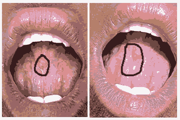
Figure 1:Pre op view.
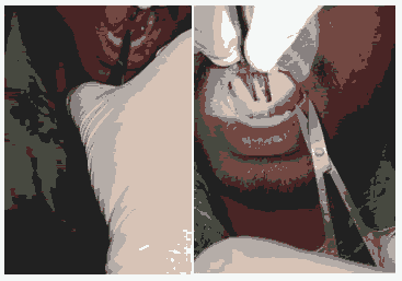
Figure 2:Intra Op view.
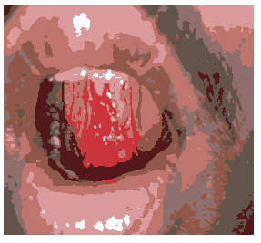
Figure 3:Post Op view.
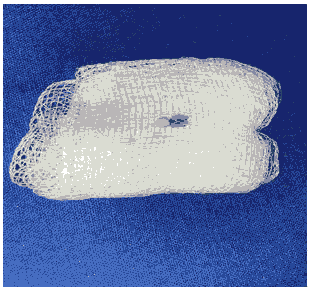
Figure 4:Salivary gland calculi.
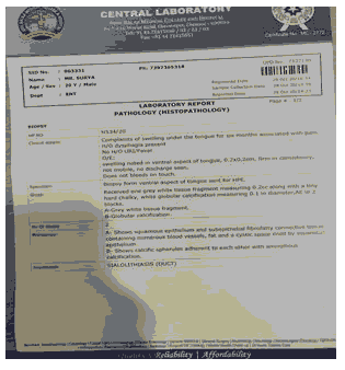
Figure 5:Histopath report.
Results and Discussion
While salivary gland stones are common in the general population, the cause of sialolith formation is unknown. The sialolith nuclei are often made up of inorganic materials including calcium phosphate and calcium carbonate, although there is evidence that the nidus can also contain bacterial matter [5]. Investigated the mineral composition of sialoliths in different parts of the ductal system and discovered that stones in Wharton's duct contained more whitlockite crystals than those in intra glandular ducts. This may be due to the higher calcium and phosphate content of Wharton's duct saliva [6].
Conclusion
With the newer treatment options of endoscope i.e. salivary Culculi lithotripsy the surgical removal may become minimally invasive but still today the long tested methods of Tran’s oral evacuation of stone especially in distal part of duct are successful with minimum hazards.
References
- Leung AK, Choi MC, Wagner GA, et al. Multiple Sialoliths and a Sialolith of Unusual Size in the Submandibular Duct: A Case Report. Oral Surg, Oral Med, Oral Pathol, Oral Radiol Endod 1999; 87:331-333.
- Lustman J, Regev E, Melamed Y, et al. A Survey on 245 Patients and a Review of the Literature. Int J Oral Maxillofac Surg 1990; 19:135-138.
- Bodner L. Giant Salivary Gland Calculi: Diagnostic Imaging and Surgical Management. Oral Surg, Oral Med, Oral Pathol, Oral Radiol, Endod 2002; 94:320-323.
- Rauch S. Disease of Salivary Grands. Thoma's Oral Pathol 1970; 997-1003
- Carr SJ. Sialolith of Unusual Size and Configuration. Report of a Case. Oral Surg, Oral Med, Oral Pathol 1965; 20:709-712.
- Teymoortash A, Buck P, Jepsen H, et al. Sialolith Cystals Localized Intraglandularly and in the Wharton's Duct of the Human Submandibular Gland: An X-ray Diffraction Analysis. Arch Oral Biol 2003; 48:233-236.
- Koch M, Zenk J, Iro H, et al. Algorithms for Treatment of Salivary Gland Obstructions. Otolaryngol Clin North Am 2009; 42:1173-1192.
- Harrison JD. Causes, Natural History, and Incidence of Salivary Stones and Obstructions. Otolaryngol Clin North Am 2009; 42:927-947.
Author Info
Mohan kumar J* and Rajasekar MK
Department of ENT, Sree Balaji Medical College and Hospital, Chrompet, Chennai, IndiaCitation: Mohan kumar J, Rajasekar MK. Swelling of the Sublingual Caruncle Area Caused by a Sialolith in the Salivary Gland Duct-A Case Report, J Res Med Dent Sci, 2022, 10(5):45-46.
Received: 21-Feb-2022, Manuscript No. 41501; , Pre QC No. 41501; Editor assigned: 23-Feb-2022, Pre QC No. 41501; Reviewed: 09-Mar-2022, QC No. 41501; Revised: 22-Apr-2022, Manuscript No. 41501; Published: 03-May-2022
