Research - (2022) Volume 10, Issue 3
Study of Geometrical Measurements of an Adult Human Canine Tooth in Both Jaws in Salah Al-Din Governorate
Naba’a Ahmed Al-Nasiri*, Saad Ahmed Mohammed and Mohammed Ahmed Abdullah
*Correspondence: Naba’a Ahmed Al-Nasiri, Department of human anatomy and histology, College of Medicine, University of Tikrit, Iraq, Email:
Abstract
Background: Variation in tooth measurements is influenced by genetic and environmental factors. Tooth size varies between and within different racial groups. Objectives: the aim of study was to improve the quality of dental care available, and to allow highly precise measurements using an approximation of the mean of each parameter for each canine tooth form. Material and Methods: 26 patients were selected, with ages ranging from 20-40 years old, for eight canine tooth measurements done with two methods: cone beam computed tomography, and extracted canine teeth measured with digital Vernier caliper. Results: result revealed that the length of the crown is longer in females than males, while the mesiodistal width is larger in males than females, and the length of the root is longer in males than females, according to sex. Conclusions: Cone beam computed tomography (CBCT) imaging allows us to measure the tooth dimensions when a patient is exposed to CBCT. Digital Vernier caliper enables to take the tooth measurement on extracted teeth. The tooth size of canine showed the greater variation of the sexual dimorphism, larger in males and smaller in females.
Keywords
Canine tooth, Measurements, CBCT, Mesiodistal width, Sexual dimorphism
Introduction
The distance between two parallel lines perpendicular to the mesiodistal axis of the tooth and tangential to the most mesial and most distal points of the crown along a parallel line to the occlusal plane is defined as the mesiodistal crown diameter, also known as tooth size, tooth crown size, or tooth mesiodistal width [1]. The conebeam computed tomography (CBCT) technology, which can be used to precisely measure the size of teeth, is the most significant advancement in imaging techniques for the maxillofacial region since the advent of the panoramic approach [2]. The capacity to create 3D pictures, the use of a noninvasive method, the decrease of orofacial structure duplication, and reduced radiation exposures and costs are all advantages of the CBCT approach [3]. Differences in tooth size or form between men and females of the same species are known as sexual dimorphism [4]. Tooth size is influenced by both the environment and heredity, and it is closely connected to sex and race. Male teeth are significantly larger than female teeth, while African teeth are larger than European teeth [5].
Many research focuses on mesiodistal width in human populations because it may be employed in forensic investigations, human evolution, biological problems, and clinical dentistry. The relationship between the mesiodistal crown diameter and arch alignment is important in clinical dentistry to determine malocclusion and crowding because a proper mesiodistal crown diameter relationship between the upper and lower teeth is required for proper inter-digitation, over-jet, and overbite in final orthodontic treatment [6].
Materials and Methods
Totally, 26 patients will be selected from Tikrit specialized dental center in Tikrit, with ages ranging from 20-40 years old. The samples were divided into two groups according to their sex.
Digital Vernier caliper measurements: First, both maxillary and mandibular canine teeth had been extracted. The digital caliper made reading the figure from the display easier, according to the manufacturer's recommendations. The display could be changed from millimeters to inches, and the display could be zeroed at the start or anytime along the slide. The digital caliper's slide may also be operated with a thumb roller or secured with a thumbscrew. The readings were shown to the tenth of a millimeter (Figures 1-3).
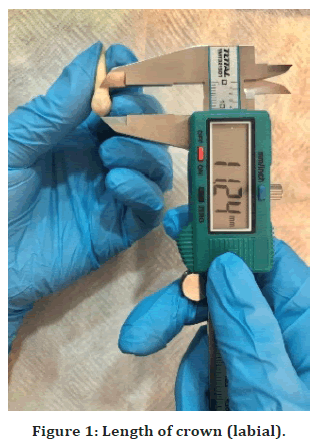
Figure 1. Length of crown (labial).
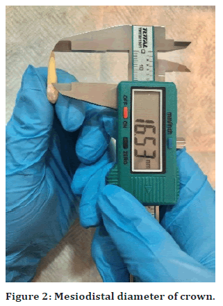
Figure 2. Mesiodistal diameter of crown.
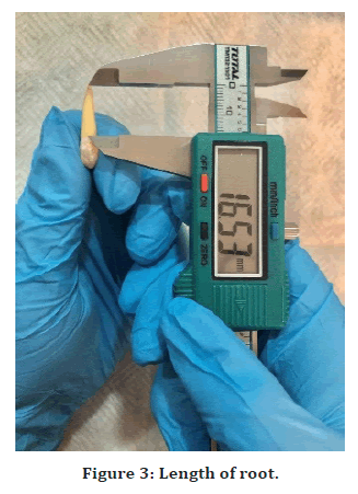
Figure 3. Length of root.
Measurements of cone beam computed tomography: All CBCT pictures were taken with a Carestream, CS 8100 3D, France. The following were the technical requirements: 15 cm circular field of view, 15.4 cm spherical imaging volume, 150mm isotropic voxel size The CBCT radiographs were obtained with the following settings: 90 kVp tube voltage, 3.20mA intensity range, and a 15-second exposure time. CBCT data was rebuilt using the system's proprietary software with 1.1mm-thick slices spaced at 0.125 mm intervals (Figures 4-6).
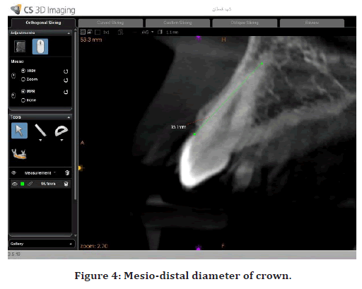
Figure 4. Mesio-distal diameter of crown.
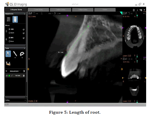
Figure 5. Length of root.
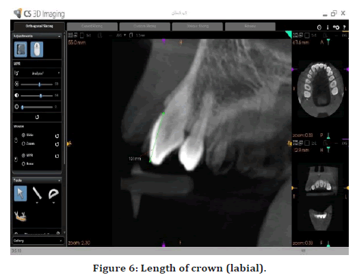
Figure 6. Length of crown (labial).
Statistical assessment
All the data collected and results were statistically analyzed, mean and standard deviation were calculated according to (ANOVA) test. Also graphs and tables were made, to determine the significance differences among the study groups by using Duncan’s multiple range test.
Results
After measuring with a (CBCT) and (Digital Vernier caliper), the results of canine tooth measurements according to sex are clarified in the Table 1 below.
| The measurement in mm | |||||
|---|---|---|---|---|---|
| No. | The name of the tooth | Sex | CL | RL | MD |
| 1 | Maxillary Left canine | Female | 8.9 | 18.2 | 8.5 |
| 2 | Maxillary Right canine | Female | 9.1 | 20.6 | 8.6 |
| 3 | Mandibular Left canine | Male | 11.1 | 22.7 | 7.7 |
| 4 | Mandibular Right canine | Male | 10.8 | 21.9 | 7.7 |
| 5 | Maxillary Right canine | Male | 10.8 | 15.1 | 7.2 |
| 6 | Maxillary Left canine | Male | 10.6 | 14.7 | 7.3 |
| 7 | Mandibular Right canine | Female | 11.8 | 14.4 | 6.9 |
| 8 | Mandibular Left canine | Female | 11.7 | 15.4 | 7.2 |
| 9 | Maxillary Right canine | Female | 10.3 | 16.8 | 7.6 |
| 10 | Maxillary Left canine | Male | 11.2 | 16.1 | 7.8 |
| 11 | Mandibular Right canine | Female | 10.2 | 16.8 | 6.6 |
| 12 | Mandibular Left canine | Male | 11.3 | 15.6 | 7.1 |
| 13 | Mandibular Right canine | Female | 10.6 | 10.7 | 5.9 |
| 14 | Mandibular Left canine | Female | 10.2 | 17.2 | 6.8 |
| 15 | Mandibular Right canine | Male | 11.6 | 17.3 | 7.4 |
| 16 | Mandibular Left canine | Male | 11.4 | 16.9 | 7.4 |
| 17 | Maxillary Left canine | Female | 10.2 | 17.8 | 7.9 |
| 18 | Maxillary Left canine | Male | 9.5 | 18.6 | 7.9 |
| 19 | Maxillary Right canine | Male | 10.3 | 19.2 | 8 |
| 20 | Maxillary Left canine | Female | 10.2 | 17.7 | 7.7 |
| 21 | Maxillary Right canine | Male | 13.8 | 13.5 | 8.7 |
| 22 | Maxillary Left canine | Male | 14.2 | 12.6 | 8.8 |
| 23 | Maxillary Left canine | Female | 11.3 | 16.6 | 8.1 |
| 24 | Maxillary Left canine | Male | 13.1 | 17.9 | 7.8 |
| 25 | Mandibular Right canine | Female | 11.3 | 18.7 | 6.9 |
| 26 | Mandibular Left canine | Female | 9.2 | 17 | 6.3 |
Table 1: The measurements of teeth in mm, CL: crown length, RL: root length, MD: mesio-distal diameter of the crown.
Statistical finding
Statistical analysis according to sex
From the results shown in the Table 2, it becomes clear that there are significant differences between males and females in the groups included in the study, it is evident that the highest value of the mean of the length of crown (Maxillary Left canine in females) did not differ from all the other groups (for the female).
| Groups | Sex | |
|---|---|---|
| Male | Female | |
| Maxillary left canine | 10.150 ± 0.98bc | 11.720 ± 1.904a |
| Maxillary Right canine | 9.700 ± 0.849c | 11.63 ± 1.89a |
| Mandibular Left canine | 10.367 ± 1.258bc | 11.267 ± 0.1528a |
| Mandibular Right canine | 10.975 ± 0.714ab | 11.200 ± 0.566a |
Table 2: Statistical analysis for length of crown (labial).
While in the group of males, there were significant differences between them. The lowest value of the mean in the group (Maxillary Right canine) was in males.
It is clear from the findings in the Table 3 below that there are significant differences between males and females in the groups studied. It is also clear that the highest value of the mean root length (Mandibular Right canine in males). There were major variations between the females in the group. Females have the lowest mean value in the population (mandibular Right canine). The Table 4 below shows that there are significant differences between males and females in the sample groups. It is also obvious that the highest value of the mean Mesio-distal diameter of crown (Maxillary Right canine in males). Although there were major variations between the males in the group, females had the lowest mean (Mandibular Right canine) in the group.
| Group | Sex | |
|---|---|---|
| Male | Female | |
| Maxillary Left canine | 15.98 ± 2.43c | 17.56 ± 0.685ab |
| Maxillary Right canine | 15.93 ± 2.94c | 18.7 ± 2.69a |
| Mandibular Left canine | 18.40 ± 3.78ab | 16.53 ± 0.987bc |
| Mandibular Right canine | 19.60 ± 3.25a | 15.15 ± 3.45c |
Table 3: Statistical analysis for length of root.
| Groups | Sex | |
|---|---|---|
| Male | Female | |
| Maxillary Left canine | 7.920 ± 0.545a | 8.050 ± 0.342a |
| Maxillary Right canine | 8.100 ± 0.707a | 7.967 ± 0.751a |
| Mandibular Left canine | 7.400 ± 0.300b | 6.767 ± 0.451c |
| Mandibular Right canine | 7.550 ± 0.212b | 6.575 ± 0.472c |
Table 4: Statistical analysis for mesio-distal diameter of crown.
Discussion and Conclusion
The mesiodistal dimensions of the teeth of the extracted teeth were measured using digital calipers, a procedure that was also utilized by [7]. The advent of CBCT enabled for the assessment of tooth geometry and measurements by using the long axis of the teeth rather than their labial surface on castings, resulting in enhanced measuring precision, as described by [8]. The slice-by-slice mode of CBCT allows for the depiction of each tooth on any selected plane, which is consistent with [9]. A research by De Angelis et al., which was similar to the current study, found a definite sex-related difference in the mesiodistal width of permanent teeth, with males having higher mesiodistal width of teeth than women [10]. According to a research by Omar H et al., the canine is the tooth with the biggest variation in size between the sexes, which is the same information in instant findings [11]. Other studies have found that sex has an impact on root length and the number of lower canine canals. The average length of the mandibular canine root in those studies was considerably longer in males than in females [12], this was agreed with existent study. Sadeghi et al., on the other hand, disagree with the findings of this study. Sadeghi et al. did not find a link between sex and tooth size [13]. The findings of this study, which show a significant influence of sex on tooth sizes in general and canines in particular, are similar with earlier findings, such as those of Eshghi et al., who found that men had a longer mean length for canines than females. In addition, Agrawal et al. found a significantly higher prevalence of sex dimorphism in canines, while Alvesalo et al. found a general influence of sex on tooth sizes, both of which are congruent with the findings of this study [14,15]. The findings of maxillary canine dimorphism in this study were comparable to those reported by Khangura et al in their investigation [16]. The average anatomical length of the crown and root of maxillary canines in present study was 10.96 mm and 16.82 mm. In mandibular canines, the average anatomical length of the crown and root was 10.9 mm and 17.05 mm, respectively, while the average anatomical length of the crown and root of maxillary canines in Indian population was 9.61 mm and 16.82 mm. In mandibular canines, the average anatomical length of the crown and root was 8.70 mm and 15.51 mm, respectively. So that only the average anatomical length of the root of maxillary canines was agreed with current study [17]. The average length of the crown was 9.61mm in the maxillary canine in this study [17] Ash and Nelson [18] reported the average length of the crown to be 10mm in the maxillary canine, which was differ slightly from present day findings. The average length of the root in maxillary canine was 16.82mm which is close to the findings of Ash and Nelson [19], (17mm), which was the same of instant results. In this study, the average length of the crown of mandibular canine was 8.70 mm, whereas the average length of the crown in Ash and Nelson’s [18] study was 11 mm. Versiani et al. [19] reported that the average length of the root of mandibular canine ranged from 12.53mm to 18.08 mm, which was not similar to the present findings. The current study revealed significant differences between males and females in the study's groups; it is clear that the greatest value of the mean of crown length (Maxillary Left canine in females) indicates that crown length is larger in females than males; however, this conclusion was disputed by [20].
The reasons for the differences in tooth sizes between men and women are sex-related differences in odontogenic timing and enamel thickness, as well as men's larger body size in comparison to women; in this context, hormonal differences and the presence of chromosome X also affect tooth sizes, it was agreed with [21]. Empirical data, on the other hand, suggests that changes in different environmental variables, such as severe starvation and nutritional improvements, can influence tooth sizes in some animals, as the current study found [22]. Tooth size varies between races as well as between people. Genetics, diet, and environmental variables all influence tooth size [23].
The canine tooth size revealed the most sexual dimorphism, being larger in males and smaller in females. Aside from genetics, numerous environmental variables, such as nutrition, illnesses, and childhood disorders, have an impact on tooth growth and can determine an individual's eventual tooth size. Other factors that influence tooth size include socioeconomic level and, in general, living standards. These discrepancies might be attributable to ethnic differences or a small sample size.
References
- Hasegawa Y, Amarsaikhan B, Chinvipas N, et al. Comparison of mesiodistal tooth crown diameters and arch dimensions between modern Mongolians and Japanese. Odontology 2014; 102:167-175.
- White SC, Pharaoh MJ. Oral radiology principles and interpretation. 7th Edn, Elsevier, st. Louis, Missouri, 2014; 185.
- Bulut DG, Kose E, Ozcan G, et al. Evaluation of root morphology and root canal configuration of premolars in the Turkish individuals using cone beam computed tomography. Eur J Dent 2015; 9:551.
- Bunger E, Jindal R, Pathania D, et al. Mesiodistal crown dimensions of the permanent dentition among school going children in Punjab population: An aid in sex determination. Int J Dent Health Sci 2014; 1:13-23.
- Altherr ER, Koroluk LD, Phillips C. Influence of sex and ethnic tooth-size differences on mixed-dentition space analysis. Am J Orthod Dentofac Orthop 2007; 132:332-9.
- Hattab FN. Mesiodistal crown diameters and tooth size discrepancy of permanent dentition in thalassemic patients. J Clin Exp Dent 2013; 5:e239.
- Abozeid H, El Shiekh M, AElsayed M. Determination of the combined mesiodistal widths of the permanent mandibular incisors and that of the maxillary and mandibular canines and premolars in a group of egyptian children in suez governorate: A cross-sectional study. Adv Dent J 2019; 1:77-85.
- Alkhatib R, Chung CH. Buccolingual inclination of first molars in untreated adults: A CBCT study. Angle Orthod 2017; 87:598-602.
- Palomo JM, Kau CH, Palomo LB, et al. Three-dimensional cone beam computerized tomography in dentistry. Dent Today 2006; 25:130.
- De Angelis D, Gibelli D, Gaudio D, et al. Sexual dimorphism of canine volume: A pilot study. Legal Med 2015; 17:163-166.
- Omar H, Alhajrasi M, Felemban N, et al. Dental arch dimensions, form and tooth size ratio among a Saudi sample. Saudi Med J 2018; 39: 86.
- Zhengyan Y, Keke L, Fei W, et al. Cone-beam computed tomography study of the root and canal morphology of mandibular permanent anterior teeth in a Chongqing population. Ther Clin Risk Manag 2016; 12:19.
- Sadeghi R, Sarlati F, Kalantari S. Relationship between crown forms and periodontium biotype. Res Dent Sci 2011; 8:1-8.
- Eshghi A, Tahririan D, Tayebi L, et al. Evaluation of the sizes of deciduous and permanent teeth in patients referring to Isfahan faculty of dentistry. Danshdhan Dahnah 2012; 722-728.
- Agrawal A, Manjunatha BS, Dholia B, et al. Comparison of sexual dimorphism of permanent mandibular canine with mandibular first molar by odontometrics. J Forensic Dent Sci 2015; 7:238.
- Khangura RK, Sircar K, Singh S, et al. Sex determination using mesiodistal dimension of permanent maxillary incisors and canines. J Forensic Dent Sci 2011; 3:81.
- Amardeep NS, Raghu S, Natanasabapathy V. Root canal morphology of permanent maxillary and mandibular canines in Indian population using cone beam computed tomography. Anatomy Res Int 2014; 2014.
- Ash MM, Nelson SJ. The permanent canines: maxillary and mandibular. In Wheeler’s Dental Anatomy, Physiology, and Occlusion. Elsevier, St. Louis, Mo, USA, 8th edition 2007; 191-174.
- Versiani MA, Pécora JD, Sousa‐Neto MD. Microcomputed tomography analysis of the root canal morphology of single‐rooted mandibular canines. Int Endod J 2013; 46:800-807.
- Pandey N, Ma M. Evaluation of sexual dimorphism in maxillary and mandibular canine using mesiodistal, labiolingual dimensions, and crown height. Indian J Dent Res 2016; 27.
- Kazzazi SM, Kranioti EF. Sex estimation using cervical dental measurements in an archaeological population from Iran. Archaeol Anthropol Sci 2018; 10:439-48.
- Silva AM, Pereira ML, Gouveia S, et al. A new approach to sex estimation using the mandibular canine index. Med Sci Law 2016; 56:7-12.
- Kaur A, Singh R, Mittal S, et al. Evaluation and applicability of Moyers mixed dentition arch analysis in Himachal population. Dent J Adv Studies 2014; 2:096-104.
Indexed at, Google Scholar, Cross Ref
Indexed at, Google Scholar, Cross Ref
Indexed at, Google Scholar, Cross Ref
Indexed at, Google Scholar, Cross Ref
Indexed at, Google Scholar, Cross Ref
Indexed at, Google Scholar, Cross Ref
Indexed at, Google Scholar, Cross Ref
Indexed at, Google Scholar, Cross Ref
Indexed at, Google Scholar, Cross Ref
Indexed at, Google Scholar, Cross Ref
Indexed at, Google Scholar, Cross Ref
Indexed at, Google Scholar, Cross Ref
Indexed at, Google Scholar, Cross Ref
Indexed at, Google Scholar, Cross Ref
Indexed at, Google Scholar, Cross Ref
Author Info
Naba’a Ahmed Al-Nasiri*, Saad Ahmed Mohammed and Mohammed Ahmed Abdullah
Department of human anatomy and histology, College of Medicine, University of Tikrit, IraqReceived: 04-Mar-2022, Manuscript No. JRMDS-22-51573; , Pre QC No. JRMDS-22-51573 (PQ); Editor assigned: 07-Feb-2022, Pre QC No. JRMDS-22-51573 (PQ); Reviewed: 21-Mar-2022, QC No. JRMDS-22-51573; Revised: 25-Mar-2022, Manuscript No. JRMDS-22-51573 (R); Published: 31-Mar-2022
