Research Article - (2022) Volume 10, Issue 10
Severity Valuation of Acute Pancreatitis with CT Severity Index and Modified CT Severity Index
Ankush Misra, M Senthil Velan and G Jayalakshmi*
*Correspondence: Dr. G Jayalakshmi, Department of Orthopaedics, Sree Balaji Medical College and Hospital, Bharath Institute of Higher Education and Research, Chennai, Tamil Nadu, India, Email:
Abstract
Introduction: The pancreatitis is one of the commonest intestinal disorder and existing as a clinically most complex disease due to the challenges in determining its severity. The severity stratification plays a main role in planning the treatment, prediction of organ failure among the acute pancreatitis patients. Though, many methods are existing, computer topography (CTSI) and modified CTSI are mainly used in hospitals. The present study aimed to analyse the disease severity among the patients admitted to the clinic.
Methods: This research was conducted on the patients who presented with acute pancreatitis diagnosed either clinically or ultrasonography (n=101, 14 were excluded in the study). All patients were subjected to CECT abdomen, according to which CT severity index and Modified CT severity index was calculated based on Revised Atlanta Score (RAC).
Results: Outcome was assessed by several factors which included duration of hospital stay, ICU admission, mortality or survival of the patient. Types of intervention tried i.e conservative or surgical. It was observed that as the severity index and score progressed for the patients, the outcome of the disease was also more morbid which was evident by the increase in revised atlanta Score of the patient. CT Severity Index (CTSI) and Modified CT Severity Index (MCTSI) increased concomitantly with Revised Atlanta Score (RAS).
Conclusion: In conclusion, it was observed that CT is an important solitary imaging modality to evaluate subjects with acute pancreatitis.
Keywords
Abdominal computed tomography, Complications, Pancreatic diseases, Pancreatic necrosis.
Introduction
Acute Pancreatitis (AP) is a complex disease with a wide range of clinical implications and challenge for its optimal diagnosis. Though the majority of the patients may recover completely, 15-20% of them develop severe disease recurrence and resulting in higher mortality rate (up to 20-30%). Diagnosing the disease severity is one of the main challengeable complications in AP patients. Presently, the disease severity has been classified in to two main phases. In first phase, the onset of systemic inflammatory response syndrome lead to the development of Multiple Organ Dysfunction Syndromes (MODS) accompanied with severe pancreatic inflammation and/or necrosis. About, 50% deaths were recorded by this type worldwide in first week of disease attack. Usually, the second phase occurred if there is no halt or immune modulation after the first phase. It is characterized by the development of compromised pancreatic necrosis or fluid aggregation with potential progression to sepsis, MODS and death. Meanwhile, the data analysis on the patients revealed that the half of pancreatic necrosis patients develops the localized organ failure and their severities normally do not affect the remote organ complications [1]. Showed that the infection rate was increased with severity and time course of the disease. Their study showed that nearly 24% patients develop infection on surgery at first week and followed by 46% and 71% on their second and third weeks respectively. It was also showed that the increase in organ failures reflected on higher mortality rates (30-100%) based on the variation in MODS [2]. Thus identification of disease severity is one of the main criteria to formulate the treatment regime and analysing the disease complications. The Severity stratification is important during the initial work-up of cases and the serum/urinary amylase concentration is a common method for diagnosis the AP patients and found to be prone for error in one-fifth of AP patients. Recently, a sophisticated method, called computer topography had been developed for finding disease stages. The CT severity index and modified CT severity index. Though, those two systems are widely used, there not adequate studies for comparing the outputs. Therefore, the aim of the present study was to assess the severity of AP using CTSI and MCTSI in patients admitted to bharath institute of higher education and research, Chennai for correlating clinical outcome measures, and to concord disease severity using Revised Atlanta Classification (RAC) method.
Materials and Methods
In the present prospective study, 101 patients were requited from department, of general surgery, bharath institute of higher education and research, Chennai during the period February 2018 to May 2019. The study was conducted after getting proper ethical permission (IRB number:LHMC/ECHR/2014/504). The patient’s data related with their demographic details, previous clinical history and laboratory assessment were reviewed along with the CT findings. Of the 101 patients, 14 showed chronic pancreatitis and excluded from the study. The CT scan was performed by trained clinicians and the assessment was done by a surgeon with 17 years of experience (median of 6 days; range 5–11 day).
Results
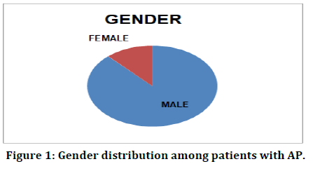
Totally, 101 patients were requited and 14 of them were excluded from the study. The remaining patients were followed up until their hospital discharge, or death or AMA. The study showed that a strong correlation between the gender and disease prevalence. In the present study, 84 patients were male and 16 of them were females (Table 1 and Figure 1) and the ratio seemed to be 5.2:1 in favour of males. The present study also revealed the disease prevalence in aging (Table 2 and Figure 2) and the predominance was observed in age groups between 31-45 and 46-60 years.
| Sex | Male | Female |
|---|---|---|
| Number | 89 | 12 |
Table 1: Gender Distribution among patients with AP.
| Age | 18-30 | 31-45 | 46-60 | 61-75 | >75 | TOTAL |
|---|---|---|---|---|---|---|
| Number | 13 | 30 | 38 | 15 | 5 | 101 |
Table 2: Age Distribution among patients with AP.
Figure 1: Gender distribution among patients with AP.
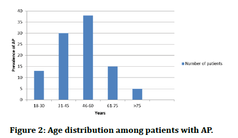
Figure 2: Age distribution among patients with AP.
Table 3 revealed the relation of the disease severity according to the Balthazar CTSI scoring system, and revised Atlanta classification. Moderate severity was found in most of the patients. The Modified CTSI scoring system also showed similar results on studied AP patients (Table 4). Balthazar grading in patients with acute pancreatitis was also studied in the present study. The mild was found higher in all of the methods followed by moderate and most severity (Table 5). Of the studied patients, 70% did not show any organ failure. 40 of them (<30%) had necrosis and 5 had (>30%) had pancreatic necrosis (Table 6, Figures 3 and 4). The comparative analysis between the CTSI and MCTSI showed that the both of the methods had given similar results (Table 7 and Figure 5).
| RAS | Total | ||||
|---|---|---|---|---|---|
| Mild | Moderate | Severe | |||
| Ctsi | Mild | 55 | 1 | 0 | 56 |
| Moderate | 11 | 19 | 10 | 40 | |
| Severe | 0 | 0 | 5 | 5 | |
| Total | 66 | 20 | 15 | 101 | |
Table 3: CT severity index and revised atlanta Score.
| Ras | Total | ||||
|---|---|---|---|---|---|
| Mild | Moderate | Severe | |||
| Modified cts1 | Mild | 55 | 2 | 0 | 57 |
| Moderate | 11 | 18 | 10 | 39 | |
| Severe | 0 | 0 | 5 | 5 | |
| Total | 66 | 20 | 15 | 101 | |
Table 4: Modified severity index.
| Severity | Balthazar CTSI | Modified CTSI | Clinical RAS |
|---|---|---|---|
| Number of cases % | Number of cases % | Number of cases % | |
| Mild | 56 | 57 | 66 |
| Moderate | 40 | 39 | 20 |
| Severe | 5 | 5 | 15 |
Table 5: Comparison of pancreatitis severity according to the balthazar CTSI, modified CTSI scoring system, and revised Atlanta classification.
| Necrosis | Organ failure | P | ||
|---|---|---|---|---|
| Absent | Transient | Persistent | ||
| Absent (n=56) | 55 | 1 | 0 | <0.001 |
| <30% necrosis (n=40) | 0 | 0 | 5 | |
| >30% necrosis (n=5) | 66 | 20 | 15 | |
Table 6: Correlations of pancreatic necrosis and organ failure.
| Outcome parameters | CTSI | P | MCTSI | P | ||||
|---|---|---|---|---|---|---|---|---|
| Mild | Moderate | Severe | <0.01 | Mild | Moderate | Severe | <0.01 | |
| Mean duration of hospital stay (days) | 7 | 8 | 14 | 7 | 8 | 14 | ||
| Organ failure | ||||||||
| Transient | 1 | 19 | 0 | 10 | 8 | 2 | ||
| Persistent | 0 | 10 | 5 | 0 | 10 | 5 | ||
| Need for intervention | 0 | 1 | 1 | 0 | 1 | 1 | ||
| Mortality | 0 | 0 | 2 | 0 | 0 | 2 | ||
Table 7: Comparison of patients with clinical severity parameters and days of hospitalization with severity grading according to CTSI and modified CTSI.
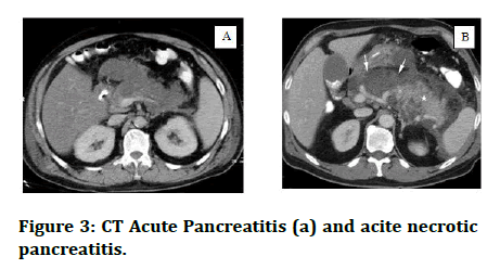
Figure 3: CT Acute Pancreatitis (a) and acite necrotic pancreatitis.
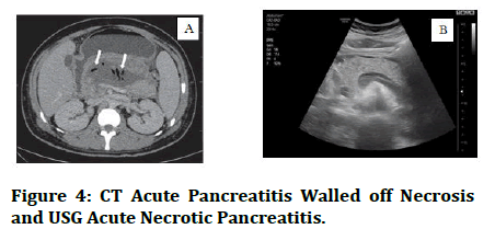
Figure 4: CT Acute Pancreatitis Walled off Necrosis and USG Acute Necrotic Pancreatitis.
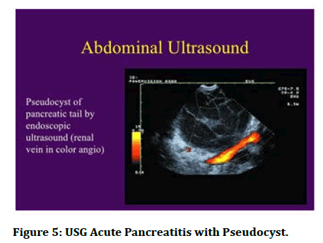
Figure 5: USG Acute Pancreatitis with Pseudocyst.
Discussion
Acute pancreatitis is commonly found in patients with the changed life style such as smokers, alcoholics etc., Hospitalization was also one the main criteria for determining its severity. The present study was aimed to analyse the disease severity using two basic methods viz., CTSI and MCTSI with their clinical outcomes based on RAC. The study showed that the presence of the substantial correspondence between the methods related with the outcome parameters including mean duration of hospital stay, manifestation of persistent of, evidence of infection, need for intervention, and mortality. Both of the methods showed better correlation in grading the disease severity. We found a strong correlation between the gender and age. The study revealed that the male were more prone for AP and its associated comorbidities. Our results were concordance with the previous studies [3,4]. The present study also showed the aging is one of the factors for disease progress and previous studies. The age group range in this study was 18 to 89 years with majority of patients in the age group of 46 to 60 years. In this study, in 18 to 30 age group, only 13 males were there; In 31 to 45 years age group, number of patients were 30; In 46 to 60 years age group number of patients were 38; In 61 to 75 years age group only 15 patients were there and more than 75 years of age, 5 patients. Several previous studies also showed such kind of similar results [5,6]. The present study had showed a strong correlation between organ failure (transient or persistent) with necrosis ration. 40 patients had less than 30% necrosis while, 5 patients only had more than 30%. The result was statistically significant (p<0.001). Similarly, [7-9] banks, freeman, and gastroenterology and isenmann, rau and beger also showed that the necrosis could be one of the main factor in determining the severity, microbial infection and mortality ration among the AP patients. A comparison was done for the patients, with clinical severity parameters and days of hospital stay with severity grading according to CTSI and Modified CTSI. Sahu, et al. [10] also showed the similar results based on their study. Further, the Mean duration of hospital stay is minimum of 7 days in mild CTSI/ MCTSI, and maximum of 14 days in severe CTSI/MCTSI. 2 patients of moderate CTSI/MCTSI needed surgical intervention which was carried in our hospital or outside in surgical gastroenterology department. Mortality was seeing in 2 patients with severe CTSI/MCTSI. Of the total 101 patients, 93 patients got discharged, 2 patients expired and 6 patients went out against medical advice.
Conclusion
In conclusion, it was observed that both CTSI and MCTSI showed noteworthy correlation with clinical outcome parameters, as well as good concordance with grading of severity as per the revised atlanta classification. CT is an important solitary imaging modality to evaluate subjects with acute pancreatitis.
Reference
- Rau B, Bothe A, Beger HG. Surgical treatment of necrotizing pancreatitis by necrosectomy and closed lavage: changing patient characteristics and outcome in a 19-year, single-center series. Surgery 2005; 138:28-39.
- Graciano AL, Balko JA, Rahn DS, et al. The pediatric multiple organ dysfunctions score (p-mods): Development and validation of an objective scale to measure the severity of multiple organ dysfunctions in critically ill children. Crit Care Med 2005; 33:1484-1491.
- Charilaou P, Agnihotri K, Garcia P, et al. Trends of Cannabis Use Disorder in the Inpatient: 2002 to 2011. Am J Med 2017; 130:678–687.
- Imrie CW, Benjamin IS, Ferguson JC, et al. A Single‐centre Double‐blind Trial of Trasylol Therapy in Primary Acute Pancreatitis. Br J Surg 1978; 65:337–341.
- Yadav D, Lowenfels AB. The epidemiology of pancreatitis and pancreatic cancer. Gastroenterology 2013; 144:1252–1261.
- Yadav D, O'Connell M, Papachristou GI. Natural history following the first attack of acute pancreatitis. Am J Gastroenterol 2012; 107:1096–1103.
- Exley AR, Leese T, Holliday MP, et al. Endotoxaemia and Serum Tumour Necrosis Factor as Prognostic Markers in Severe Acute Pancreatitis. Gut 1992; 33:1126–1128.
- Banks PA, Freeman ML. Practice guidelines in acute pancreatitis. Am J Gastroenterol 2006; 101:2379–2400.
- Isenmann R, Rau B, Beger HG. Early Severe Acute Pancreatitis: Characteristics of a New Subgroup. Pancreas 2001; 22:274–278.
- Sahu B, Abbey P, Anand R, et al. Severity assessment of acute pancreatitis using ct severity index and modified ct severity index: Correlation with clinical outcomes and severity grading as per the Revised Atlanta Classification. Indian J Radiol Imaging 2017; 27:152-160.
Author Info
Ankush Misra, M Senthil Velan and G Jayalakshmi*
Department of Orthopaedics, Sree Balaji Medical College and Hospital, Bharath Institute of Higher Education and Research, Chennai, Tamil Nadu, IndiaCitation: Ankush Misra, M Senthil Velan, G Jayalakshmi, Severity Valuation of Acute Pancreatitis with CT Severity Index and Modified CT Severity Index, J Res Med Dent Sci, 2022, 10 (10): 062-066.
Received: 29-Jul-2022, Manuscript No. JRMDS-22-66697; , Pre QC No. JRMDS-22-66697(PQ); Editor assigned: 01-Aug-2022, Pre QC No. JRMDS-22-66697(PQ); Reviewed: 16-Aug-2022, QC No. JRMDS-22-66697; Revised: 30-Sep-2022, Manuscript No. JRMDS-22-66697(R); Published: 10-Oct-2022

