Case Report - (2022) Volume 10, Issue 5
Right Sided Supernumerary Kidney with Pyelonephritis: A Rare Presentation
VVSS Sagar1*, Sourya Acharya1, Sunil Kumar1, Amol Bhawane2, Samarth Shukla3, Chitturi Venkata Sai Akhil4 and K. B. Harshith Gowda5
*Correspondence: VVSS Sagar, Department of General Medicine, Datta Meghe Institute of Medical Sciences, Wardha, Maharashtra, India, Email:
Abstract
Renal malformations are one of the most frequent congenital abnormalities. A supernumerary kidney is unusual among these. Due of the abnormality's rarity, the real incidence is uncertain. The first instance was documented in 1965, and there are now less than 100 cases identified. The majority of the time, this ailment is asymptomatic, but when it is, it usually manifests itself in the fourth decade of life. We discuss a case of a 67 years old woman who presented with a right supernumerary kidney with pyelonephritis fused to the native kidney, which is an infrequent occurrence.Keywords
Supernumerarykidney,Pyelonephritis,Congenitalanomalies,Fusedkidney, MalformationIntroduction
Urogenital abnormalities are widespread, contributing for 33% of all congenital malformations. The most unusual renal anomaly is a supernumerary kidney, which can be encapsulated, completely distinct from the native kidney or tethered to it by a connective tissue sheath. The supernumerary kidney is generally situated caudally to the same side kidney and is associated with a bifid or less often a double ureter. When there are more than two kidneys, this anatomic variation is considered to exist with the additional kidney having separate collecting system, vascular system, and parenchyma with a discrete capsule. Supernumerary kidneys are often associated with urolithiasis, pyonephrosis or hydronephrosis. Rarely Wilm’s tumor and adenocarcinoma may occur [1].
Case Presentation
A 67 years old female was brought in a drowsy state to the casualty with the complaints of multiple episodes of loose stools, right lower abdominal pain and swelling over bilateral lower limbs since 2 days. Patient has history of 2 episode of high grade fever which was intermittent in nature associated with chills. No history of cough, abdominal pain, vomiting, chest pain, breathlessness, loss of consciousness, headache, neck pain, seizures. No history of systemic hypertension, bronchial asthma, diabetes mellitus, tuberculosis. Patient general condition was poor, febrile with 1020 F, tachycardia of 122/min, blood pressure 100/70 mm of Hg. Bilateral crackles present on auscultation, central nervous system examination revealed drowsy, stuporous, delayed response, normal tone, power 3/5 in all limbs, deep tendon reflexes absent, bilateral plantars extensor [2].
Laboratory investigations suggestive of Hb 6.5, MCV 86.3, WBC count 103800 per cu. mm, platelet count 2,69,000 per cu. mm, urine examination revealed plenty of pus cells, urea 197 mg/dl, creatinine 7.5 mg/dl, sodium 120 mmol/l, potassium 5.5 mmol/l, alkaline phosphate 141 U/L, SGOT 49 U/L, SGPT 19 U/L, total protein 4.3 g/dl, albumin 2.0 g/dl, total bilirubin 1.2 mg/dl. Bone marrow was not done as patient had leukemoid reaction and peripheral smear suggestive of no blast cells, normal basophils, no precursors and only few occasional metamyelocytes and hence did not suspect chronic myeloid leukemia [3].
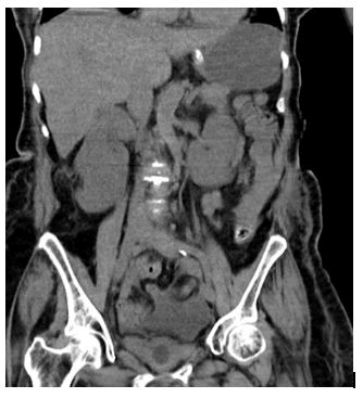
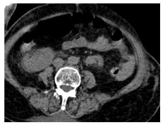
Neuroimaging such as CT brain without contrast was done suggestive of normal study of brain, no haematoma or oedema. Usg abdomen pelvis was done suggestive of slightly raised bilateral cortical echotexture of both kidneys with maintained corticomedullary differentiation [4]. CT KUB was done suggestive of right sided supernumeraray kidney fused to lower pole of upper native kidney having renal pelvis facing anterolaterally with extensive perinephric fat stranding? Pyelonephritis with bilateral pleural effusion with subsegmental atelectasis and free fluid seen in pelvis (Figures 1A and 1B).
Figure 1A: Showing coronal image of KUB demonstrating fusion of accessory kidney with the right kidney with evidence of perinephric fat stranding.
Figure 1B: Showing axial section shows facing of the right sided hilum of kidney anterolaterally with extensive fat stranding.
Patient was started on sustained low efficiency daily dialysis for 3 sessions. Patient was started on inotropes as patient was in shock. Patient was started on furosemide infusion and taken on non-invasive ventilation in view of pulmonary oedema. Patient was started on injectable antibiotics such as meropenem 500 mg thrice a day, linezolid 600 mg twice a day, doxycycline 100 mg twice a day, colistin 2 MIU twice a day [5].
Discussion
The supernumerary kidney is indeed a very atypical congenital defect, with just around 100 instances documented in the literature. The majority of the time, this ailment goes unnoticed, but when it does, it usually shows up around the fourth decade of life. Pain, a palpable abdominal lump, and fever are the most often reported symptoms. Other symptoms such as urinary incontinence may also be present in certain situations. Imaging, such as Computed Tomography (CT), Magnetic Resonance Imaging (MRI), or ultrasonography, is used to make the diagnosis. One additional kidney is found in the majority of cases with supernumerary kidney. The additional kidney is usually located on the same side and caudal to the left kidney and is usually smaller than the original kidney. During the 5th-7th weeks of pregnancy, an abnormal division of the nephrogenic cord into two distinct metanephric blastemas is considered to cause the additional kidney. This procedure results in two kidneys with partially or completely duplicated ureteral buds forming an extra kidney. These can arise when there are two independent collecting systems or when one ureter empties into the other. Supernumerary kidneys have also been identified in conjunction with an ectopic ureter sometimes draining into vaginal canal. Urinary incontinence will be present in these circumstances [6].
There are various hypotheses on how a supernumerary kidney develops: a) ureteral bud bifurcates and penetrates independently into the metanephric blastema which later develops and divide into two kidneys b) two ureteral buds independently penetrates the metanephrogenic blastema and c) linear infarcts cause disintegration of the metanephrogenic blastema [7]. The patient was diagnosed by CT by chance in our situation. In individuals with a fused supernumerary kidney, the duplex kidney should be contemplated. In the duplex kidney, there is whole or partial duplication of the collecting system with a normal parenchyma. A supernumerary kidney has its own vascular and capsule components. On an ultrasound, separating the artery and the ureter may be challenging. In this case, CT is superior to US and is an appropriate imaging modality [8].
Various urogenital (vaginal atresia, horseshoe kidney, ectopic opening of urethra, duplication of urethra) are associated with supernumerary kidney [9]. Other associated defects include imperforate anus, neural tube defects, coarctation of aorta, VSD. In our case, there was no associated oddity. It is critical to be aware that numerous abnormalities may be associated with these situations and to thorough inspection of the patient is essential to detect further anomalies [10].
Conclusion
Supernumerary kidneys are quite uncommon. In two ways, this instance is considerably more peculiar. First, the additional kidney is situated on the right side of the body. Second, fusion to lower pole of native one. There were no associated congenital malformations in our patient. His unexplained right lower quadrant discomfort, on the other hand, might be attributable to an anatomic variance with a documented symptom of pain, fever and vomitings were due to septicaemia evidenced by pyelonephritis in imaging.
References
- Stigers J. The Reference Manual of Pediatric Dentistry: Definitions, Oral Health Policies, Recommendations, Endorsements, Resources. Pediatr Dent 2019.
- Ettinger RL, Chalmers J, Frenkel H. Dentistry for persons with special needs: how should it be recognized. J Dent Educ 2004;68:803-806.
[Croosref] [Google Scholar] [Indexed]
- Dolan TA. Professional education to meet the oral health needs of older adults and persons with disabilities. Spec Care Dentist 2013;33:190-197.
[Croosref] [Google Scholar] [Indexed]
- Ahmad MS, Razak IA, Borromeo GL. Undergraduate education in special needs dentistry in Malaysian and Australian dental schools. J Dent Educ 2014;78:1154-1161.
[Croosref] [Google Scholar] [Indexed]
- Scully C, Dios PD, Kumar N. Special Care in Dentistry E-Book.Handbook of Oral Healthcare. Elsevier sci 2006.
- Uma Maheswari TN, Nivedhitha MS, Ramani P. Expression profile of salivary micro RNA-21 and 31 in oral potentially malignant disorders. Braz Oral Res 2020;34:2.
- Avinash CKA, Tejasvi MLA, Maragathavalli G, et al. Impact of ERCC1 gene polymorphisms on response to cisplatin based therapy in Oral Squamous Cell Carcinoma (OSCC) patients. Indian J Pathol Microbiol 2020;63:538.
- Chaitanya NC, Muthukrishnan A, Rao KP, et al. Oral Mucositis Severity Assessment by Supplementation of High Dose Ascorbic Acid During Chemo and/or Radiotherapy of Oro-Pharyngeal Cancers-A Pilot Project. Indian J Pharm Educ Res 2018;52:532-539.
- Chaturvedula BB, Muthukrishnan A, Bhuvaraghan A, et al. Dens invaginatus: a review and orthodontic implications. Br Dent J 2021;230:345-350.
- Patil SR, Maragathavalli G, Ramesh DNS, et al. Assessment of Maximum Bite Force in Pre-Treatment and Post Treatment Patients of Oral Submucous Fibrosis: A Prospective Clinical Study. J Hard Tissue Biol 2021;30:211-216.
- PradeepKumar AR, Shemesh H, Nivedhitha MS, et al. Diagnosis of Vertical Root Fractures by Cone-beam Computed Tomography in Root-filled Teeth with Confirmation by Direct Visualization: A Systematic Review and Meta-Analysis. J Endod 2021;47:1198-214.
- Ezhilarasan D, Lakshmi T, Subha M, et al. The ambiguous role of sirtuins in head and neck squamous cell carcinoma. Oral Dis 2021.
[Croosref][Google Scholar][Indexed]
- Patel A. Motivational Factors for Treating Patients with Special Health Care Needs. Virginia Commonwealth University 2015.
- Dao LP, Zwetchkenbaum S, Inglehart MR. General Dentists and special needs patients: does dental education matter. J Dent Educ 2005;69:1107-1115.
- Wolff AJ, Barry Waldman H, Milano M, et al. Dental student's experiences with and attitudes toward people with mental retardation. J Am Dent Assoc 2004;135:353-357.
- Kuthy RA, Heller KE, Riniker KJ. Students opinions about treating vulnerable populations immediately after completing community-based clinical experiences. J Dent Educ 2007;71(5):646-54.
- McKenzie CT, Mitchell SC. Dental Students' Attitudes About Treating Populations That Are Low-Income Rural, Non-White, and with Special Needs: A Survey of Four Classes at a U.S. Dental School. J Dent Educ 2019;83 (6):669-678.
- Milnes AR, Tate R, Perillo E. A survey of dentists and the services they provide to disabled people in the Province of Manitoba. J Can Dent Assoc 1995;61(2):149-152,155-158. n
- Salama FS, Kebriaei A, Durham T. Oral care for special needs patients: a survey of Nebraska general dentists. Pediatr Dent 2011;33(5):409-414.
- Baird WO, McGrother C, Abrams KR, et al. Access to dental services for people with a physical disability: a survey of general dental practitioners in Leicestershire, UK. Community Dent Health 2008;25(4):248-252.
- Smith G, Rooney Y, Nunn J. Provision of dental care for special care patients: the view of Irish dentists in the Republic of Ireland. J Ir Dent Assoc 2010;56(2):80-84.
- Loeppky WP, Sigal MJ. Patients with special health care needs in general and pediatric dental practices in Ontario. J Can Dent Assoc 2006;72 (10):915.
- Chadha G, Panchmal GS, Shenoy RP, et al. Attitude of dentists towards providing oral health care to Patients with Special Health Care Needs (PSHCN) in Mangalore, India. Int J Oral Care Res 2015;3:1-7. [Croosref]
[Google Scholar][Indexed]
- Holzinger A, Lettner S, Franz A. Attitudes of dental students towards patients with special healthcare needs: Can they be improved. Eur J Dent Educ 2020;24 (2):243-251.
- Gambhir RS, Dhaliwal JS, Aggarwal A, et al. Covid-19: a survey on knowledge, awareness and hygiene practices among dental health professionals in an Indian scenario. Rocz Panstw Zakl Hig 2020;71 (2):223-229.
- Nasser Z, Fares Y, Daoud R, et al. Assessment of knowledge and practice of dentists towards Coronavirus Disease (COVID-19): a cross-sectional survey from Lebanon. BMC Oral Health. 2020;20 (1):281.
Author Info
VVSS Sagar1*, Sourya Acharya1, Sunil Kumar1, Amol Bhawane2, Samarth Shukla3, Chitturi Venkata Sai Akhil4 and K. B. Harshith Gowda5
1Department of General Medicine, Datta Meghe Institute of Medical Sciences, Wardha, Maharashtra, India2Department of Nephrology, Datta Meghe Institute of Medical Sciences, Wardha, Maharashtra, India
3Department of Pathology, Datta Meghe Institute of Medical Sciences, Wardha, Maharashtra, India
4Department of General Paediatrics, Datta Meghe Institute of Medical Sciences, Wardha, Maharashtra, India
5Department of General Radiodiagnosis, Datta Meghe Institute of Medical Sciences, Wardha, Maharashtra, India
Citation: VVSS Sagar, Sourya Acharya, Sunil Kumar, Amol Bhawane, Samarth Shukla, Chitturi Venkata SaiAkhil4,HarshithGowdaKB,Right Sided Supernumerary Kidney with Pyelonephritis: A Rare Presentation, J Res Med Dent Sci, 2022, 10(5):288-292.
Received: 23-Feb-2022, Manuscript No. 50494; , Pre QC No. 50494; Editor assigned: 25-Feb-2022, Pre QC No. 50494; Reviewed: 11-Mar-2022, QC No. 50494; Revised: 25-Apr-2022, Manuscript No. 50494; Published: 06-May-2022


