Case Report - (2022) Volume 10, Issue 12
Rice Bodies in Juvenile Rheumatoid Arthritis
Manickavasagam M*, Mervin Rosario and Lionel John
*Correspondence: Manickavasagam M, Department of Orthopaedics, Sree Balaji Medical College and Hospital, India, Email:
Abstract
Rice bodies, named after their macroscopic likeness to polished grains of rice, were first described in the literature more than a century ago in association with Tuberculous arthritis. More recently, their presence has been linked to chronic inflammatory processes, notably rheumatoid arthritis. Though the macroscopic and microscopic appearance of rice bodies and their clinical associations and significance have been discussed in the rheumatology literature, few cases have been documented in the pediatric population. In addition, a few descriptions exist of the imaging characteristics of rice bodies but, to our knowledge, none of them about children. Our case shows an atypical presentation of juvenile rheumatoid arthritis with rice bodies as a painful soft-tissue mass. A 4 yr. old boy presented with a painful swelling of right knee for 2 months, MRI was done and rice bodies were seen in response to synovial inflammation in MRI and surgical evacuation which resulted in gush of synovial fluid and intraarticular bodies which is confirmed in biopsy.
blog
blog
blog
blog
blog
blog
blog
blog
blog
blog
blog
blog
blog
blog
blog
blog
blog
blog
blog
blog
blog
blog
blog
blog
blog
blog
blog
blog
blog
blog
blog
blog
blog
blog
blog
blog
blog
blog
blog
blog
blog
blog
blog
blog
blog
blog
blog
blog
blog
Keywords
Rheumatoid arthritis, Range of motion, Trauma, Fever
Introduction
Rice bodies–named after their macroscopic likeliness to polished grains, have been vastly associated with Tuberculous arthritis but of late studies show they are related to rheumatoid arthritis, our case shows an atypical presentation of juvenile rheumatoid arthritis with rice bodies as soft tissue mass [1-5].
Case Report
A 4-year-old otherwise healthy boy presented to the orthopaedic clinic with a 2-month history of painful swelling of the right knee. He had no history of trauma, fever, or systemic symptoms. On physical examination, a large suprapatellar mass was present. This mass was soft, with uniform consistency and well circumscribed borders. On palpation mild tenderness was present. No associated erythema was noted. Range of motion was slightly decreased.
Results and Discussion
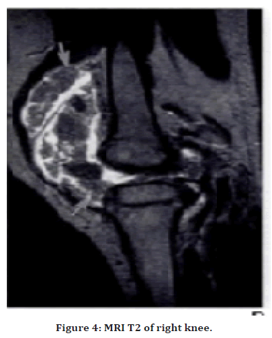
Rice bodies are a nonspecific response to synovial inflammation. First described in association with tuberculous arthritis in 1895 by Riese. Rice bodies are more frequently encountered in noninfectious arthritis, particularly rheumatoid arthritis. With incidence of 72% in adults One Theory postulates that rice bodies arises from micro infarction of the synovium with sloughing of infarcted tissue into the joint space .Whereas another theory states that they arise from encasement of entrapped cells in the fibrinous exudate found in inflammatory synovial fluid. The presence of blood vessels in occasional specimens, as in our case, have raised the possibility that rice bodies may originate from detached fragments of hypertrophied synovial villi. The findings of MRI in our juvenile case as mentioned above is not so common and it makes peculiar to the case. The differential diagnosis of the imaging findings includes pigmented villonodular synovitis and synovial osteochondromatosis. The lack of hemosiderin deposition should allow exclusion of pigmented villonodular synovitis. The cartilaginous components in synovial osteochondromatosis normally shows increased signal intensity on T2-weighted MRI, but the more fibrous component of rice bodies gives them a darker signal allowing differentiation from synovial osteochondromatosis.
Investigations and management
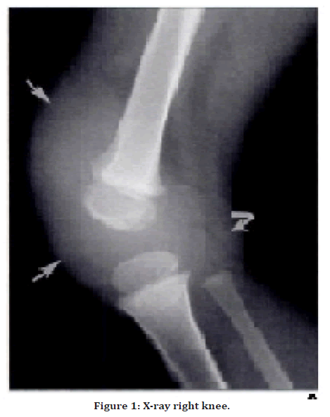
MRI report shows X-Ray right knee (Figure 1) soft tissue opacity in the suprapatellar bursa that extended into the joint space.

Figure 1. X-ray right knee.
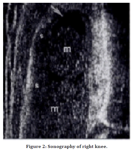
Sonography (Figure 2) shows large mass with associated effusion filling the suprapatellar bursa.

Figure 2. Sonography of right knee.
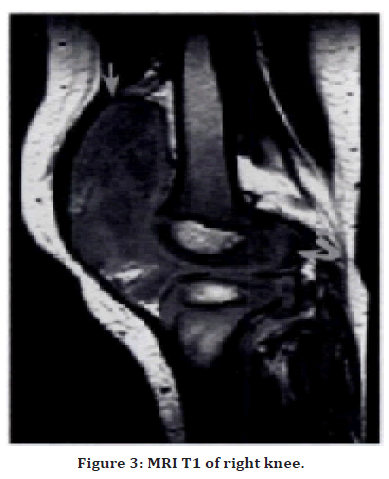
MRI, T1(Figure 3) large joint effusion with a plication in the suprapatellar bursa seen

Figure 3. MRI T1 of right knee.
MRI, T2 (Figure 4) Heterogeneous mass with a nodular appearance filling the knee joint and suprapatellar bursa.

Figure 4. MRI T2 of right knee.
Contrast MRI: Hypertrophied synovium was found throughout the knee.
Surgical excision of the joint capsule produced a gush of synovial fluid and multiple intraarticular bodies. Biopsy done on synovium reveals intra-articular bodies with variable components of collagen, cartilage. Scattered Inflammatory cells were also seen with blood vessel all consistent with rice bodies. Lab investigations like ESR, ANA, RF were all negative. Final results reveal seronegative mono-articular juvenile rheumatoid arthritis with rice bodies was diagnosed.
Conclusion
In summary, our case demonstrates a presentation of juvenile rheumatoid arthritis with rice bodies as a painless soft-tissue knee mass. Although the presence of rice bodies has shown no association with disease severity or duration, diagnosis is important because removal provides symptomatic relief to the patient although rice bodies are apparently uncommon with juvenile rheumatoid arthritis, it is important to be aware of their imaging characteristics because they may provide a clue to the underlying disease process.
References
- Riese H. The rice corpuscles in tuberculous synovial sacs. German J Surg 1895; 42:1-99.
- Chung C, Coley BD, Martin LC. Rice bodies in juvenile rheumatoid arthritis. Am J Roentgenol 1998; 170:698-700.
- Griffith JF, Peh WC, Evans NS, et al. Multiple rice body formation in chronic subacromial/subdeltoid bursitis: MR appearances. Clin Radiol 1996; 51:511-514.
- Tan CH, Rai SB, Chandy J. MRI appearances of multiple rice body formation in chronic subacromial and subdeltoid bursitis, in association with synovial chondromatosis. Clin Radiol 2004; 59:753-757.
- Tsifountoudis I, Kapoutsis D, Tzavellas AN, et al. Lipoma arborescens of the knee: Report of three cases and review of the literature. Case Rep Med 2017; 2017.
Indexed at, Google Scholar, Cross Ref
Indexed at, Google Scholar, Cross Ref
Indexed at, Google Scholar, Cross Ref
Author Info
Manickavasagam M*, Mervin Rosario and Lionel John
Department of Orthopaedics, Sree Balaji Medical College and Hospital, Chennai 600044, IndiaReceived: 24-Nov-2022, Manuscript No. jrmds-22-81001; , Pre QC No. jrmds-22-81001(Q); Editor assigned: 26-Nov-2022, Pre QC No. jrmds-22-81001(Q); Reviewed: 12-Dec-2022, QC No. jrmds-22-81001(Q); Revised: 16-Dec-2022, Manuscript No. jrmds-22-81001(R); Published: 23-Dec-2022
