Research - (2022) Volume 10, Issue 7
Retrospective Analysis on Type of Cavity Preparation Done for Class II Mesio Occlusal Composite Restorations
*Correspondence: Pradeep S, Department of Conservative Dentistry and Endodontics, Saveetha Dental College, Saveetha Institute of Medical and Technical sciences (SIMATS), Saveetha University, India, Email:
Abstract
Objective: Controversy exists regarding cavity preparation for restoration of interproximal caries in posterior teeth in terms of preserving the tooth structure. This study aimed to determine the type of cavity preparation in class II mesio occlusal composite restoration. Materials and methods: Study samples of 800 cases were obtained from the data of 86000 patients between March 2020 and March 2021. Statistical software used for analysis was the SPSS (statistical package for the social sciences) which is designed by IBM and the statistical tests used were frequency tables along with bar graphs to analyse and compare the obtained results. The obtained data was tabulated in excel systematically. Data was then entered in the SPSS analysis software and descriptive analysis and correlation statistics performed. The obtained results were tabulated and graphically represented. Results: Results from the study revealed that occurrence of class II cavity was higher among the male population 53.4% when compared with female population 46.6%.Most of the class 2 cavity preparation was done in the maxillary region 66.8% and in the mandibular region is 31.3%.Conservative like cavity preparation was higher 77% and conventional preparation is 14.7% and box only preparation is 6.9% and in slot preparation is 1.7%. Conclusion: Further evaluation based on bigger sample size, multi-location studies with details on the type of cavity design could be helpful. The findings of the study showed that Conservative like cavity preparation was commonly occurring in the male population and maxillary posterior region has a higher incidence of conservative preparation.
Keywords
Class II cavity, Composite restoration, Mesio occlusal, Design
Introduction
In the clinical setting, clinicians often encounter teeth that have lost most of their structure due to trauma, caries or cavity preparation [1]. Restorative dentists try to overcome this problem by using composite resins [2].Composite resins are suitable Material in class 2 restoration and benefit from chemical bonding to tooth structure. By chemically bonding to the cavity walls, composite resins reinforce the remaining tooth structure and have a good success rate [3].A preparation design that widens toward the occlusal surface is routinely performed in adhesive dental procedures. This preparation design greatly facilitates the placement of the restorative composite into tight areas, effectively reducing voids at the margins of the final restoration [4].
Class II carious lesions occur on proximal surfaces of premolars and molars. They may occur in combination with occlusal (Class I) caries or they may occur alone [5]. In situations where the presence of caries is on the occlusal as well as the proximal surface, a two-surface cavity is prepared.
However, in order to gain access to the proximal carious lesion, the dentist frequently has to break through an otherwise healthy marginal ridge. This is referred to as “convenience form” since there is no other way to reach such lesions for thorough removal of caries [6]. In addition, since the proximal box has only three walls, accessory retentive measures may be incorporated to prevent horizontal sliding of the proximal portion of the restoration out of the box [7].On some teeth, such as maxillary molars for example, when an oblique ridge is not involved, a dovetail outline is followed on the occlusal portion of the Class II cavity to provide retention against horizontal displacement of the proximal portion of the restoration [8].
When a Class II carious lesion exists without involvement of the occlusal, a slot cavity is prepared which is essentially the proximal portion of the Class II preparation [9]. Extensive loss of crown of a tooth leads to problems with retention subsequent restorations. In posterior teeth these can be alleviated by the use of dentine pins [10]. In more extensive cases where there is loss of one or more cusps additional means of retention, such as placement of pins, might be warranted [11]. The survival of composite restorations depends on the type of restoration, number of bonded surfaces, size of the cavity and type of restored tooth [12].Our team has extensive knowledge and research experience that has translate into high quality publications [13–31]. The aim of the study is to determine the type of cavity preparation done in class II mesio occlusal composite restoration.
Materials and Methods
Study setting
This study was carried out in a university setting which consists of subjects predominantly South Indian population. Advantages of the study include available data and similar ethnicity. Disadvantages of this study is the fact that it is a unicentric study and the geographic locations trends are not assessed. Approval of the study is by the ethical board of Saveetha University. Number of people involves 3 reviewers. A Guide, Researcher and a reviewing expert.
Sampling
This is a retrospective study in which the samples were considered from the time period of March 2020 to March 2021. Case sheets reviewed for the research include patients with class 2 MO composite restoration. Cross verification of the required samples was done by the reviewing expert. Measures were taken to minimize the sampling bias. These are inclusion of only clear and readily available data followed by simple random sampling. Both internal and external validation was also obtained to carry out the study.
Data collection/tabulation
Data required for this study was procured by reviewing the patient records of about 86000 patients visiting the dental college. The samples were collected from March 2020 to March 2021. Dental Information Archiving Software is the database system used in college to record all the details of the patient, which includes their demographic data, photographs, diagnosis and treatment reports The required data i.e., patients with class 2 MO cavity preparation were collected and entered in a methodical manner in an excel sheet for the tabulation of data and further statistical analysis data was validated by 1-2 external reviewers and all the nonspecific, unclear or incomplete data were excluded from the study.
Analytics
Statistical software used for analysis is the SPSS (statistical package for the social sciences) which is designed by IBM and the statistical test used was frequency tables along with bar graphs to analyse and compare the obtained results. Independent variables include ethnicity and age. Dependent variables include Gender, Teeth no, Cavity design.
Results
Out of total sample size (800 cases),Results from the study reveals that occurrence of class II cavity was higher among the male population 53.4% when compared with female population 46.6% (Figure 1); Most of the class 2 cavity preparation was done in the maxillary region 66.8% and in mandibular region is 31.3% (Figure 2); Conservative like cavity preparation was higher 77% and conventional preparation is 14.7% and box only preparation is 6.9% and in slot preparation is 1.7% (Figure 3). Further assessment of the type of cavity preparation revealed that in 40.3% of the male population Conservative like cavity design was prepared, 7.6% of Conventional cavity preparation, 4.4% of Box preparation and 1.1% of Slot preparation. The correlation between the Gender and the type of Cavity preparation revealed that Pearson Chi-Square Value-0.01; p<0.05. Hence statistically significant (Figure 4). An assessment of the tooth region with the type of cavity preparation revealed that 53.1% of Conservative type of cavity design was prepared in the maxillary region and 23.9 in the mandibular region. The correlation between the Tooth region and the type of cavity preparation revealed that Pearson Chi-Square Value-0.02;p>0.05. Hence statistically significant (Figure 5).
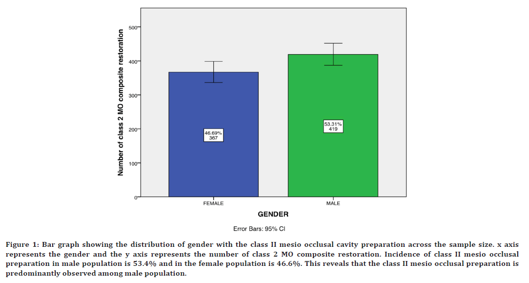
Figure 1. Bar graph showing the distribution of gender with the class II mesio occlusal cavity preparation across the sample size. x axis represents the gender and the y axis represents the number of class 2 MO composite restoration. Incidence of class II mesio occlusal preparation in male population is 53.4% and in the female population is 46.6%. This reveals that the class II mesio occlusal preparation is predominantly observed among male population.
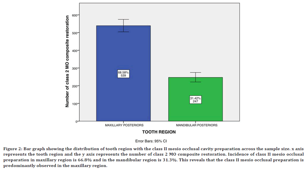
Figure 2. Bar graph showing the distribution of tooth region with the class II mesio occlusal cavity preparation across the sample size. x axis represents the tooth region and the y axis represents the number of class 2 MO composite restoration. Incidence of class II mesio occlusal preparation in maxillary region is 66.8% and in the mandibular region is 31.3%. This reveals that the class II mesio occlusal preparation is predominantly observed in the maxillary region.
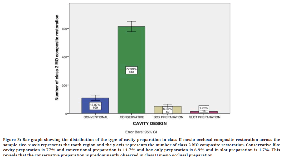
Figure 3. Bar graph showing the distribution of the type of cavity preparation in class II mesio occlusal composite restoration across the sample size. x axis represents the tooth region and the y axis represents the number of class 2 MO composite restoration. Conservative like cavity preparation is 77% and conventional preparation is 14.7% and box only preparation is 6.9% and in slot preparation is 1.7%. This reveals that the conservative preparation is predominantly observed in class II mesio occlusal preparation.
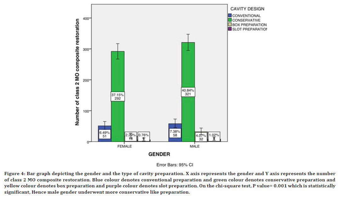
Figure 4. Bar graph depicting the gender and the type of cavity preparation. X axis represents the gender and Y axis represents the number of class 2 MO composite restoration. Blue colour denotes conventional preparation and green colour denotes conservative preparation and yellow colour denotes box preparation and purple colour denotes slot preparation. On the chi-square test, P value= 0.001 which is statistically significant, Hence male gender underwent more conservative like preparation.
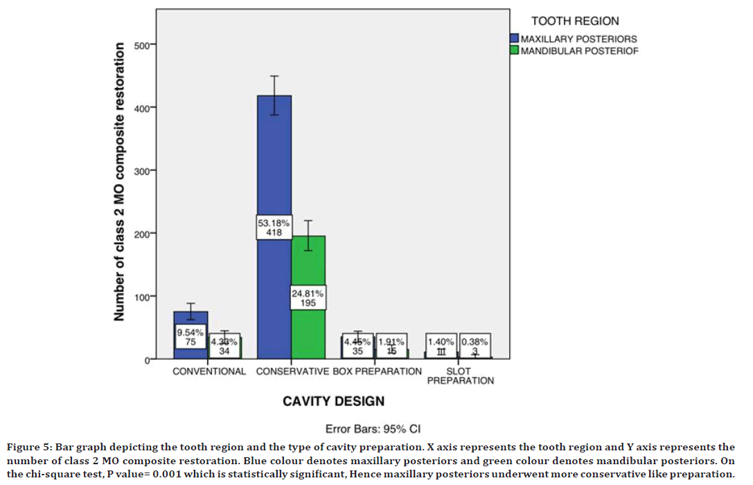
Figure 5. Bar graph depicting the tooth region and the type of cavity preparation. X axis represents the tooth region and Y axis represents the number of class 2 MO composite restoration. Blue colour denotes maxillary posteriors and green colour denotes mandibular posteriors. On the chi-square test, P value= 0.001 which is statistically significant, Hence maxillary posteriors underwent more conservative like preparation.
Discussion
FEM studies by Line et al. and Khera et al. only MO cavities were evaluated and only conservative like cavity preparation has higher success rate then slot or pin type of cavity preparation [32,33]. Further work was subsequently reported by Gabel et al, Brown et al. and Weiland et al. The conclusions of these studies were that the conservative like cavity preparation should enhance the properties of the restorative material in such a way that those unfavorable are properly compensated; the conservative like cavity preparation allows the operator to work efficiently in such a way that a mechanically sound preparation is obtained. A study by Naubi at al. facial slot Class II cavity preparation saves time, conserves tooth structure, offers better esthetics, does not alter occlusal relationships, may preserve a natural proximal contact and enjoys greater patient acceptability than traditional approaches. This restoration is particularly well suited to situations where interproximal relationships are compromised because of misalignment of teeth. Proximal part of the cavity that is formed in this process is referred to as the box type cavity design. It has a floor (gingival) and walls (buccal, lingual and axial). The floor is ideally slightly larger than the occlusal opening, to provide retention against vertical displacement (undercut effect). Potentially, a large Class II cavity may involve all five surfaces of molars. This occurs when there are carious lesions on both proximal surfaces and when the occlusal caries is extending buccally and lingually through the grooves. It is extremely important to place retention grooves at the line angles since in the absence of the occlusal portion these become the only means of retention against horizontal displacement [34–36].
However, there were a few limitations encountered in this study. This study contained some data that were unclear of certain reporting parameters such data were not considered. Another limitation was the geographic limitation i.e., assessment of predominantly South Indian population. Further this study is an unicentric study. Future research should focus on panel data to better understand the cavity design in class II mesio occlusal composite restorations. The scope of this study is the incidence of the type of cavity preparation in clinical practice.
Conclusion
Further evaluation based on bigger sample size, multilocation studies with details on the type of cavity design could be helpful. The findings of the study showed that Conservative cavity preparation was commonly performed in the male population and in maxillary posteriors.
Acknowledgement
The authors would like to acknowledge the help and support rendered by the department of conservative & endodontics and information technology of Saveetha dental college and hospitals and the management for the constant assistance with the research.
Funding
The present project is supported/funded/sponsored by Saveetha Institute of Medical and Technical Sciences, Saveetha Dental College and Hospitals, Saveetha University contributed by Southern Engineering Co Ltd.
Conflict of Interest
None.
Reference
- Manhart J, Chen HY, Hickel R. The suitability of packable resin-based composites for posterior restorations. J Am Dent Assoc 2001; 132:639–645.
- Orahood JP, Cochran MA, Swartz M, et al. In vitro study of marginal leakage between temporary sealing materials and recently placed restorative materials. J Endod 1986; 12:523–527.
- Moorthy A, Hogg CH, Dowling AH, et al. Cuspal deflection and microleakage in premolar teeth restored with bulk-fill flowable resin-based composite base materials. J Dent 2012; 40:500–505.
- Elsharkasi MM, Platt JA, Cook NB, et al. Cuspal deflection in premolar teeth restored with bulk-fill resin-based composite materials. Oper Dent 2018; 43:1-9.
- Keerthana T, Ramesh S, Deepak S. Comparative analysis of success rate in class II cavities with direct and indirect restorations-A retrospective analysis. Int J Dentistry Oral Sci 2021; 8:1526-1532.
- Anand VS, Kavitha C, Subbarao CV. Effect of cavity design on the strength of direct posterior composite restorations: An empirical and FEM Analysis. Int J Dent 2011; 2011.
- Zhu SW, Chen GP. A finite element analysis of the residual stress distribution in the interphase of restoration-tooth structure. Adv Mat Res 2011; 211:710-714.
- Chen GP, Zhu SW, Tao JL. Shrinkage stress analysis of the restored-tooth structure by the finite element method. Adv Mat Res 2011; 146:1792-1796.
- Bohaty BS, Ye Q, Misra A, et al. Posterior composite restoration update: Focus on factors influencing form and function. Clin Cosm Inv Dent 2013; 5:33.
- Ortega VL, Pegoraro LF, Conti PC, et al. Evaluation of fracture resistance of endodontically treated maxillary premolars, restored with ceromer or heat‐pressed ceramic inlays and fixed with dual‐resin cements. J Oral Rehabil 2004; 31:393-397.
- Hannig C, Westphal C, Becker K, Attin T. Fracture resistance of endodontically treated maxillary premolars restored with CAD/CAM ceramic inlays. J Prosthet Dent 2005; 94:342–349.
- Abdel-Aziz M. Effect of pulpal extension on the fracture resistance of endodontically treated molars restored with different CAD/CAM ceramic onlays. Egyptian Dent J 2016; 62:1357-1367.
- Muthukrishnan L. Imminent antimicrobial bioink deploying cellulose, alginate, EPS and synthetic polymers for 3D bioprinting of tissue constructs. Carbohydrate Polymers 2021; 260:117774.
- PradeepKumar AR, Shemesh H, Nivedhitha MS, et al. Diagnosis of vertical root fractures by cone-beam computed tomography in root-filled teeth with confirmation by direct visualization: A systematic review and meta-analysis. J Endod 2021; 47:1198.
- Chakraborty T, Jamal RF, Battineni G, et al. A review of prolonged post-COVID-19 symptoms and their implications on dental management. Int J Int J Environ Res Public Health 2021; 18:5131.
- Muthukrishnan L. Nanotechnology for cleaner leather production: A review. Environ Chem Lett 2021; 19:2527-2549.
- Teja KV, Ramesh S. Is a filled lateral canal-A sign of superiority? J Dent Sci 2020; 15:562.
- Narendran K, Jayalakshmi S, Ms N, et al. Synthesis, characterization, free radical scavenging and cytotoxic activities of phenylvilangin, a substituted dimer of embelin. Indian J Pharm Sci 2020; 82.
- Reddy P, Krithikadatta J, Srinivasan V, et al. Dental caries profile and associated risk factors among adolescent school children in an urban south-Indian City. Oral Health Prev Dent 2020; 18:379–386.
- Sawant K, Pawar AM, Banga KS, et al. Dentinal microcracks after root canal instrumentation using instruments manufactured with different NiTi alloys and the SAF system: A systematic review. Applied Sci 2021; 4984.
- Bhavikatti SK, Karobari MI, Zainuddin SL, et al. Investigating the antioxidant and cytocompatibility of Mimusops elengi Linn extract over human gingival fibroblast cells. Int J Environ Res Public Health 2021; 18:7162.
- Karobari MI, Basheer SN, Sayed FR, et al. An in vitro stereomicroscopic evaluation of bioactivity between Neo MTA Plus, Pro Root MTA, Biodentine & glass ionomer cement using dye penetration method. Materials 2021; 14:3159.
- Rohit Singh T, Ezhilarasan D. Ethanolic extract of Lagerstroemia speciosa (L.) pers., induces apoptosis and cell cycle arrest in HepG2 cells. Nutrition Cancer 2020; 72:146-156.
- Romera A, Peredpaya S, Shparyk Y, et al. Bevacizumab biosimilar BEVZ92 versus reference bevacizumab in combination with FOLFOX or FOLFIRI as first-line treatment for metastatic colorectal cancer: A multicentre, open-label, randomised controlled trial. Lancet Gastroenterol Hepatol 2018; 3:845-855.
- Raj RK. β‐Sitosterol‐assisted silver nanoparticles activates Nrf2 and triggers mitochondrial apoptosis via oxidative stress in human hepatocellular cancer cell line. J Biomed Mater Res Part A 2020; 108:1899-1908.
- Vijayashree Priyadharsini J. In silico validation of the non-antibiotic drugs acetaminophen and ibuprofen as antibacterial agents against red complex pathogens. J Periodontol 2019; 90:1441.
- Priyadharsini JV, Girija AS, Paramasivam A. In silico analysis of virulence genes in an emerging dental pathogen A. baumannii and related species. Arch Oral Biol 2018; 94:93-98.
- Uma Maheswari TN, Nivedhitha MS, Ramani P. Expression profile of salivary micro RNA-21 and 31 in oral potentially malignant disorders. Braz Oral Res 2020; 34:e002.
- Gudipaneni RK, Alam MK, Patil SR, et al. Measurement of the maximum occlusal bite force and its relation to the caries spectrum of first permanent molars in early permanent dentition. J Clin Pediatr Dent 2020; 44:423–428.
- Chaturvedula BB, Muthukrishnan A, Bhuvaraghan A, et al. Dens invaginatus: A review and orthodontic implications. Br Dent J 2021; 230:345-350.
- Kanniah P, Radhamani J, Chelliah P, et al. Green synthesis of multifaceted silver nanoparticles using the flower extract of Aerva lanata and evaluation of its biological and environmental applications. Chem Select 2020; 5:2322-2331.
- Khera SC, Goel VK, Chen RC, et al. Parameters of MOD cavity preparations: A 3-D FEM study, Part II. Oper Dent 1991; 16:42–54.
- Scribante A, Cacciafesta V, Sfondrini MF. Effect of various adhesive systems on the shear bond strength of fiber-reinforced composite. Am J Orthod Dentofacial Orthop 2006; 130:224-227.
- Brown Jr WE. A mechanical basis for the preparation of class II cavities for amalgam fillings in deciduous molars. J Am Dent Assoc 1949; 38:417-423.
- Rudolphy MP, Van Loveren C, Van Amerongen JP. Grey discolouration for the diagnosis of secondary caries in teeth with class II amalgam restorations: An in vitro study. Caries Res 1996; 30:189-193.
- Demarco FF, Corrêa MB, Cenci MS, et al. Longevity of posterior composite restorations: not only a matter of materials. Dent Mater 2012; 28:87-101.
Indexed at, Google Scholar, Cross Ref
Indexed at, Google Scholar, Cross Ref
Indexed at, Google Scholar, Cross Ref
Indexed at, Google Scholar, Cross Ref
Indexed at, Google Scholar, Cross Ref
Indexed at, Google Scholar, Cross Ref
Indexed at, Google Scholar, Cross Ref
Indexed at, Google Scholar, Cross Ref
Indexed at, Google Scholar, Cross Ref
Indexed at, Google Scholar, Cross Ref
Indexed at, Google Scholar, Cross Ref
Indexed at, Google Scholar, Cross Ref
Indexed at, Google Scholar, Cross Ref
Indexed at, Google Scholar, Cross Ref
Indexed at, Google Scholar, Cross Ref
Indexed at, Google Scholar, Cross Ref
Indexed at, Google Scholar, Cross Ref
Indexed at, Google Scholar, Cross Ref
Indexed at, Google Scholar, Cross Ref
Indexed at, Google Scholar, Cross Ref
Indexed at, Google Scholar, Cross Ref
Indexed at, Google Scholar, Cross Ref
Indexed at, Google Scholar, Cross Ref
Indexed at, Google Scholar, Cross Ref
Indexed at, Google Scholar, Cross Ref
Indexed at, Google Scholar, Cross Ref
Indexed at, Google Scholar, Cross Ref
Indexed at, Google Scholar, Cross Ref
Indexed at, Google Scholar, Cross Ref
Indexed at, Google Scholar, Cross Ref
Indexed at, Google Scholar, Cross Ref
Author Info
Department of Conservative Dentistry and Endodontics, Saveetha Dental College, Saveetha Institute of Medical and Technical sciences (SIMATS), Saveetha University, Chennai, IndiaReceived: 08-Jun-2022, Manuscript No. JRMDS-22-64996; , Pre QC No. JRMDS-22-64996 (PQ); Editor assigned: 10-Jun-2022, Pre QC No. JRMDS-22-64996 (PQ); Reviewed: 24-Jun-2022, QC No. JRMDS-22-64996; Revised: 29-Jun-2022, Manuscript No. JRMDS-22-64996 (R); Published: 06-Jul-2022
