Research - (2022) Volume 10, Issue 8
Retrospective Analysis of Incidence of Dental Caries in Anterior Teeth between Males and Females
Revathi B and Anjaneyulu*
*Correspondence: Anjaneyulu, Department of Conservative Dentistry and Endodontics, Saveetha Dental college, Saveetha Institute Of Medical and Technical Sciences, Poonamallee High Road, Chennai, India, Email:
Abstract
Aim: To analyse the incidence of dental caries in anterior teeth between males and females. Introduction- In India, the prevalence of dental caries is reported to be 50-60%. Dental caries are preventable, above all in the early stages. Because of insufficient awareness on prevalence of dental caries and its preventive measures, the condition is still found to be sustaining in communities of both genders. Hence the current study analyses the incidence of dental caries in anterior teeth between males and females. Materials and methods: The study consists of 5713 individual’s case sheets with the age range of 18 to 70 years. Datas were collected from patient’s databases using DIAS. The data was further analyzed for class III mesial and distal caries and class IV mesial and distal caries. The collected data was tabulated and imported to SPSS. Results- Males are affected more than females. Class III mesial caries is most common followed by class IV mesial, class III distal and class IV distal. Tooth number 11 and 21 was highly prone to dental caries among anterior teeth. Males are highly affected with class IV mesial caries and females with class III mesial caries (significant; P<0.001). 11 and 21 is highly prone to class IV mesial caries followed by class III mesial, class IV distal and class III distal caries (significant; P<0.001) Conclusion: This study signifies the importance of educating the patient about oral hygiene practices and prevention methods through assessment of anterior tooth caries.
Keywords
Incidence, Dental caries, Anterior teeth, Males, Females
Introduction
Dental caries is the most common epidemic situation present worldwide. It occurs within the mouth by creating the imbalance in the pH of the saliva, caries promoting oral micro flora, thereby altering the oral environment. Dental caries is a condition where the overall wellbeing is affected including systemic condition of an individual. Many general health conditions have oral manifestations that increase the risk of oral disease which, in turn, is a risk factor for many systemic diseases, such as diabetes and cardiovascular diseases [1]. In India, the prevalence of dental caries is reported to be 50-60% [2].
Socioeconomic inequalities tend to be higher in countries where there are minimal public dental care programs than in countries with some degree of public dentalcare programs [3]. Dental caries are preventable, above all the causative actors in the early stages of detection [4]. Because of insufficient awareness on prevalence of dental caries and its preventive measures, the condition is still found to be sustaining in communities of both genders.
Among all the dental caries, caries affecting the anterior teeth are more concerned by patients than those affecting the posterior teeth [5]. Due to compromise in aesthetics, the fear of tooth loss urges the patient to report to dental care. If at all, the anterior edentulous space is present, the overall confidence of the patient tends to drag down on their daily life activities.
Hence this study emphasizes mainly towards the incidence of anterior teeth caries thereby aids to build a more complete profile of oral health conditions of the South Indian population and be a complement to the analysis of global trends of the disease. Our team has done some extensive knowledge and research experience that has translated into high quality publications [6–25]. Also multiple previous literature concentrated mainly on the posterior teeth caries than anterior teeth. Hence the current study analyses the incidence of dental caries in anterior teeth between males and females.
Materials and Methods
Study design
This was a retrospective study for a period of 4 months from December 2020 to March 2021. The study consists of 5713 individual’s case sheets which were collected from DIAS. The data was further analyzed for class III mesial and distal caries and class IV mesial and distal caries. The obtained data was cross verified by another reviewer.
Inclusion criteria
✔ Patients with age between 18 to 70 years.
✔ Patient with class III and class IV caries.
Exclusion criteria
✔Any Restorations, endodontically treated teeth, any tooth abnormality and history of tooth bleaching or radiation therapy in that particular tooth. ✔ Case sheets without intraoral photographs.
Statistical analysis
Data was analyzed using the Statistical Packages of Social Sciences, version 17 (IBM Corporation). Prevalence of class III and class IV caries among males and females were done using descriptive statistics. Association of type of anterior teeth caries between males and females was analyzed using chi square test with 5% level of significance. Statistical significance was set at P less than or equal to 0.05.
Results and Discussion
From the study, Males (48.5%) are affected more than females (51.4%) (Figure 1). Class III mesial caries (35.3%) is most common followed by class IV mesial (33.7%), class III distal (17.9%) and class IV distal (12.9%) (Figure 2). Tooth number 11(29.8%) and 21(29.7%) was highly prone to dental caries among anterior teeth followed by 12 (12.5%), 22(11.9 %), 23 (3.94%) (Figure 3). The association between gender and type of caries was found to be significant with P<0.001. Males are highly affected with class IV mesial caries (21.6%) and females with class III mesial caries (22.7%) (Figure 4). The association between tooth number and type of caries was found to be significant with P<0.001. Tooth number 11 and 21 is highly prone to class IV mesial caries followed by class III mesial, class IV distal and class III distal caries (Figure 5).
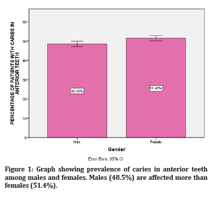
Figure 1: Graph showing prevalence of caries in anterior teeth among males and females. Males (48.5%) are affected more than females (51.4%).
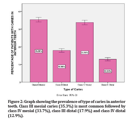
Figure 2:Graph showing the prevalence of type of caries in anterior teeth. Class III mesial caries (35.3%) is most common followed by class IV mesial (33.7%), class III distal (17.9%) and class IV distal (12.9%).
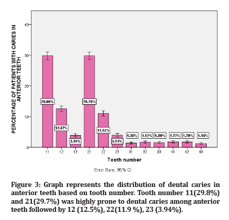
Figure 3:Graph represents the distribution of dental caries in anterior teeth based on tooth number. Tooth number 11(29.8%) and 21(29.7%) was highly prone to dental caries among anterior teeth followed by 12 (12.5%), 22(11.9 %), 23 (3.94%).
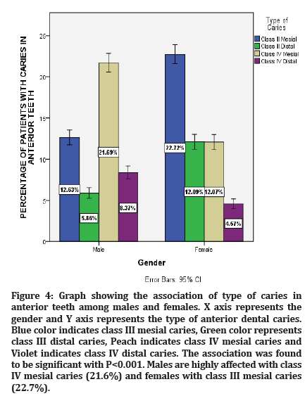
Figure 4:Graph showing the association of type of caries in anterior teeth among males and females. X axis represents the gender and Y axis represents the type of anterior dental caries. Blue color indicates class III mesial caries, Green color represents class III distal caries, Peach indicates class IV mesial caries and Violet indicates class IV distal caries. The association was found to be significant with P<0.001. Males are highly affected with class IV mesial caries (21.6%) and females with class III mesial caries (22.7%).
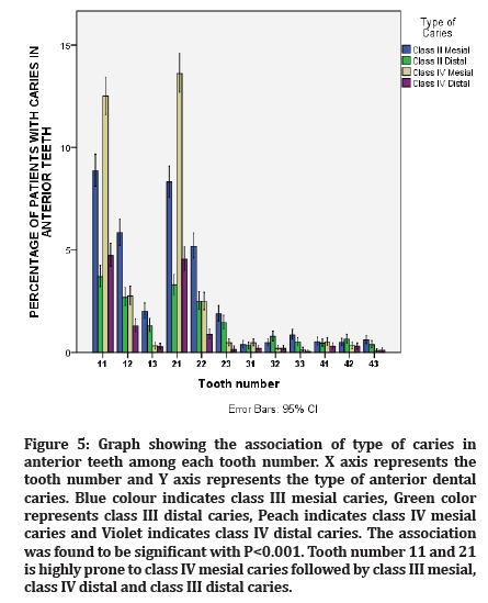
Figure 5:Graph showing the association of type of caries in anterior teeth among each tooth number. X axis represents the tooth number and Y axis represents the type of anterior dental caries. Blue colour indicates class III mesial caries, Green color represents class III distal caries, Peach indicates class IV mesial caries and Violet indicates class IV distal caries. The association was found to be significant with P<0.001. Tooth number 11 and 21 is highly prone to class IV mesial caries followed by class III mesial, class IV distal and class III distal caries.
From the present study, 48.5% of the males and 51.4% of the females were affected with dental caries in anterior teeth. In accordance with the result, a study conducted by Talabani, et al. reported females showed higher prevalence of anterior teeth caries than males [26]. In contrast, a study conducted by Hedge et al., showed 43.5% prevalence in females and 56.4% in males during the year 2014-2015 among the Mangalore population [27]. The incidence or prevalence of dental caries vary among males and females based on the population the study was conducted. Previous studies concluded that higher prevalence of caries in anterior teeth is due to early eruption of teeth, oral habits and prolonged exposure to a cariogenic environment.
In the present study, class III mesial caries was found to be highly prevalent followed by class IV mesial, class III distal and class IV distal. Class III caries were found to be most common in anterior teeth compared to class IV caries which is in line with the study conducted by Santia, et al. who reported that class IV caries is the least prevalent among anterior teeth [28]. One of the significant etiologies leading to class IV caries is trauma, whereas class III caries is highly associated with food impaction, improper oral hygiene and brushing frequency which is increasingly attributed among the general population [29].
On analyzing the tooth number with anterior caries, maxillary anterior are highly affected than mandibular anterior. That is tooth number 11 and 21 were highly affected with anterior caries followed by 12 and 22. Earlier studies reported that this can be due to close approximation of submandibular gland and sublingual gland to mandibular anterior thereby preventing the process of demineralization [30].
Significant association was found between gender and type of caries. Males are highly affected with class IV caries whereas females are affected with class III caries. A study conducted by Hedge et al. no significant association was found between gender and caries among the Mangalore population [27]. In contrast, a study by Lukacs, et al. mentioned that higher caries prevalence among females can be due to their earlier eruption of dentition, therefore longer exposure of teeth to the cariogenic oral environment, frequent snacking during food preparation and imbalance of hormonal levels than males [31]. Also significant association was found between gender and the tooth which is affected. Both males and females have a high incidence of caries in maxillary anterior compared to mandibular anterior. Teeth surfaces which are susceptible to caries play an important role since the proximal surfaces are not selfcleansing, they act as retention areas hence promoting the caries [31].
Conclusion
From the current study, it is proved that males are more affected than females with anterior dental caries. Class III caries are found to be more common than class IV caries. Maxillary anterior teeth are more prone to caries than mandibular teeth. Thus the incidence of anterior tooth caries aids in establishing accurate oral health examination. This study also signifies the importance of educating the patients about the oral hygiene practices and prevention methods. Further studies can be done by including a wider population to promote the level of dental care to the next step.
References
- Burt BA, Kolker JL, Sandretto AM, et al. Dietary patterns related to caries in a low-income adult population. Caries Res 2006; 40:473–480.
- Thomas S, Raja RV, Kutty R, et al. Pattern of caries experience among an elderly population in south India. Int Dent J 1994; 44:617–622.
- Montero MJ, Douglass JM, Mathieu GM. Prevalence of dental caries and enamel defects in connecticut head start children. Pediatr Dent 2003; 25:235–239.
- Shah N, Sundaram KR. Impact of socio-demographic variables, oral hygiene practices, oral habits and diet on dental caries experience of Indian elderly: A community-based study. Gerodontology 2004; 21:43–50.
- Baldissera RA, Corrêa MB, Schuch HS, et al. Are there universal restorative composites for anterior and posterior teeth? J Dent 2013; 41:1027–1035.
- Muthukrishnan L. Imminent antimicrobial bioink deploying cellulose, alginate, EPS and synthetic polymers for 3D bioprinting of tissue constructs. Carbohydrate Polymers 2021; 260:117774.
- PradeepKumar AR, Shemesh H, Nivedhitha MS, et al. Diagnosis of vertical root fractures by cone-beam computed tomography in root-filled teeth with confirmation by direct visualization: A systematic review and meta-analysis. J Endod 2021; 47:1198-1214.
- Chakraborty T, Jamal RF, Battineni G, et al. A review of prolonged post-COVID-19 symptoms and their implications on dental management. Int J Environ Res Public Health 2021; 18.
- Muthukrishnan L. Nanotechnology for cleaner leather production: A review. Environ Chem Letters 2021; 19:2527-2549.
- Teja KV, Ramesh S. Is a filled lateral canal–A sign of superiority?. J Dent Sci 2020; 15:562.
- Narendran K, MS N, Sarvanan A. Synthesis, characterization, free radical scavenging and cytotoxic activities of phenylvilangin, a substituted dimer of embelin. Indian J Pharm Sci 2020; 82:909-912.
- Reddy P, Krithikadatta J, Srinivasan V, et al. Dental caries profile and associated risk factors among adolescent school children in an Urban South-Indian city. Oral Health Prev Dent 2020; 18:379–386.
- Sawant K, Pawar AM, Banga KS, et al. Dentinal microcracks after root canal instrumentation using instruments manufactured with different NiTi alloys and the SAF system: A systematic review. Appl Sci 2021; 11:4984.
- Bhavikatti SK, Karobari MI, Zainuddin SLA, et al. Investigating the antioxidant and cytocompatibility of Mimusops elengi Linn extract over human gingival fibroblast cells. Int J Environ Res Public Health 2021; 18:7162.
- Karobari MI, Basheer SN, Sayed FR, et al. An in vitro stereomicroscopic evaluation of bioactivity between Neo MTA Plus, Pro Root MTA, biodentine & glass ionomer cement using dye penetration method. Materials 2021; 14.
- Rohit Singh T, Ezhilarasan D. Ethanolic extract of Lagerstroemia speciosa (L.) Pers., induces apoptosis and cell cycle arrest in HepG2 cells. Nutr Cancer 2020; 72:146–156.
- Ezhilarasan D. MicroRNA interplay between hepatic stellate cell quiescence and activation. Eur J Pharmacol 2020; 885:173507.
- Romera A, Peredpaya S, Shparyk Y, et al. Bevacizumab biosimilar BEVZ92 versus reference bevacizumab in combination with FOLFOX or FOLFIRI as first-line treatment for metastatic colorectal cancer: A multicentre, open-label, randomised controlled trial. Lancet Gastroenterol Hepatol 2018; 3:845–855.
- Raj R K. β‐Sitosterol‐assisted silver nanoparticles activates Nrf2 and triggers mitochondrial apoptosis via oxidative stress in human hepatocellular cancer cell line. J Biomed Materials Res 2020; 108:1899-1908.
- Vijayashree Priyadharsini J. In silico validation of the non-antibiotic drugs acetaminophen and ibuprofen as antibacterial agents against red complex pathogens. J Periodontol 2019; 90:1441–1448.
- Vijayashree Priyadharsini J, Smiline Girija AS, Paramasivam A. In silico analysis of virulence genes in an emerging dental pathogen A. baumannii and related species. Arch Oral Biol 2018; 94:93–98.
- Uma Maheswari TN, Nivedhitha MS, Ramani P. Expression profile of salivary micro RNA-21 and 31 in oral potentially malignant disorders. Braz Oral Res 2020; 34:e002.
- Gudipaneni RK, Alam MK, Patil SR, et al. Measurement of the maximum occlusal bite force and its relation to the caries spectrum of first permanent molars in early permanent dentition. J Clin Pediatr Dent 2020; 44:423–428.
- Chaturvedula BB, Muthukrishnan A, Bhuvaraghan A, et al. Dens invaginatus: A review and orthodontic implications. Br Dent J 2021; 230:345–350.
- Kanniah P, Radhamani J, Chelliah P, et al. Green synthesis of multifaceted silver nanoparticles using the flower extract of Aerva lanata and evaluation of its biological and environmental applications. Chem Select 2020; 5:2322–2331.
- Al Zahawi AR, Mahmood MA, Talabani RM, et al. The prevelence and causes of dental non carious cervical lesion in the sulaimani population (cross-sectional study). J Dent Med Sci 2015; 14:94.
- Aparna M, Sreekumar S, Thomas T, et al. Assessment of dental caries experience among 5-16 year old school going children of Mangalore, Karnataka, India: A cross-sectional study. Ann Essences Dent 2018; 10:12-17.
- Ferreira Zandoná A, Santiago E, Eckert GJ, et al. The natural history of dental caries lesions: A 4-year observational study. J Dent Res 2012; 91:841–846.
- Petersen PE. Sociobehavioural risk factors in dental caries-international perspectives. Community Dent Oral Epidemiol 2005; 33:274–279.
- Urzua I, Mendoza C, Arteaga O, et al. Dental caries prevalence and tooth loss in chilean adult population: First national dental examination survey. Int J Dent 2012; 2012:810170.
- Lukacs JR, Largaespada LL. Explaining sex differences in dental caries prevalence: Saliva, hormones, and “life-history” etiologies. Am J Hum Biol 2006; 18:540–555.
Indexed at, Google Scholar, Cross Ref
Indexed at, Google Scholar, Cross Ref
Indexed at, Google Scholar, Cross Ref
Indexed at, Google Scholar, Cross Ref
Indexed at, Google Scholar, Cross Ref
Indexed at, Google Scholar, Cross Ref
Indexed at, Google Scholar, Cross Ref
Indexed at, Google Scholar, Cross Ref
Indexed at, Google Scholar, Cross Ref
Indexed at, Google Scholar, Cross Ref
Indexed at, Google Scholar, Cross Ref
Indexed at, Google Scholar, Cross Ref
Indexed at, Google Scholar, Cross Ref
Indexed at, Google Scholar, Cross Ref
Indexed at, Google Scholar, Cross Ref
Indexed at, Google Scholar, Cross Ref
Indexed at, Google Scholar, Cross Ref
Indexed at, Google Scholar, Cross Ref
Indexed at, Google Scholar, Cross Ref
Indexed at, Google Scholar, Cross Ref
Indexed at, Google Scholar, Cross Ref
Indexed at, Google Scholar, Cross Ref
Indexed at, Google Scholar, Cross Ref
Indexed at, Google Scholar, Cross Ref
Indexed at, Google Scholar, Cross Ref
Indexed at, Google Scholar, Cross Ref
Indexed at, Google Scholar, Cross Ref
Author Info
Revathi B and Anjaneyulu*
Department of Conservative Dentistry and Endodontics, Saveetha Dental college, Saveetha Institute Of Medical and Technical Sciences, Poonamallee High Road, Chennai, IndiaCitation: Revathi B, Anjaneyulu, Retrospective Analysis of Incidence of Dental Caries in Anterior Teeth between Males and Females, J Res Med Dent Sci, 2022, 10 (8): 123-127.
Received: 07-Jul-2022, Manuscript No. jrmds-22-68879; , Pre QC No. jrmds-22-68879(PQ); Editor assigned: 09-Jul-2022, Pre QC No. jrmds-22-68879(PQ); Reviewed: 26-Jul-2022, QC No. jrmds-22-68879; Revised: 02-Aug-2022, Manuscript No. jrmds-22-68879 (R); Published: 09-Aug-2022
