Research - (2021) Volume 9, Issue 8
Relationship among Intercanine width, Intermolar Width and Arch Length in Upper and Lower Arch for Dentistry Students of Thi-Qar University
Sameerah Jameel Tarfa* and Nabra F Salih
*Correspondence: Sameerah Jameel Tarfa, College of Dentistry, University of Thi-Qar, Iraq, Email:
Abstract
The arch dimensions are very important and useful to the clinician in all fields of dentistry. In orthodontics, intercanine width, intermolar width, and arch length are all important parameters for diagnostic and treatment planning. The goal of this study was to figure out the link between intercanine width (ICW), intermolar width (IMW), and arch length (VMD) in each upper and lower arch in a class I normal occlusion, as well as the relationship between the upper and lower arches of the same measures. 30 students (15 males and 15 females) with class I normal occlusion (20-22 years old) were chosen from Thi-Qar University/College of Dentistry in Iraq. Intercanine width (ICW), intermolar width (IMW), vertical canine distance (VCD), and vertical molar distance (VMD) were all measured. Spearman's correlation coefficient was used in order to determine the correlation among variables. Intercanine width (ICW), intermolar width (IMW), as well as vertical canine distance (VCD) and vertical molar distance (VMD) of both upper and lower arches, revealed a strong association. Upper intercanine width (UICW) and upper vertical molar distance (UVMD) as well as lower vertical molar distance (LVMD) have a modest association. The lower intercanine width (LICW), on the other hand, exhibited no significant relationships. The relationship between upper and lower vertical molar distances was extremely significant (LVMD).
Keywords
VCD, LVMD, ICW, IMW, UICW, LVMD
Introduction
The knowledge of standard dimensions for the dental arch in human populations is especially useful to clinicians in various fields of dentistry, such as orthodontics, prosthodontics, and oral surgery, and it is of great interest to anthropologists who study dental arch growth and development in relation to various environmental, genetic, and physical factors for different populations [1].
Determination of the normal occlusion is the first step of understanding the normal as well as abnormal (malocclusion). Therefore, it is an essential step for clinical diagnosis, orthodontic management and public health preventive program [2].
The teeth in these arches are placed in such a way that they are in harmony with their counterparts in the same arch as well as those in the opposing arch [3].
Aside from the effect of the orofacial musculature labially, buccally, and lingually, the size and relationship of the dental arches is significant in maintaining correct occlusion of the teeth [4].
Arch shape, arch size, and dimension are all critical considerations in case evaluation, diagnosis, and treatment planning.
Arch width, arch length, and arch depth are the three dimensions that make up an arch. Intercanine width, interpremolar width, and intermolar width are all used to determine arch width.
In treatment planning, the link between intercanine width (ICW), intermolar width (IMW), and arch length (AL) is critical. Transverse expansion causes an increase in intercanine and intermolar width, as well as a shift in arch length and arch perimeter [5].
Materials and Methods
Study cast of 30 adults (15 males and females); age ranged (20-22 yrs.) was selected according to special criteria from the orthodontic department in Thi-Qar university college of dentistry. The study sample is summarized in Table 1.
| Number of cases | Number of casts | |
|---|---|---|
| Male | 15 | 15 |
| Female | 15 | 15 |
| Total | 30 | 30 |
Table 1: Characteristics of the study sample.
The sample has a complete set of permanent teeth, including class I molar occlusion, and class I canine occlusion [6]. Also free of local factors that compromise the integrity of the dental arches (congenital missing, retained deciduous, and supernumerary teeth) [1,7].
The teeth are normally shaped, normal vertical and horizontal dental relationships, less than well-aligned arches 2mm of space or crowding in each arch, no rotation or displaced tooth, and no midline shift.
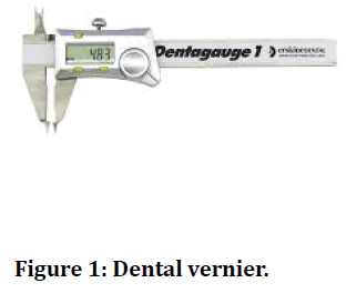
The instruments and supplies used are sharp lead pencils (0.5-pencil marker), Dental vernier (Dentaurum) Figure 1, and millimetre ruler (15 cm).

Figure 1. Dental vernier.
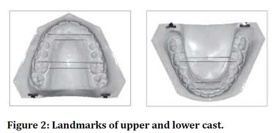
With a pointed pencil (0.5 pencil marker), some toothrelated spots visible in an occlusal view were meticulously marked bilaterally in the maxillary and mandibular study casts to facilitate the identification of the landmarks that will be used for measuring the dimensions of dental arch. To guarantee that the points were accurately positioned on the casts of the study.
Measurements were taken from (30 upper and 30 lower) dental casts, the dental Vernier was used to measure the dimensions of the dental arch.
Dental cast analysis
The following landmarks were used in the maxillary and mandibular study models (Figure 2)

Figure 2. Landmarks of upper and lower cast.
• Incisal point: The point in the halfway between the two central incisal borders of t. [8-10].
• Canine point: The cusp tip of the permanent canines on the right and left sides [8,11-13].
• Mesiobuccal cusp tip of the first molars: The mesiobuccal cusp tips of the right and left first permanent molars [1,2,4,7].
Dental arch dimensions
Dental arch width
• Inter canine distance: The linear distance between one canine's cusp point and the cusp tip of the other [1,4,14,15].
• Inter first molar distance: The liner distance between the from mesiobuccal cusp tip of one first permanent molar and the mesiobuccal cusp tip of the other first permanent molar [2,4,14,15].
Dental arch length
• Vertical Molar Distance (VMD): The vertical distance between the incisal point perpendicular to a line joining the mesial surface of the first permanent molars [7,15].
• Vertical Canine Distance (VCD): The vertical distance from the incisal point and a line joining the cusp tips of the first permanent canines [2,4,7,16].
Inter examiner calibration
The inter examiner calibration was done between two readers for the all measurements for the whole samples. The statistical comparison by (t-test) showed that there was no significant difference (P>0.05) between the examiner readings (Table 2).
| Seq | Variables | Mean | t-test | Sig. | |
|---|---|---|---|---|---|
| 1st Record | 2nd Record | ||||
| 1. | Max. ICW | 34.27 | 34.27 | 1 | NS |
| 2. | Max. IMW | 52.03 | 51.92 | -1.458 | NS |
| 3. | Max. VCD | 7.92 | 8.07 | -0.565 | NS |
| 4. | Max.VMD | 26.13 | 26.15 | -0.765 | NS |
| 5. | Mand. ICW | 25.66 | 25.83 | -0.375 | NS |
| 6. | Mand. IMW | 44.49 | 44.83 | -1.381 | NS |
| 7. | Mand. VCD | 5.01 | 4.75 | -1.063 | NS |
| 8. | Mand. VMD | 21.29 | 21.3 | 0.044 | NS |
| All dimensions in mm | |||||
| NS: Non-Significant at P>0.05. | |||||
| Max: Maxillary, Mand: Mandibular, ICW: Inter Canine Width, IMW: Inter Molar Width, VCD: Vertical Canine Distance, VMD: Vertical Molar Distance. | |||||
Table 2: Inter examiner test for measurements made on study casts.
Results
The following results of the computerized statistical analysis of all the dental arch dimensions that have been derived from measuring the study models are grouped into:
Descriptive statistics
The descriptive statistics were applied to the all variables of the maxillary and mandibular dental arch entire sample dimensions including; mean, minimum, maximum, standard deviation, and standard error. The mean of dental arch compared with the mandibular arch (Table 3).
| No. | Minimum | Maximum | Mean | Std. Error | Std. Deviation | |
|---|---|---|---|---|---|---|
| UICW | 30 | 29.86 | 50.25 | 34.2785 | 0.67129 | 3.67682 |
| UIMW | 30 | 42.99 | 59.22 | 51.9822 | 0.65566 | 3.59121 |
| UVCD | 30 | 3.68 | 10.89 | 7.8868 | 0.30367 | 1.66327 |
| UVMD | 30 | 21.6 | 29.62 | 26.062 | 0.41902 | 2.29509 |
| LICW | 30 | 22.82 | 29.73 | 25.7827 | 0.26891 | 1.47287 |
| LIMW | 30 | 35.36 | 50.23 | 44.8888 | 0.56558 | 3.0978 |
| LVCD | 30 | 2.33 | 8.25 | 4.9353 | 0.26504 | 1.45167 |
| LVMD | 30 | 16.9 | 24.45 | 21.3037 | 0.33033 | 1.80929 |
Table 3: Descriptive of upper and lower arch.
The relations between upper and lower arches for the same measurements
A high significant correlation was observed between UICW, UIMW and LIMW, while a significant correlation was found between UVCD and UVCD and LVCD. A high significant correlation was observed between UVMD and LVMD (Table 4).
| UICW | UIMW | UVCD | UVMD | LICW | LIMW | LVCD | LVMD | |
|---|---|---|---|---|---|---|---|---|
| UICW | 1 | .712** | 0.142 | 0.26 | 0.560** | 0.636** | 0.051 | 0.333 |
| UIMW | 0.712** | 1 | -0.105 | 0.112 | 0.607** | .804** | -0.156 | 0.069 |
| UVCD | 0.142 | -0.105 | 1 | .639** | 0.306 | 0.018 | .372* | 0.503** |
| UVMD | 0.26 | 0.112 | 0.639** | 1 | 0.487** | -0.012 | 0.301 | 0.799** |
| LICW | 0.560** | 0.607** | 0.306 | 0.487** | 1 | 0.578** | 0.047 | 0.375* |
| LIMW | 0.636** | 0.804** | 0.018 | -0.012 | 0.578** | 1 | -0.106 | 0.041 |
| LVCD | 0.051 | -0.156 | .372* | 0.301 | 0.047 | -0.106 | 1 | 0.372* |
| LVMD | 0.333 | 0.069 | .503** | 0.799** | 0.375* | 0.041 | 0.372* | 1 |
| L: Lower, U: Upper. | ||||||||
| **: Correlation is significant at the 0.01 level. | ||||||||
| *: Correlation is significant at the 0.05 level. | ||||||||
Table 4: Spearman correlation between upper and lower arch.
The relations within the same arch
Between UICW and UIMW, as well as between UVCD and UVMD, we found a high significant association, whereas among LVCD & LVMD, there was a substantial association. Between LICW and LIMW, there was a strong significant association (Table 4).
Other relations
A high significant correlation was observed between UICW and LIMW, as well as between UIMW and LICW, UVCD and LVMD, UVMD and LICW, while a significant correlation was found between LICW and LVMD (Table 4).
Discussions
The relation between upper and lower arches for the same measurements
On applying the correlation test, all the measurements of the upper arch found to have a highly significant correlation (p ≤ 0.01) with their antagonist in the lower arch, except the VCD showed insignificant correlation only (p ≤ 0.05). In fact, this is expected relations; since the upper dental arch occupy the lower in normal class ? occlusion. Sillman et al. [17] carried out a longitudinal study from birth to early adulthood and evaluated changes in various dental arch dimensions.
Less increase in the anterior arch length in the mandibular dental arches than in maxillary with the period of growth extend to 10 years and then no evidence of significant changes was observed. Mohammad et al. [18] did not confirm with the finding of Sillman et al. [17] concerning the maxillary anterior arch length. From the deciduous to the permanent dentition, increase in the arch length occurs primarily by increase in the labial position of the permanent incisors [8]. AL-Zubair [1] found a significant correlation between upper and lower arch length.
The relations within the same arch
Within the same arch the correlation was found to be highly significant (p ≤ 0.01) between the ICW and IMW for both lower and upper arches, while the VCD and the VMD found to have insignificant relation in lower arch (p ≤ 0.05), and highly significant in the upper arch (p ≤ 0.01). Hojensgaard et al. [19] showed that the asthmatic children have significantly greater molar vertical distance than the normal children especially in the maxillary dental arch which was associated with the increase in the inclination of the maxillary incisors.
Osborn et al [20] studied the effect of the lip pumper on the mandibular dental arch dimension, the result showed increase in the molar vertical distance by 1.2 mm and was attributed to the anterior tipping of the mandibular incisors. AL-Zubair [1] found in their study a very high correlation between inter canine width and inter molar width in both upper and lower arches.
Other relations
A highly significant correlation (p ≤ 0.01) was found between UICW and LIMW, the same finding with LICW and UIMW, LICW showed a significant relation (p ≤ 0.05) with LVMD and highly significant relation (p ≤ 0.01) with UVMD, while UVCD had a highly significant relation (p ≤ 0.01) with LVMD only. It seems to be the lower intercanine width (LICW) had a significant relation with arch length for both upper and lower arches (VMD) and this come in line with Radnzic et al. [21] who carried out an investigation to determine the relation between dental crowding and dental arch dimension, the result showed highly significant decrease of the molar vertical distance in the crowded arches than no crowded arches. Upper arch lengths and upper inter canine widths, as well as lower arch lengths and lower inter canine widths, showed a clear connection, but upper arch lengths and upper inter molar widths and lower arch lengths and lower inter molar widths showed a poor connection.
The same finding was reached by this study except for the relation between the upper arch length (UVMD) and the upper inter canine distance (UICD) which was found to have an insignificant correlation with each other (p ≤ 0.05). Fried et al. [22] and Germane et al. [23], utilizing a mathematical model of the dental arch, discovered a link between VMD and ICW. They found that the increase in arch perimeter due to intercanine growth was midway between that of incisors and molars. Tibana et al [24] discovered a substantial link between UICW and LVMD within the same arch, but only a minor association between ICW and VMD. The current research discovered a weak link between UICW and UVMD, as well as UICW and LVMD. This is consistent with AL-findings [1].
The dental casts of 197 Spanish adult patients were chosen in a research by Paulino et al. [25]. A computerized approach was used to measure ICW, IMW, and VMD on each dental cast.
The results revealed a strong association between ICW and VMD for both the upper and lower arches, which is consistent with Ramadan's et al. findings [15], as well as the findings of our investigation for the lower arch solely.
References
- AL-Zubair N. Maxillary and mandibular dental arch dimensions and forms in a sample of Yemeni population aged (18-26) years with class ? normal occlusion. Master Thesis, College of Dentistry, Baghdad University- Iraq 2002.
- AL-Sarraf HA. Maxillary and mandibular dental arch dimension in children aged 12-15 years with class ? normal occlusion "Cross sectional study". Master Thesis, Mosul University, Mosul-Iraqi 1996.
- Akapata AS, Jackson D. Overjet values of children and young adults in Lagos. Oral Epidermiol 1979; 7:174-177.
- AL-Kodimi NHA. Dental arch dimensions in Yemeni sample aged (10-15) years with normal occlusion and class ? with anterior crowding in both arches. (Comparative study). Master Thesis, Baghdad University- Iraq 2003.
- Ash MM. Wheelers dental anatomy, physiology and occlusion W.B. Saunders Company.:1993; 325-282.
- Houston WJ, Stephens CD, Tulley WJ. A textbook of orthodontics. Wright PSG. 1996; 119.
- Nader MS. Facial and arch form and dimensions in a sample of 16-21 years old Palestinians class ? occlusion. Master Thesis. Baghdad University-Iraq 2003.
- Younes SA. Maxillary arch dimensions in Saudi and Egyptian population sample Am J Orthod Dentofacial Orthop 1984; 85:83-87.
- Diwan R, Elahi JM. A comparative study between three ethnic groups to derive some standards for maxillary arch dimensions. J Oral Rehabilitation 1990; 17:43-48.
- Mack PJ. Maxillary arch central incisor dimensions in Nigerian and British population sample. B Dent J 1981; 9:67-70.
- Staley RN, Stuntz WR, Peterson L. A comparison of arch widths in adults with normal occlusion and adults with class ?? division ? occlusion. Am J Orthod Dentofac Orthop 1985; 88:163-169.
- Ho KK, Kerr JS. Arch dimensional changes during and following fixed appliance therapy. Br J Ortho 1987; 14:293-297.
- Ismail AM, Hossain N, Hatem S. Maxillary arch dimensions in Iraqi population sample. Iraqi Dent J 1996; 8:111-120.
- Bishara SE, Jakobsen JR, Treder J, et al. Arch width changes from 6 week to 45 years of age. Am J Orthod. Dentofac Orthop 1997; 111:401-409.
- Ramadan OZ. Relation between photographic facial measurements and lower dental arch measurement in adult Jordanian males with class ? normal occlusion. Master Thesis, Mosul University Iraq 2000.
- Borgan BE. Dental arch dimensions analysis among Jordanian school children. Master Thesis. Cairo University-Egypt 2001.
- Sillman JH. Dimensional changes of dental arches: Longitudinal study from birth -25 years. Am J Orthod Dentofac Orthop 1964; 50:824-842.
- Mohammad IS. Maxillary arch dimensions: across sectional study between 9-17 years. Master Thesis, Baghdad University-Iraq 1993.
- Hojensgard E, Wenzel A. Dentoalveolar morphology in children with asthma and perennial rhinitis. Eur J Orthodontics 1987; 9:256-270.
- Osborn WS, Nanda RS, Currier F. Mandibular arch perimeter changes with lip bumper treatment. Am. J. Orthod Dentofac Orthop 1991; 99:527-532.
- Radnzic D. Dental crowding and its relationship to mesiodistal crown diameters and arch dimensions. Am J Orthod Dentofac Orthop 1988; 94:50-56.
- Fried KH. Palate-tongue reliability. Angle Orthod 1971; 61:308-323.
- Germanne Y, Lindauer SJ, Rubinstein LK, et al. Increase in arch perimeter due to orthodontic expansion. Am J Orthod Dentofac Orthop 1991; 100:421-427.
- Tibana RH, Palagi LM, Miguel JA. Changes in dental arch measurements of young adults with normal occlusion. A longitudinal study. Angle Orthodontist 2004; 74:618-23.
- Paulino V, Parades V, Candia J, et al. Prediction of arch length based on intercanine width. Eur J Orhtodont 2008; 30:295-98.
Author Info
Sameerah Jameel Tarfa* and Nabra F Salih
College of Dentistry, University of Thi-Qar, IraqCitation: Sameerah Jameel Tarfa, Relationship among Intercanine width, Intermolar Width and Arch Length in Upper and Lower Arch for Dentistry Students of Thi-Qar University, J Res Med Dent Sci, 2021, 9(8): 126-130
Received: 27-Jul-2021 Accepted: 11-Aug-2021
