Research - (2022) Future Prospects of clinical and Medical Research
Quantum Dots for Detection and Treatment of Breast Cancer
Manish Kumar Gupta1*, Naresh Kumar2, Pallavi Srivastava3 and Jayanand4
*Correspondence: Manish Kumar Gupta, Department of Pharmaceutical Chemistry, SGT College of Pharmacy, SGT University, Gurugram, Haryana, India, Email:
Abstract
Nanotechnology is a research and development ϔied concerned with creating 'things' - mostly materials and devices - on the scale of molecules and atoms. The use of matter on a nuclear, atomic, and supramolecular scale for industrial purposes is known as nanotechnology. Quantum dot structures are a product designed by nanotechnologists, and it is an emerging ϔied with various applications. Breast cancer is diagnosed with the help of these structures. Due to their unique optical & electrical features, QDs are widely used as in vitro & in vivo ϔuorophores in sub-atomic, cell, & in vivo imaging. Points of interest in differentiating metastases are revealed by quantum-dots-based innovation. With the advancement of these dot structure mix and adjustment, the impact of QDs on tumor metastasis research will become ever more signiϔicnt as the science progresses. As a result, we will be able to establish a link between nanotechnology and cancer biology, as well as develop novel techniques to treat and diagnose the deadly disease.
Keywords
Breast cancer, Diagnosis, Nanotechnology, Treatment, Quantum dots
Introduction
Nanotechnology
Nano science & nanotechnology are the study & application of small objects, & they can be used to a broad range of science subjects, including physics, physical science, materials science, and design. Nanotechnology is touted as having the potential to increase energy productivity, aid in environmental cleanup, and treat serious medical diseases. It is expected to have the ability to significantly increase fabrication creation at significantly lower costs. Nanotechnology's results will be smaller, less expensive, lighter, and more practical, and will use less energy and crude materials to manufacture, according to proponents of the technology.
Nanotechnology's applications include: The first commercial applications of nanotechnology appeared in the 2000s; however, most of them are limited to the mass use of uninvolved nanomaterials.
Nanotech advocates for the use of TiO2 in sunscreen, cosmetics, & certain foods; silver nano-particles in packaging of food, clothes, disinfectants, & home apparatus, such as carbon Nano-tubes for stain-resilient composites; & cerium oxide as a fuel stimulus. Nanotechnology is being used in developing countries to help treat diseases and prevent medical concerns. Nanomedication is the umbrella word for this type of nanotechnology. Nanotech is also used or advanced for a variety of mechanical and purges processes. Desalination, water filtering, groundwater treatment, wastewater treatment, & other Nano-remediation are all examples of refinement and natural clean-up applications. Nanotechnology has already proven to be a useful tool in medical research. Nanotechnology could also lead to less expensive, more durable drug-delivery platforms in the future [1], [2].
Quantum dot structures
Quantum Dots (QDs) are human-made nanoscale crystals with the ability to transport electrons. When UV light strikes these semiconducting nanoparticles, they emit a rainbow of colors. These synthetic semiconductor nanoparticles have found uses in composites, solar cells, and fluorescent organic names. Quantum structures are tiny nanostructures with a wide range of properties depending on their material and shape. They can be used as dynamic materials in single-electron semiconductors, for example, due to their unique electrical characteristics.
The properties of these dot structures are determined by their size, shape, arrangement, and structure, such as whether they are strong or empty. A reliable assembly technology that leverages quantum dots' capabilities for wide-ranging applications in areas such as catalysis, hardware, photonics, data storage, imaging, medication, or detection should be capable of manufacturing large quantities of nano-crystals with the same boundaries. Because certain organic particles are capable of atomic recognition and self-assembly, nano-crystals could become an important structural component for selfassembled functional nano-devices. The molecule-like energy conditions of QDs also contribute to unusual optical features, such as a molecule size ward frequency of fluorescence, which is used in the development of optical tests for natural and clinical imaging. Until now, the use of colloidal QDs in bio analytics & bioidentification has revealed the broadest series of applications for colloidal QDs.
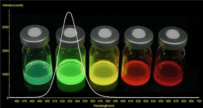
Despite the fact that the first quantum specks drew attention to their latent capacity, improving fundamental features, particularly colloidal dependability in a saltcontaining arrangement, took a lot of effort. Initially, quantum specks were used in forgery circumstances, and these particles would have just encouraged in 'real' examples, as human blood. These difficulties have been addressed, and QDs have started a variety of applications in cancer biology (Figure 1) [3], [4].
Figure 1: Vials of quantum dots producing vivid colors.
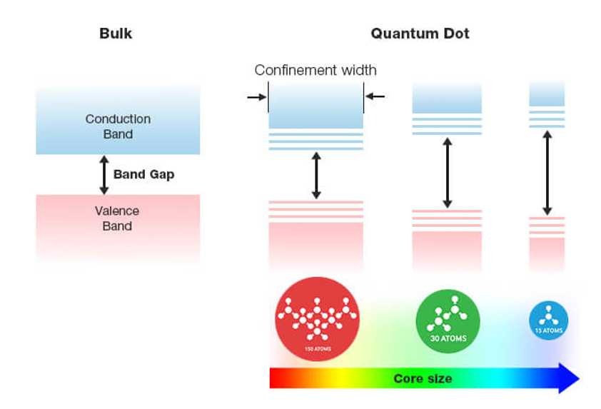
Working of Quantum dot structures: Quantum dots are semiconductor nanoparticles that illuminate a certain color when light is shone on them. The color they emit is dependent on the nanoparticle's size. When UV light is shined on the QDs, certain electrons acquire enough energy to break free from the atoms. This allows them to migrate around the nanoparticle, constructing a conductance band wherein electrons are able to flow over a material & transmit electricity. When the electrons return to the atom's outer orbit, the atom becomes more stable. The energy differential between the conductance band & the valence band determines the color of the light. The larger the nanoparticle, the greater the energy difference between the valence & conductance bands, resulting in a deeper blue coloring. The energy differential between the valence band & the conductance band is lower with larger nanoparticles, which causes the glow to shift towards red.
Quantum specks can be made from a variety of semiconductor materials, such as cadmium selenite, cadmium sulphide, and indium arsenide. The properties of quantum specks can be found in nanoparticles of these or any other semiconductor substance. Quantum dot formations glow due to a hole between the valence band and the conductance band, which is present in all semiconductor materials. QDs may be able to improve the efficiency of sun-based cells. A photon of light creates one electron in regular sun-based cells. Both silicon quantum spots and lead sulphide quantum specks have been shown to produce two electrons for a single photon of light in tests. Using QDs in solar cells ominously improve their ability to generate electric force in this way. Analysts are also investigating the use of quantum dabs in displays for applications extending from mobile phone to TVs that uses less energy than present displays. The green, red, & blue hues required to make the full range of hues would be available by placing distinct size QDs in every pixel of a display screen (Figure 2)[4]–[6].
Figure 2: Transition of electron between Valence Band and Conduction Band in quantum dots.
Breast Cancer and Related Genes
Breast cancer is a kind of cancer that originates in the breast cells. This tumor starts when certain breast cells start to grow abnormally, as per medical specialists. These cells proliferate at a higher pace than healthy cells and continue to expand, producing a lump or mass. Cells in our breast may migrate (metastasize) to our lymphatic system or other parts of our body. Cells in the milkproducing ducts are the most frequent cause of breast cancer. Invasive lobular carcinoma is a form of cancer that begins in the glandular tissue called lobules,although it may also begin in other cells or tissue in the breast.
BRCA1 and BRCA2: An acquired mutation in the BRCA1 or BRCA2 quality is the most well-known cause of innate breast malignant growth. These characteristics aid the production of polypeptides that repair damaged DNA in normal cells. Strange cell development, which can lead to cancer, can be triggered by transformed forms of these traits.
p53: The p53 gene is also a tumor suppressor gene that produces a tumor suppressor protein that stops cells with damaged DNA from growing by causing cell cycle arrest or inducing apoptosis. Li-Fraumeni Syndrome is caused by inherited mutations in this gene.
ATM: It creates tumor suppressor proteins that activate enzymes that repair broken DNA strands, ensuring the genetic material of the cell remains stable. Inheriting one faulty copy of this gene has been associated to breast cancer.
L-myc: This gene generates transcription factor and is a proto-oncogene. Excessive cell proliferation and tumor development are caused by mutations in this gene, which is connected to the growth of breast cancer.
HER2: This gene generates the HER2 protein, which is a type of receptor found on breast cells. This regulates a cell's growth, proliferation, and repair mechanisms. Overexpression of receptors is caused by a mutation in this gene, resulting in excessive cell replication and uncontrolled cell growth.
PTEN: This gene also creates tumor suppressor proteins, which supply instructions for the synthesis of a cell cycle regulator enzyme. A mutation in this gene causes aberrant cells to proliferate and develop rapidly, resulting in the creation of a tumor. This gene has been discovered to be mutated in 20-25 percent of breast cancer patients.
Other genes: Inherited breast cancers can also be caused by other gene alterations. These gene mutations are far less prevalent, and they don't increase the risk of breast cancer nearly as much as the BRCA1 and BRCA2 genes. ATM, TP-53, CHEK-2, CDH1, PALB-2, and other genes are among them.
Literature Review
Dina et al. in through their paper concluded that the most frequent cancer in the world is breast cancer. The biopsy of a SLN is used to phase axillary lymph nodes. For SLN mapping, organic dyes & radio-colloid are currently employed; however, they expose patients to ionizing radiation, are unstable during surgery, & cause local tissue damage. QDs could be utilized to map the SLN without requiring a biopsy. For early-stage breast cancer, surgical elimination of the primary tumor is the best therapeutic option, however due to issues delineating tumor margins, cancer cells frequently survive, leading to recurrences. Image-guided tumor excision with functionalized QD could allow imagining of cancer cells. QDs are photo-stable and penetrate farther into tissues.
The possibility of QD buildup is raised by the slow removal of QD. Nonetheless, positive results from recent in vivo research and a first in-human study with cadmium-free QD imply that it has a lot of potential for cancer diagnosis and treatment [4].
Chun-Wei Peng and Yan Li in their research highlighted the known applications of Quantum dots for the detection & cure of numerous types of tumors. QD-based probes have shown promising results in cellular and in vivo molecular screening due to their superior photophysical properties of semiconductor nano-crystal Quantum Dots (QDs). More and more studies have shown that QDs-based technology may become an optimistic method in cancer research. Progress in QD synthesis and modification will make studying tumor metastasis a more critical issue in the future [3].
Discussion
Application of QDs in the field of breast cancer
Detection of correlation between HER2 gene amplification & protein expression: The HER2 oncogene is expressed or amplified in 15 to 20% of breast tumors, and has been linked to a poor prognosis and responsiveness to trastuzumab therapy. Breast cancer patients' HER2 gene status influences their eligibility for trastuzumab therapy, and a high proportion (41-56%) of these patients respond to targeted therapy. Several studies have linked enhanced HER2 expression to an increase in chromosome 17 copy number rather than HER2 gene amplification. The frequency of chromosome 17 aneuploidy linked with conflicting results was demonstrated by comparing immunohistochemistry & fluorescence in situ hybridization results in both invasive ductal and invasive lobular carcinomas.
Because HER2 overexpression is one of the leading origins of breast tumor, an accurate screening of its degree of protein manifestation is critical. Though, due to a lack of quantification, current approaches for assessing HER2 expression levels are insufficient. This could lead to a discrepancy between HER2 tests and therapies. As a result, a new and more effective diagnostic procedure is required. To quantify the degree of HER2 expression, a new immune-histochemical (IHC) approach combining QDs with trastuzumab & single-particle imaging was developed.
IHC with QDs (IHCQD) was used to explore the correlation between IHC with 3,3′diaminobenzidine, IHCQD, & fluorescence in situ hybridization in tissues from 36 breast tumor individuals with full data. The IHCQD score was computed using the amount of QDconjugated trastuzumab molecules that bound precisely to a tumor cell. In 37 cases, the IHCQD score was proportionately linked with the FISH score of HER2 gene copy quantity (R = 0.83). The IHCQD score with cutoff level was precisely close with the FISH mark with a cutoff value of 3 when HER2 positivity remained considered being positive.
Furthermore, in HER2 positive cases, the IHC-QDs total & the time to progression of trastuzumab treatment were well associated (R = 0.68). The association between FISH score and TTP, on the other hand, was not seen. We created a precise quantitative IHC approach based on trastuzumab-conjugated QDs & single-particle screening examination, & propose that IHCQDs score could be used as a predictor of trastuzumab therapy.
HER2 polypeptides are immunostained through a principal & a non-principal antibody connected with HRP in traditional IHCDAB. The principal antibodies utilized for treatment pathology diagnostics identify the intracellular domain of the HER2 polypeptide. Trastuzumab detects the HER2 extra-cellular region. As a result, trastuzumab's epitope varies from that of analytic antibodies. Trastuzumab was monomerized & then combined with QDs in IHCQDs. Various shortened versions of HER2, such as p95HER2, have been described to deficiency the extracellular domain. Trastuzumabconjugated QDs are unable to attach to HER2 truncated versions. Furthermore, MUC4 overexpression has been shown to seal the HER2 protein's surface. These effects on HER2's extracellular domain prohibit trastuzumab from interacting with the protein, but not with a diagnostic antibody. The gap between diagnostics and therapeutic efficacy is caused by epitope differences between antibodies[8].
Diagnosis of breast cancer: Scientists & researchers looked into a new technique for labeling HER2 on the outer layer of cancerous cells, also known as HER2/neu, abundantly expressed in about 26%–32% of invasive breast tumor & play a key role in prognosis & cure choice.
Other trials using QDs to detect HER2 for breast growth diagnosis exist whole. Scientists and proponents of nanotechnology have also reported on the use of multicolor QDs for measurable & concurrent profiling of numerous biomarkers in integral breast tumor cells & clinical cases, as well as a comparison of the new QDsbased molecular sketching equipment to traditional western blotting & fluorescence in situ hybridization (FISH). The multicolor bio-conjugates were employed to identify five clinically relevant tumor markers in breast tumor cells MCF-7 & BT474 [3], [4], [9].
Using QD-Abs profiling, a measurable link between HER2 gene intensification & HER2 polypeptide expression was discovered. This research demonstrates that combined QDs could be used to sense low echelons of HER2 protein expression. To circumvent the limitations of those studies' practical application, researchers recently employed QDs conjugated with antibody for HER2 status valuation in breast tumor. 701 patients with aggressive breast tumor were enrolled in the trial, with three men and 698 women. QDs-immunohistochemistry analytical system was used to detect HER2 expression in breast tumor in an automated, delicate, easy & quantitative manner.
The QDs-based methodology is more subtle, precise, & cost-effective than traditional IHC, especially in situations of IHC (2+), indicating that this new method has clinical potential, and particularly in developing countries [10].
Because QD emission peaks are much narrower than organic dye emission peaks, more QD emissions can be resolved within the visible spectrum than ordinary fluorophores. QDs may be utilized for cancer detection and therapy with excellent precision when conjugated with diagnostic & therapeutic drugs. The multiplexed quantification of three major cancer indicators, cancer antigen 125, carcino-embryonic antigen (CEA), & Her-2/Neu (C-erbB-2) was achieved using semiconductor nanoparticle QDs integrated into a modular, microfluidic biosensor. Both serum and entire saliva specimens were used to show the functionality of the combined sample treating, analyte seizure, & detection modalities. To conclude the sandwich-type immunoassay, nano-bio-chips with a fluorescence transduction signal with QD-labeled sensing antibody were used in conjunction with antigen capture using a micro porosity agarose bead array sustained within a microfluidics ensemble. When QD probes were used in this tiny biosensor configuration, signal amplification was 30 times of standard molecular fluorophores, and reported restrictions of detection were reduced by roughly 2 orders of scale compared to enzyme-linked immunosorbent assays. Measurements via the nano-biochip technology correlate to established methods with A2 = 0.95 & A2 = 0.96 for saliva & serum, respectively, according to assay validation studies. In combination with next-generation fluorophores, this integrated nanobio- chip examination system anticipates to be a subtle, multiplexed tool for critical diagnostic & prognostic applications [6].
Immunofluorescence imaging of HER2 and ER: Breast tumor is a complex tumor, & a better understanding of it is essential for better treatment outcomes. For molecular pathology, the utilization of QD-based immunefluorescent nanotechnology (QD-IHC) has the ability to enhance tumor heterogeneity identification. This potential is investigated using QD-IHC detection of HER2 & ER. In breast cancer tissue microarrays, QD-IHC can exhibit breast cancer heterogeneity more clearly & sensitively than traditional IHC. Furthermore, imaging ER & HER2 at the same time may aid in understanding their interactions throughout the formation of heterogeneous breast cancer.
MUC1 is a glycoprotein present over interfaces of most epithelial cells that has been shown to be a useful biomarker for early cancer detection. Scientists & nanotechnologists developed an aptamer-based, quantifiable identification strategy for MUC1 using a three-part DNA hybridization process with QD-marking. When the three particularly designed DNA strands are mixed in the absence of MUC1 peptides, strong fluorescence is observed; however, because the MUC1 peptide attaches to the aptamer thread in such a manner that the quencher & fluorescence reporter may be introduced, there is a steady drop in fluorescence strength when MUC1 peptides were present.
The revealing boundary for MUC1 with this unique technique is in the nanomolar range, and a linear reply for the estimated range seen in blood serum may be created. This research added to our understanding of MUC1’s aptamer/analyte binding site/mode, enhancing the possibility of refining this identification technology for the primary detection of various forms of epithelial and breast malignancies in large populations.
Triple negative breast cancer therapy using quantum dot-based micelle conjugation: Theranostic for EGFRoverexpressing cancer has been developed using a QDbased micelle coupled with an EGFR nano-body & loaded with an anti-cancer medication, amino-flavone (AF). The indium phosphate core-zinc sulphide shell QDs fluorescence in the near-infrared (NIR) allowed for in vivo nanoparticle bio distribution investigations. In EGFR-abundant expressing MDA-MB-468 triple-negative breast tumor cells, the anti-EGFR nano-body 7D12 combination increased cellular absorption and cytotoxicity of the QD-based micelles. In an orthotopic triple-negative breast tumor xenograft mouse model, the AF-enclosed Nb-combined micelles collected in tumors at higher quantities than the non-targeted micelles, resulting in more efficient tumor regression. In addition, no systemic toxicity was detected with the therapies. As a result, our QD-based Nb-combined micelle could be a useful theranostic nano-platform for EGFR-abundant expressing malignancies like TNBCs.
Photodynamic and radiation therapies using quantum dots: Semiconductor QDs & nanoparticles made of lipids, metals, or polymers have shown promise in the early detection & treatment of cancer. Cadmiumcontaining semiconductors are routinely used to make quantum dots with unique optical characteristics. Cadmium is a possibly hazardous metal, & the toxicity of QDs to living cells & humans has yet to be thoroughly studied. As a result, researchers are looking for less hazardous compounds with similar targeting and optical capabilities. The study of luminescence nanoparticles as light foundations for tumor healing, on the other hand, is quite intriguing. Despite advancements in neurosurgery & radiotherapy, the forecast for patients with malicious gliomas has remained mostly unchanged in recent decades.
To decrease harm to adjacent tissues, cancer treatment necessitates precise precision in delivering ionizing radiation. Some recent investigation has concentrated on producing photosensitizing QDs for the generation of radicals when visible light is absorbed.
Despite the point that visible light is harmless, this method is only appropriate for treating superficial tumors. Ionizing radiation penetrates far deeper, giving it a significant advantage in the treatment of internal organ tumors. The use of QDs and nanoparticles to produce electrons & radicals in photodynamic & radiation therapies, as well as their blend, is an example of this notion. Photodynamic Treatment (PDT) is a non-invasive therapeutic method that has shown to be effective. Photosensitizer (PS) medicines & an external light source are required components of PDT, and cytotoxic reactive oxygen species are produced, which kill tumor cells. Despite being a new prospective anticancer therapy method, PDT’s success is limited due to photosensitizers’ low water solubility, which restricts its general use. Nano-platforms centered on PS embedded in nanomaterials can be used for targeted PDT with fewer side effects & higher efficiency [5].
Targeted silencing of HER2/neu gene: Short interfering RNA (siRNA) gene silencing is quickly becoming a popular way to investigate gene role in mammalian cells. Though different ways have been successful in delivering siRNA, tracing their conveyance & measuring their transfection efficacy has proven difficult without an appropriate tracking mediator. As a result, scheming an effectual and self-tracking RNA interference transfection mediator is a difficulty. Chitosan nanoparticles with enclosed QDs were created & used to transport HER2/neu siRNA, according to Nanotechnology Advocates. The inclusion of fluorescent QDs in the chitosan NPs can be used to manage the conveyance & transfection of siRNA using such a design. With chitosan/QD NP surface tagged with HER2 antibody attacking the HER2 receptors on SKBR3 cells, selective delivery of HER2 siRNA to HER2-overexpressing SKBR3 breast tumor cells were demonstrated. The conjugated siRNA’s gene-silencing possessions were additionally confirmed using luciferase & HER2 ELISA tests. Future gene silencing experiments in vivo will be easier to track with these self-tracking siRNA delivery NPs. Quantum dot structures can also aid to reduce the overall cytotoxicity of a mammalian cell.
Antimonene quantum dots for cancer therapy: Photothermal therapy (PTT) has proved to have a lot of promise in cancer treatment. However, creating photothermal agents (PTAs) based on nanomaterials (NMs) with acceptable photo-thermal conversion efficiency (PTCE) & biocompatibility remains a significant difficulty. A unique liquid exfoliation approach was used to create a new cohort of PTAs based on 2-D Antimonene QDs. The addition of polyethylene glycol (PEG) to the surface of AMQDs greatly improved biocompatibility & constancy in physiological media.
The PTCE of the PEG-coated AMQDs was 45.6 percent that is greater than many other NMs-based PTAs such graphene, gold, molybdenum, & black phosphorus (BP). NIR-induced fast degradability was also a unique property of the AMQD-based PTAs. The PEG-coated AMQDs displayed significant NIR-induced tumor destruction capabilities in both in vitro and in vivo experiments. Through the development of an entirely new PTA platform, this work is expected to extend the usage of 2D Antimonene in biomedical solicitations. The NIR-induced rapid degradability of the AMQDs-based PTAs was also an exceptional property. The PEG-coated AMQDs displayed considerable NIR-triggered tumor ablation capacity in both in vitro and in vivo tests. Through the growth of a fully new PTA platform, this work is anticipated to extend the effectiveness of 2D Antimonene in biomedical solicitations.
Conclusion
In the subject of cancer biology, nanotechnology has enormous potential. Quantum dots (QDs) are man-made Nano size gems capable of moving electrons, according to nanotechnology researchers and oncologists. When UV light strikes these semiconducting nanoparticles, they emit light in a variety of colors. Composites, sunlightbased cells, and fluorescent organic names have all been found to use these counterfeit semiconductor nanoparticles. They are, however, utilized to diagnose breast cancer in the field of cancer biology.
These designed probes are good carriers for delivering chemotherapeutic medications and genes for modifying tumor cells in the targeted region of our breast, despite being an alternative to imaging. QDs have enormous potential for detecting a variety of cancer indicators and thereby sorting out the complicated gene expression profiles of malignancies, allowing for more precise clinical diagnosis. QDs have unquestionably become a vital tool for targeting, visualizing, and performing specialized therapeutic tasks in today's world.
The study of QDs in cancer visualization will considerably increase their clinical applicability in the near future, particularly with linked QDs in attacking metastasis & measurable evaluation of molecular targets. Linked QDs could currently attack solid tumor tissues with mature vasculature, but micro-metastasis in the absence of wellbuilt vasculature is difficult to identify. As a result, the surface of QDs must be improved to allow for efficient extravasation, micro-metastasis, and the initiation of antigen binding to tumor antigens. It's challenging to develop an internal (or external) standard for grading the fluorescence strength on tumors, which characterizes the degree of molecular targets, in order to quantify molecular objectives in micro-metastasis.
Furthermore, the surface of QDs must be fine-tuned to reduce RES uptake while increasing tumor-specific acceptance. Because metastases can arise in the RES system, particularly in the liver & lymph nodes, nonspecific RES absorption will result in inaccurate results. One way to improve QD tumor directing is to modify their surface to make them long-circulating in the circulation. A prominent technique for attaining extended circulation & decreasing RES uptake is to alter the surface coating, for as by inserting huge molecular-weight PEG molecules to adorn QDs. QDs will have a substantial impact on cancer treatment.
References
- Tanaka T, Decuzzi P, Cristofanilli M, et al. Nanotechnology for breast cancer therapy. Biomed Microdevices2009;11(1):49â??63.
[Crossref], [GoogleScholar], [Indexed]
- Bayda S, Adeel M, TuccinardiT, et al. The history of nanoscience and nanotechnology: From chemical-physical applications to nanomedicine. Molecules 2019;25(1):112.
[Crossref], [GoogleScholar], [Indexed]
- Li Y, Peng CW. Application of quantum dots-based biotechnology in cancer diagnosis: Current status and future perspectives. J Nanomater2010:11.
[Crossref], [GoogleScholar], [Indexed]
- Radenkovic D, Kobayashi H, Remsey-Semmelweis E, et al. Quantum dot nanoparticle for optimization of breast cancer diagnostics and therapy in a clinical setting. Nanomed Nanotechnol Biol Med2016;12(6):1581â??1592.
[Crossref], [GoogleScholar], [Indexed]
- Juzenas P, Chen W, Sun YP, et al. Quantum dots and nanoparticles for photodynamic and radiation therapies of cancer. Adv Drug Deliv Rev2008;60(15):1600â??1614.
[Crossref], [GoogleScholar], [Indexed]
- Taneja P, Singh AK. The development of quantum dot-based detection system for the diagnosis of breast cancer. Crit Rev2020;7(1):728-733.
[Crossref], [GoogleScholar], [Indexed]
- Nifontova G, Ramos-Gomes F, Baryshnikova M, et al. Cancer cell targeting with functionalized quantum dot-encoded polyelectrolyte microcapsules. Front Chem2019;7:34.
[Crossref], [GoogleScholar], [Indexed]
- Makroo RN, Chowdhry M, Kumar M, et al. Correlation between HER2 gene amplification and protein overexpression through fluorescence in situ hybridization and immunohistochemistry in breast carcinoma patients. Indian J Pathol Microbiol2012;55(4):481â??484.
[Crossref], [GoogleScholar], [Indexed]
- Sun G, Xing W, Xing R, et al. Targeting breast cancer cells with a cuins2/ZnS quantum dot-labeled Ki-67 bioprobe. Oncol Lett2018;15(2):2471â??2476.
[Crossref], [GoogleScholar], [Indexed]
- Nounou MI, ElAmrawy F, Ahmed N, et al. Breast Cancer: Conventional Diagnosis and Treatment Modalities and Recent Patents and Technologies. Basic Clin Res 2015;9(Suppl 2):17â??34.
[Crossref], [GoogleScholar], [Indexed]
Author Info
Manish Kumar Gupta1*, Naresh Kumar2, Pallavi Srivastava3 and Jayanand4
1Department of Pharmaceutical Chemistry, SGT College of Pharmacy, SGT University, Gurugram, Haryana, India2Department of Microbiology, RIMT University, Mandi Gobindgarh, Punjab, India
3Department of Biotechnology, Sanskriti University, Mathura, Uttar Pradesh, India
4School of Biomedical Engineering, Shobhit Institute of Engineering & Technology, Meerut, India
Received: 02-May-2022, Manuscript No. JRMDS-22-58462; , Pre QC No. JRMDS-22-58462(PQ); Editor assigned: 04-May-2022, Pre QC No. JRMDS-22-58462(PQ); Reviewed: 14-May-2022, QC No. JRMDS-22-58462; Revised: 14-May-2022, Manuscript No. JRMDS-22-58462(R); Published: 21-Jun-2022


