Research - (2021) Volume 9, Issue 8
Prevalence of Vasculitis in Chronic Venous Insufficiency
*Correspondence: Gokuld Yatheendranathan, Department of General Surgery, Bharath Institute of Higher Education and Research, India, Email:
Abstract
A prevalence rate of 2.3% was noted with a study size of 130 patients was observed in this study. Statistically it was found that there seemed to be no significant relationship between Vasculitis and presence of type 2 diabetes mellitus in the study population in this study. Patients who were diagnosed with Vasculitis, (33.3%) patient was a hypertensive, however two of them (66.7%). It was also noted that among the 130 patients studied, 57 of them were smokers against 73 who were non-smokers and among the 57 smokers only 3 had developed Vasculitis. Only class 5 and 6 (healed and active ulcer respectively) were taken into account. Among the 130 patients studied only three patients were found to have elevated ESR levels.
Keywords
Diabetes mellitus, Smokers, VasculitisIntroduction
Vasculitis is a heterogeneous group of disorder that causes inflammation in the blood vessels, destroys it and also causes necrosis of the vessel walls. It affects both arteries and veins. It can even turn to be life threatening and affects multiple organs. The etiology of the disorder is still not clear but it can be associated with the environmental factors such as drugs, UV light, chemicals, smoking etc.. Hence this study aims to analyse the prevalence of Vasculitis in patients who were diagnosed with chronic venous insufficiency [1-3].
Methodolgy
Patients (130 No’s) who were diagnosed with chronic venous insufficiency with a chronic non healing ulcer were taken for the study.
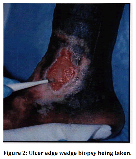
Patients were diagnosed to have Vasculitis based on certain diagnostic criteria such as the presence of elevated C-reactive protein, Erythrocyte Sedimentation Rate, fever, elevated total counts and on the basis of biopsy from the ulcer and of the vein. Erythrocyte Sedimentation Rate, Wound swab for Culture & Sensitivity, Venous Doppler, Urine Analysis, and Anti HCV Antigen were analyzed.
Results
Among the 130 patients only 3 patients had Vasculitis; In the 130 patients enrolled, 76.9% of the study group were males and 23.1% were females, all affected 3 were male, smokers, aged above 40 and , all three of them werediagnosed to have Type 2 Diabetes Mellitus. patients enrolled, 23.1% of them were diagnosed to have hypertension while 76.9% of the patients did not have hypertension.
Among the 3 patients who were found to have Vasculitis, two (66.7%) of them did not have hypertension against one (33.3%) who was diagnosed to have hypertension. Statistically the role of hypertension in Vasculitis was found to be insignificant.
Short saphenous vein pathology was found to be present in only 36 patients (27.7%), however among all the 3 patients who were found to have Vasculitis, none of them were found to have pathology with the Short Saphenous System.
Only 5 patients (3.8%) had a painful ulcer at the time of presentation, while the remaining 125 patients (96.2%) did not complain of any pain.
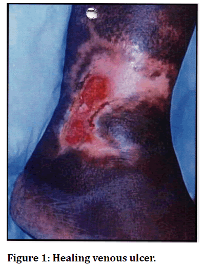
Among the patients who had Vasculitis, all three of the patients were found to have a painful ulcer at the time of presentation. All 3 patients had elevated levels of c reactive proteins (Figures 1 and Figure 2).

Figure 1. Healing venous ulcer.

Figure 2. Ulcer edge wedge biopsy being taken.
Discussion and Conclusion
All of the patients diagnosed with Vasculitis were smokers, however it was also noted that among the 130 patients studied, 57 of them were smokers against 73 who were non-smokers. In a paper published by G. R. Struthers, et al suggested that there was a significant relationship between smoking and Vasculitis the difference in our study is due to the fact that among the 57 smokers only 3 had developed Vasculitis thereby reducing its statistical significance [1-7]. All the patients with Vasculitis presented with painful ulcers. Among the 130 patients studied only 5 had painful ulcers. Statistically this was found to be highly significant in favour of Vasculitis presenting with painful ulcers and 19 shown to have elevated levels of leukocytes. Those 3 patients were started on low dose steroids and after improvement; surgery was performed eventually after healing of the ulcer.
References
- Mozes G, Carmichael JW, Gloviczki P. In: Gloviczki P, Yao JST, Editor. Handbook of venous disorders: guidelines of the American venous forum. 2nd Edn. London: Arnold; 2001.
- Cotton LT. Varicose veins: Gross anatomy and development. Br J Surg 1961; 48:589-597.
- Browse N, Burnand K, Thomas M. Diseases of the veins: Pathology, diagnosis, and treatment.london: Edward Arnold 1988.
- Leu H J, Vogt M, Pfrunder H. Morphological alterations of non-vancose and vancose veins. Cardiol l 979; 74:435-444.
- Somjen G M. Anatomy of the superficial venous system. Dermatol Surg 1995; 21:35-45.
- Thomson H. The surgical anatomy of the superficial and perforating veins of the lower limb. Ann R Coll Surg Engl 1979; 61:198-205.
- Cotton LT. Varicose veins: Gross anatomy and development. Br J Surg 1961; 48:589-597.
Author Info
Department of General Surgery, Bharath Institute of Higher Education and Research, Selaiyur, Chennai- 600073 Tamil Nadu, IndiaCitation: Gokuld Yatheendranathan, Prevalence of Vasculitis in Chronic Venous Insufficiency, J Res Med Dent Sci, 2021, 9(8): 87-86
Received: 14-Jul-2021 Accepted: 09-Aug-2021
