Research - (2021) Volume 9, Issue 9
Prevalence of Secondary Caries Among Different Restorations
Chris Noel Timothy* and S Delphine Priscilla Antony
*Correspondence: Chris Noel Timothy, Department of Conservative dentistry and Endodontics, Saveetha Dental College, Saveetha Institute of Medical and Technical Sciences, Saveetha University Tamilnadu, India, Email:
Abstract
Introduction: Secondary caries is an unfortunate occurrence that occurs immediately adjacent to the restoration, usually caused by inadequate extension of the restoration, microleakage or improper excavation of caries from the original lesion. Aims: The aim of this study was to assess the prevalence of secondary caries among different restorations. Materials and methods: This were a comparative, descriptive study, where all the data of the patients who reported to the dental clinics in saveetha dental college, SIMATS, Chennai, India, was obtained from the dental information archiving software (DIAS). Patient records were collected between March 2020 and March 2021. Data was collected and tabulated. The collected data was further analysed, recorded in Microsoft Excel software and was subjected to statistical analysis using IBM SPSS statistics analyser v.23.0. Results and discussion: The total sample size of the current study was 126 cases. In this study, we observed that the restoration most frequently associated with secondary caries was composite. We also observed that the secondary caries occurred more frequently in females in the age group of 36 to 45 years. Conclusion: Within the limitations of the current study, we observed that the most common restoration involved with secondary caries was composite and the most common age group which presented with secondary caries was 36 to 45 years.
Keywords
Secondary caries, Restoration, Composite, Amalgam, Full veneer crowns
Introduction
Secondary caries is an unfortunate occurrence that occurs immediately adjacent to the restoration, usually caused by inadequate extension of the restoration, microleakage or improper excavation of caries from the original lesion. Secondary caries occurs along an old restoration over a period. Bacterial accumulation or contamination occur due to improper isolation, inoculation, and micro cracks. Demineralisation of the tooth structure presents as a radiolucency below the restoration radiographically. The control of micro cracks, use of topical fluoride, proper oral hygiene and regular check-ups can aid in the reduction of secondary caries.
In other studies of similar nature, it was found that the use of fluoride releasing restorative materials such as GIC reduced the incidence of secondary caries and the replacement of restoration when compared to conventional treatment modalities [1]. It was found that marginal ditching, especially in the case of class 2 restorations, regardless of the type of restoration, microbial accumulation was seen. Mutans streptococci and lactobacilli were most commonly present and are the leading cause of secondary caries. Under the ditches, a large amount of bacterial accumulation was observed [2]. It was observed in a recent study that, among the general population, the prevalence of secondary caries was highest in composite [3]. Secondary caries was also found to be the leading cause for the replacement of amalgam restoration [4]. Proper clinical diagnosis and radiological assessment is essential to properly treat secondary caries and to prevent recurrence.
In previous studies, it was found that the major reason for replacement of a restoration was due to the presence of secondary caries [5]. In a practice-based study, it was found under clinical observation that gingival location was the primary site of origin for secondary caries in both composite and amalgam restorations [6]. It was also stated in another study that the presence of marginal staining could serve as an effective predictor for secondary caries, thereby preventing excessive loss of tooth structure [7,8]. Replacement or repair of an existing restoration must be carefully planned, to prevent leakage. It was found in a study that the bond strength is severely affected when a new material is placed over an old restoration or over unprepared tooth structure [2,9]. In another study, it was found that the replacement of all types of restorations in permanent and primary teeth was consistently about 50 percent, indicating a severe lack of awareness and precautions to be taken for the prevention of secondary caries [10–13].
Previously our team had a rich experience in working on various research projects across multiple disciplines [14– 29]. Now the growing trend in this area motivated us to pursue this project. This research is needed to gain better understanding of the various restorations and the prevalence of secondary caries among them. This will also help in the understanding of micro leakages and ways to prevent them. On review of literature, it was observed that there was a limited number of clinical research based on the prevalence of secondary caries among different restorations and gaining an understanding on how to better treat a patient. The aim of the current study is to assess the prevalence of secondary caries among different restorations.
Materials and Methods
This research study was defined as a descriptive study where all the patient’s data who reported to saveetha dental college and hospitals, SIMATS, Chennai, India and were diagnosed with secondary caries were obtained from the dental information archiving software (DIAS).
This study setting was an institutional setting, and the research study was conducted in the undergraduate and postgraduate dental clinics of saveetha dental college. This setting came with various pros and cons. The pros included the presence of a larger population and an abundant availability of data. Some of the cons included the study taking place in an unicentred setting and possessing a very limited demographic. The dependent variables in this study included the type of restoration and the presence or absence of secondary caries. The independent variables include the age of subject and gender of the subject. The selection of the study population was performed at random. This population was selected from the patients who visited the undergraduate and postgraduate dental clinics in saveetha dental college. The approval to undertake this research study had been approved by the ethical board of saveetha university (applied). n=126 cases were reviewed, and cross verification was performed by an additional reviewer. The minimisation of sample bias was performed by an additional reviewer, acquiring all the data from within the university and as an additional measure, simple random sampling was performed. There was a presence of high internal and low external validity. Sample collection was performed from March 2020 to March 2021.
The data was then arranged in a methodical manner using Microsoft Excel software and was tabulated based on 3 parameters namely, age of subject, gender of subject and the type of restoration. The data was validated by an additional reviewer. Any incomplete or censored data that was present in the collected data was excluded from the study.
Statistical analysis of the compiled data was performed using IBM SPSS statistical analyser. Chi square test was done for statistical analysis. The inclusion criteria for this study were outpatients with secondary caries irrespective of their age or gender. The exclusion criteria included outpatients who did not have the presence of secondary caries.
Results and Discussion
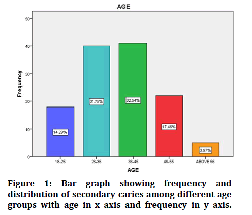
The data was collected and sorted based on the 3 parameters mentioned previously. Table 1 and Figure 1 explains about the distribution of secondary caries among different age groups. The total sample size of this study was 126 cases out of which, the most common age group of 36-45 years consisted of 32.5%, followed by 26 to 35 years with 31.7%, 46-55 years with 17.4%, 18 to 25 years with 14.2% and patients above 56 years with 3.9%. We also found that the age group of 36 to 45 years were 8.3 times more than the patients older than 56 years of age. In another study it was observed that the mean age of the patients with secondary caries was 36.7 years suggesting that the results of our study are in concordance with literature [30].
Table 1: Age.
| Age | Frequency | Percent | Valid percent | Cumulative percent | |
|---|---|---|---|---|---|
| Valid | 18-25 | 18 | 14.3 | 14.3 | 14.3 |
| 26-35 | 40 | 31.7 | 31.7 | 46 | |
| 36-45 | 41 | 32.5 | 32.5 | 78.6 | |
| 46-55 | 22 | 17.5 | 17.5 | 96 | |
| Above 56 | 5 | 4 | 4 | 100 | |
| Total | 126 | 100 | 100 | ||
Figure 1: Bar graph showing frequency and distribution of secondary caries among different age groups with age in x axis and frequency in y axis.
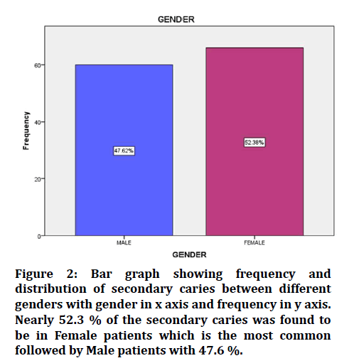
Table 2 and Figure 2 demonstrated the frequency and distribution of secondary caries between different genders. We observed that out of 126 cases, more prevalence was observed among females with 52.3% followed by males with 47.6%. This may be associated with differences in tooth morphology and oral hygiene practices between genders, but further research is required to prove this. In a recent study conducted by Ponnudurai Arangannal et al, it was found that the highest prevalence of secondary caries was observed in women, suggesting that the results of the current study are in concordance with literature [31].
Table 2: Gender.
| Gender | Frequency | Percent | Valid percent | Cumulative percent | |
|---|---|---|---|---|---|
| Valid | Male | 60 | 47.6 | 47.6 | 47.6 |
| Female | 66 | 52.4 | 52.4 | 100 | |
| Total | 126 | 100 | 100 | ||
Figure 2: Bar graph showing frequency and distribution of secondary caries between different genders with gender in x axis and frequency in y axis. Nearly 52.3 % of the secondary caries was found to be in Female patients which is the most common followed by Male patients with 47.6 %.
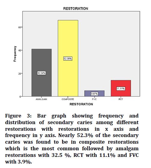
Table 3 and Figure 3 demonstrated the frequency and distribution of secondary caries among different restorations. Here we found that, among 126 restorations, the most common restoration affected with secondary caries was composite with 52.3% followed by amalgam with 32.5%, RCT (root canal treatment) with 11.1% and FVC with 3.9%. We observed that the prevalence of secondary caries in composite was 13.4 times higher compared to FVC. The reason could be due to the poor bond between the restoration and the tooth structure, microleakage, improper removal of caries, improper curing, etc. on review of literature, it was found that, in a study conducted by Nedeljkovic, Ivana, et al, there was significantly higher rate of microleakage compared to amalgam in composite restorations (60μm) and that composite restorations showed the highest rate of secondary caries (59.8%). In another study by A Mjo�� r, Ivar et al, composite restorations showed a higher rate of replacement due to secondary caries compared to amalgam restorations (80:20) suggesting that the results of the current study are in concordance with literature [32–35]. Our institution is passionate about high quality evidence-based research and has excelled in various fields [36–42]. We hope this study adds to this rich legacy.
Table 3: Restoration.
| Frequency | Percent | Valid percent | Cumulative percent | ||
|---|---|---|---|---|---|
| Valid | Amalgam | 41 | 32.5 | 32.5 | 32.5 |
| Composite | 66 | 52.4 | 52.4 | 84.9 | |
| FVC | 5 | 4 | 4 | 88.9 | |
| RCT | 14 | 11.1 | 11.1 | 100 | |
| Total | 126 | 100 | 100 | ||
Figure 3: Bar graph showing frequency and distribution of secondary caries among different restorations with restorations in x axis and frequency in y axis. Nearly 52.3% of the secondary caries was found to be in composite restorations which is the most common followed by amalgam restorations with 32.5 %, RCT with 11.1% and FVC with 3.9%.
Study limitations: presence of a smaller sample size, along with the study being a unicentered study with a limited demographic and a lack of variety in the data collected.
Future scope: this study could pave the way for new research with development of newer materials which show better physical properties and chemical properties, less microleakage and overall better prognosis.
Conclusion
Within the limitations of the current study, the most common restoration involved with secondary caries was composite which occurred predominantly in females. The most common age group which presented with secondary caries was 36 to 45 years.
References
- Mjör IA, Toffenetti F. Secondary caries: A literature review with case reports. Quintessence Int 2000; 31:165–79.
- Kidd EA, Joyston-Bechal S, Beighton D. Marginal ditching and staining as a predictor of secondary caries around amalgam restorations: a clinical and microbiological study. J Dent Res 1995; 74:1206–11.
- Nedeljkovic I, De Munck J, Vanloy A, et al. Secondary caries: Prevalence, characteristics, and approach. Clin Oral Investig 2020; 24:683–91.
- Pimenta LAF, de Lima Navarro MF, Consolaro A. Secondary caries around amalgam restorations. J Prosthetic Dent 1995; 74:219–22.
- Mjör IA, Moorhead JE, Dahl JE. Reasons for replacement of restorations in permanent teeth in general dental practice. Int Dent J 2000; 50:361–366.
- Mjör IA. The location of clinically diagnosed secondary caries. Quintessence Int 1998; 29:313–317.
- Shen C, Mondragon E, Gordan VV, et al. The effect of mechanical undercuts on the strength of composite repair. J Am Dent Assoc 2004; 135:1406–1412.
- Shen C, Speigel J, Mjör IA. Repair strength of dental amalgams. Oper Dent 2006; 31:122–126.
- Fusayama T. New concepts in operative dentistry: Differentiating two layers of carious dentin and using an adhesive resin. Quintessence Publishing Company 1980; 180.
- Healey HJ, Phillips RW. A clinical study of amalgam failures. J Dent Res 1949; 28:439–446.
- Jokstad A, Mjör IA. Analyses of long-term clinical behavior of class-II amalgam restorations. Acta Odontol Scand 1991; 49:47–63.
- Fusayama T, Hosoda H, Iwamoto T. An improved self-curing acrylic restoration and comparison with silicate cement restorations. J Prosthet Dent 1964; 14:537–553.
- Lavelle CL. A cross-sectional longitudinal survey into the durability of amalgam restorations. J Dent 1976; 4:139–143.
- Sathish T, Karthick S. Wear behaviour analysis on aluminium alloy 7050 with reinforced SiC through taguchi approach. J Materials Res Technol 2020; 9:3481-3487.
- Varghese SS, Ramesh A, Veeraiyan DN. Blended module-based teaching in biostatistics and research methodology: A retrospective study with postgraduate dental students. J Dent Educ 2019; 83:445–50.
- Samuel SR, Acharya S, Rao JC. School interventions-based prevention of early-childhood caries among 3-5-year-old children from very low socioeconomic status: Two-year randomized trial. J Public Health Dent 2020; 80:51–60.
- Venu H, Raju VD, Subramani L. Combined effect of influence of nano additives, combustion chamber geometry and injection timing in a DI diesel engine fuelled with ternary (diesel-biodiesel-ethanol) blends. Energy 2019; 174:386-406.
- Samuel MS, Bhattacharya J, Raj S, et al. Efficient removal of chromium(VI) from aqueous solution using chitosan grafted graphene oxide (CS-GO) nanocomposite. Int J Biol Macromol 2019; 121:285–92.
- Venu H, Subramani L, Raju VD. Emission reduction in a DI diesel engine using exhaust gas recirculation (EGR) of palm biodiesel blended with TiO2 nano additives. Renewable Energy 2019; 140:245-63.
- Mehta M, Deeksha, Tewari D, et al. Oligonucleotide therapy: An emerging focus area for drug delivery in chronic inflammatory respiratory diseases. Chem Biol Interact 2019; 308:206–215.
- Sharma P, Mehta M, Dhanjal DS, et al. Emerging trends in the novel drug delivery approaches for the treatment of lung cancer. Chem Biol Interact 2019; 309:108720.
- Malli Sureshbabu N, Selvarasu K, V JK, et al. Concentrated growth factors as an ingenious biomaterial in regeneration of bony defects after periapical surgery: A report of two cases. Case Rep Dent 2019; 2019:7046203.
- Muthukrishnan S, Krishnaswamy H, Thanikodi S, et al. Support vector machine for modelling and simulation of heat exchangers. Thermal Sci 2020; 24:499-503.
- Krishnaswamy H, Muthukrishnan S, Thanikodi S, et al. Investigation of air conditioning temperature variation by modifying the structure of passenger car using computational fluid dynamics. Thermal Sci 2020; 24:495-498.
- Gheena S, Ezhilarasan D. Syringic acid triggers reactive oxygen species-mediated cytotoxicity in HepG2 cells. Hum Exp Toxicol 2019; 38:694–702.
- Vignesh R, Sharmin D, Rekha CV, et al. Management of complicated crown-root fracture by extra-oral fragment reattachment and intentional reimplantation with 2 years review. Contemp Clin Dent 2019; 10:397–401.
- Ke Y, Al Aboody MS, Alturaiki W, et al. Photosynthesized gold nanoparticles from Catharanthus roseus induces caspase-mediated apoptosis in cervical cancer cells (HeLa). Artif Cells Nanomed Biotechnol 2019; 47:1938–46.
- Vijayakumar Jain S, Muthusekhar MR, Baig MF, et al. Evaluation of three-dimensional changes in pharyngeal airway following isolated lefort one osteotomy for the correction of vertical maxillary excess: A prospective study. J Maxillofac Oral Surg 2019; 18:139–146.
- Jose J, Subbaiyan H. Different treatment modalities followed by dental practitioners for ellis class 2 fracture–A questionnaire-based Survey. Open Dent J 2020; 4:59–65.
- Foster LV. Validity of clinical judgements for the presence of secondary caries associated with defective amalgam restorations. Br Dent J 1994; 177:89–93.
- Arangannal P, Mahadev SK, Jayaprakash J. Prevalence of dental caries among school children in Chennai, based on ICDAS II. J Clin Diagn Res 2016; 10:ZC09–12.
- Nedeljkovic I, Teughels W, De Munck J, et al. Is secondary caries with composites a material-based problem? Dent Mater 2015; 31:e247–77.
- Mjör IA. Clinical diagnosis of recurrent caries. J Am Dent Assoc 2005; 136:1426–1433.
- Bernardo M, Luis H, Martin MD, et al. Survival and reasons for failure of amalgam versus composite posterior restorations placed in a randomized clinical trial. J Am Dent Assoc 2007; 138:775–783.
- Demarco FF, Corrêa MB, Cenci MS, et al. Longevity of posterior composite restorations: not only a matter of materials. Dent Mater 2012; 28:87–101.
- Vijayashree Priyadharsini J. In silico validation of the non-antibiotic drugs acetaminophen and ibuprofen as antibacterial agents against red complex pathogens. J Periodontol 2019; 90:1441–1448.
- Ezhilarasan D, Apoorva VS, Ashok Vardhan N. Syzygium cumini extract induced reactive oxygen species-mediated apoptosis in human oral squamous carcinoma cells. J Oral Pathol Med 2019; 48:115–21.
- Ramesh A, Varghese S, Jayakumar ND, et al. Comparative estimation of sulfiredoxin levels between chronic periodontitis and healthy patients-A case-control study. J Periodontol 2018; 89:1241–1248.
- Mathew MG, Samuel SR, Soni AJ, et al. Evaluation of adhesion of Streptococcus mutans, plaque accumulation on zirconia and stainless steel crowns, and surrounding gingival inflammation in primary molars: randomized controlled trial. Clin Oral Investig 2020; 24:3275–80.
- Sridharan G, Ramani P, Patankar S, et al. Evaluation of salivary metabolomics in oral leukoplakia and oral squamous cell carcinoma. J Oral Pathol Med 2019; 48:299–306.
- J PC, Marimuthu T, Devadoss P, Kumar SM. Prevalence and measurement of anterior loop of the mandibular canal using CBCT: A cross sectional study. Clin Implant Dent Relat Res 2018; 20:531–534.
- Ramadurai N, Gurunathan D, Samuel AV, et al. Effectiveness of 2% Articaine as an anesthetic agent in children: Randomized controlled trial. Clin Oral Investig 2019; 23:3543–3550.
Author Info
Chris Noel Timothy* and S Delphine Priscilla Antony
Department of Conservative dentistry and Endodontics, Saveetha Dental College, Saveetha Institute of Medical and Technical Sciences, Saveetha University Tamilnadu, Chennai, IndiaCitation: Chris Noel Timothy, S Delphine Priscilla Antony,Prevalence of Secondary Caries Among Different Restorations , J Res Med Dent Sci, 2021, 9(9): 190-194
Received: 23-Aug-2021 Accepted: 13-Sep-2021


