Case Report - (2021) Volume 9, Issue 11
Preauricular Epidermal Cyst-A Case Report
MK Rajasekar and Nithya Balasubramanian*
*Correspondence: Nithya Balasubramanian, Department of ENT, Sree Balaji Medical College and Hospital, Chennai, Tamil Nadu, India, Email:
Abstract
Epidermal cysts are benign tumours which can occur anywhere in the body. They are generally seen on the scalp, face, neck, dorsal and ventral trunk. Here we see a case of epidermal cyst in the pre-auricular region and not originating from a pre-auricular sinus and discuss the clinical presentations, imaging and treatment. Patient underwent surgery for excision of the cyst.
Keywords
Epidermal cysts, Benign tumours, Ectoderm, Squamous epithelium
Introduction
Epidermal cysts are inclusion cysts, originating from ectoderm lined by squamous epithelium. Because epidermoid cysts are uncommon in the head and neck, they are frequently misdiagnosed. Soft nodular lesions with a sessile base characterise epidermal cysts. Cutaneous cysts are divided into three categories based on their shape and differentiation pattern: (1) Stratified squamous epithelium, (2) Non-stratified squamous epithelium, and (3) non-stratified squamous epithelium, (3) Absence of epithelium.
The majority of lesions are sporadic; however familial inheritance is possible, particularly in people who have several lesions. Multiple epidermoid cysts can appear as sporadically scattered lesions throughout the body or as cluster lesions in the retro auricular area, which is typical.
Epidermoid cysts are fluid-filled protrusions that arise from the follicular infundibulum and are found immediately beneath the skin's surface. Lesions frequently appear on their own. Injury-induced epithelial implantation, on the other hand, is regarded an etiologic factor. As a result, it's also known as an epidermal inclusion cyst. Furthermore, epidermal cells penetrate deep into the skin and grow as a result of surgery or skin problems cysts. Because the cyst wall is lined with stratified squamous epithelium, keratin layers will peel off and accumulate inside the cyst. An epidermoid is usually caused by a blockage of the follicular opening [1-7].
Case History
A 51 year old male presented to the Department of ENT, Sree Balaji Medical College and Hospital, with a history of swelling over the pre-auricular region for 2 months. The swelling was associated with pain. Local examination of the swelling showed that the size of the swelling was around 1x1 cm, soft and cystic in consistency, mobile and associated with tenderness.
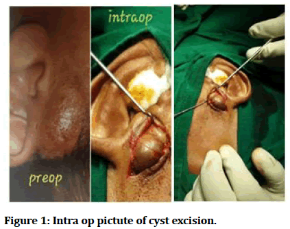
Patient was taken up for surgery after doing necessary investigations and after obtaining clearance from anaesthesia. The sample taken from the pre-auricular region was sent for histopathology (Figure 1).
Figure 1: Intra op pictute of cyst excision.
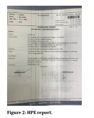
The results of the histopathology showed that it was a cystic lesion lined by stratified squamous epithelium and granulation tissue with foreign body type of giant cell reaction enclosing keratinous material (Figure 2).
Figure 2: HPE report.
Discussion
The histopathological features and clinical importance of many forms of cutaneous cysts containing fluid or semisolid material have been characterised. Some cysts have an epithelial cell wall that is either stratified squamous epithelium or unstratified squamous epithelium. True cysts are what they're called. Pseudo cysts, on the other hand, are cysts that are not bordered by epithelium and are instead surrounded by connective or granulation tissue. In general, cutaneous cysts are spherical, domeshaped, projecting, or deeply situated dermal or subcutaneous papules or nodules that can be found in a variety of places. Histopathological investigation is usually used to confirm the diagnosis.
An epidermoid cyst is a form of cutaneous cyst with an epithelial lining that looks like the epidermis (wall). Keratin is produced by the cyst's lining. Because there are no sebaceous glands within the cyst lining, the name sebaceous cyst, which was previously used as a synonym for epidermoid cyst, is incorrect. The growth of surface epidermoid cells within the dermis causes it. In fundibular cyst, epidermal cyst, epidermal inclusion cyst, and epidermoid inclusion cyst are all common synonyms.
Dermoid and epidermoid cysts are both referred to as "dermoid," though they are two distinct things. Both are cystic choristomas that are filled with keratin, cholesterol clefts, or degraded blood components and are created by keratinizing squamous epithelium; however, real dermoid cysts contain skin appendages on their walls, whereas epidermoid cysts do not. Dermoid cysts are widespread and are usually identified in infancy or early childhood. They are usually found superficially or in the anterior orbit, mould bone, and only rarely cause bone lysis. Epidermoid cysts, on the other hand, can appear anywhere on the body and are frequently discovered later in life. If they are in the orbit, they usually create a relationship with the bone. Although these cysts are recognized as benign lesions, rare malignancy can arise. These cysts may progress slowly for years [8-13].
Conclusion
Here we see that the patient presented with an infected pre-auricular cyst not associated with a pre-auricular sinus which a variant from the normal presentation.
Because epidermoid cysts are uncommon in the head and neck, they are frequently misdiagnosed. The purpose of this case report is to show how epidermoid and dermoid cysts can present as a differential diagnosis for head and neck tumours.
References
- Baykal C, Yazganoglu KD. Clinical atlas of skin tumors. Springer Science Business Media 2014.
- https://evolve.elsevier.com/cs/product/9780323680745?role=student
- Zito P, Schar R. Epidermoid (Sebaceous cyst). Treasure Island (FL) Stat Pearls Publishing 2019.
- Wollina U, Langner D, Tchernev G, et al. Epidermoid cysts–a wide spectrum of clinical presentation and successful treatment by surgery: a retrospective 10-year analysis and literature review. Open Access Macedonian J Med Sci 2018; 6:28.
- Blanco G, Esteban R, Galarreta D, et al. Orbital intradiploic giant epidermoid cyst. Arch Ophthalmol 2001; 119:771-773.
- https://www.elsevier.com/books/head-and-neck-imaging-2-volume-set/som/978-0-323-05355-6
- Dutta M, Saha J, Biswas G, et al. Epidermoid cysts in head and neck: our experiences, with review of literature. Indian J Otolaryngol Head Neck Surg 2013; 65:14-21.
- Janarthanam J, Mahadevan S. Epidermoid cyst of submandibular region. J Oral Maxillofac Pathol 2012; 16:435.
- de Mendonça JC, Jardim EC, Dos Santos CM, et al. Epidermoid cyst: Clinical and surgical case report. Annals Maxillofac Surg 2017; 7:151.
- Pires-Gonçalves L, Silva C, Teixeira M, et al. Testicular epidermoid cyst-Ultrasound and MR typical findings with macroscopy correlation. Int Braz J 2011; 37:534-536.
- Bin Manie MA, Al-Qahtani KH, Al Ammar A, et al. Epidermoid cyst of the suprasternal region: A rare case report. Br J Otorhinolaryngol 2020; 86:133-135.
- Lincoski CJ, Bush DC, Millon SJ. Epidermoid cysts in the hand. J Hand Surg 2009; 34:792-796.
- Takemura N, Fujii N, Tanaka T. Epidermal cysts: The best surgical method can be determined by ultrasonographic imaging. Clin Exp Dermatol 2007; 32:445-447.
Author Info
MK Rajasekar and Nithya Balasubramanian*
Department of ENT, Sree Balaji Medical College and Hospital, Chennai, Tamil Nadu, IndiaCitation: MK Rajasekar, Nithya Balasubramanian, Preauricular Epidermal Cyst-A Case Report, J Res Med Dent Sci, 2021, 9(11): 107-108
Received: 06-Oct-2021 Accepted: 01-Nov-2021


