Research - (2022) Volume 10, Issue 1
Phyto-assisted Synthesis and Assessment of Anti-Inflammatory and Antioxidant Properties of Copper and Graphene Oxide Nano Composite Reinforced with Amla Extract-An In-Vitro Study
Gaurav N Ketkar1, Sankari Malaiappan1* and Rajeshkumar S2
*Correspondence: Sankari Malaiappan, Department of Periodontics, Saveetha Dental College and Hospital, Saveetha Institute of Medical and Technical Sciences, India, Email:
Abstract
Background: conventional methods used for synthesis of nanoparticles use toxic chemicals, thus leading to toxicity in the environment. Hence, we need to shift to “green synthesis”. Previously done have shown excellent antimicrobial properties of the copper and graphene oxide nanocomposite reinforced with amla extract. Hence, this study was conducted to assess the anti-inflammatory and antioxidant properties of copper and graphene oxide nanocomposite reinforced with amla extract. Copper causes cell wall lysis by leakage of intracellular substances hence is an excellent antimicrobial agent, graphene oxide for its barrier and structural strength hence these were chosen to create a nanocomposite. Aim: Aim of the study was phyto-assisted preparation of nano copper with nano graphene oxide nanocomposite and evaluation of its anti-inflammatory and antioxidant properties. Material and methods: Anti-inflammatory and antioxidant properties of the nanocomposite were assessed using Bovine Serum Albumin (BSA) and DPPH Assay respectively at 10 µL, 20 µL, 30 µL, 40µL, 50 µL. Results: Values for anti-inflammatory property of nanoparticles were higher than the standard values at 40µL, 50 µL concentrations. Percentage of inhibition was highest at 40 µL (86%) and 50 µL (84.6%). The values for antioxidant property of nanoparticles were found to be higher than the standard values at concentrations except at 40µL, 50 µL. Percentage of inhibition was highest at 20 µL (86.2%). Conclusion: Within the limits of the study, it can be concluded that copper and graphene oxide nanocomposite has exceptional anti-inflammatory and antioxidant properties and further can be incorporated in dental material or can be used to coat suture materials to improve their properties.
Keywords
Copper, Characterisation, Graphene oxide, Green synthesis, Nanoparticle, Nanocomposite, Antiinflammatory, Antioxidant properties
Introduction
Nanotechnology is an emerging technology and has led to a new revolution in every field of science. Nanoparticles have gained a lot of importance in the research community in recent years. This technology has been used in the fields of optics, electronics, and biomedical and materials sciences [1]. Potent antimicrobial, anticancer, antioxidant agents, drug, and gene delivery, etc are some of the highlighted advantages of the nanoparticles in the recent years [2-4]. Nanotechnology deals with nanoparticles that are atomic or molecular aggregates characterized by size less than 100 nm. These are basic elements derived by modifying their atomic and molecular properties [5,6].
Several conventional methods are used for synthesis of nanoparticles like chemical reduction [7], laser ablation [8], inert gas condensation [9,10], sol-gel method [11]. Even though less time is utilized for synthesizing large quantities of nanoparticles using conventional physical and chemical methods, toxic chemicals are required as capping agents to maintain stability, thus leading to toxicity in the environment. “Green synthesis” offers numerous benefits of eco friendliness and compatibility for biomedical applications, where toxic chemicals are not used for the synthesis protocol. The use of agricultural wastes [9] or plants and their parts [10], has emerged as an alternative to chemical synthetic procedures because it does not require elaborate processes such as intracellular synthesis and multiple purification steps or the maintenance of microbial cell cultures.
Copper nanoparticles (CuNP) are superior owing to their nontoxicity, biocompatibility, use in drug and bactericidal activity [12,13]. Contact killing property of copper was studied widely in recent years. Studies have shown that increased bacterial intracellular oxidative stress in the bacterial cell wall due to release of ions from the copper surface which results in bacterial cell lysis [14]. Synthesis of copper nanoparticles is highly technique sensitive due to its high incidence of oxide layer formation on the nanoparticle surface which will result in reduced antibacterial property [15].
Graphene oxide is known for its excellent mechanical strength, electrical conductivity and most importantly the barrier properties, also easy step-down preparation of graphene oxide nanoparticles makes it one of the most efficient carriers of nanoparticles in any nanocomposite. It is an atomically-thin, 2-dimensional (2D) sheet of sp2 carbon atoms in a honeycomb structure [16 ].
The objective of this study was to use amla fruit extract to synthesize copper and graphene oxide nanoparticles and to evaluate its anti-inflammatory and antioxidant properties as its excellent potency against oral aerobes was already proven in the previous studies [16]. Also the nanocomposite showed minimal cytotoxic effects.
Material and Methods
Preparation of amla extract
Freshly collected organic amla fruits were thoroughly washed multiple times in distilled water. Seed was taken out and the pulp was cut into small pieces using a sterile knife and was grounded into small particles by means of a mortar and pestle. Amla extract was prepared by 1 grams of amla pulp with 100 ml distilled water to make 1 molar solution of amla extract [16].
Synthesis of copper and graphene oxide (Cu-GO) nanocomposite
Nanocomposite synthesis was done by mixing 50 ml of both 1M solutions of copper and graphene oxide nanoparticles as mentioned in the previous steps. The nanocomposite solution was stirred overnight on an orbital shaker followed by a magnetic heated stirrer till colour change was observed. UV-vis spectrometric readings were taken hourly to check the synthesis of copper-graphene oxide nano composite. The resultant mixture was centrifuged and CuGO nanocomposite was obtained [16].
Anti-inflammatory activity
Test group
10 μL, 20 μL, 30 μL, 40 μL and 50 μL of the nanocomposite solution was taken in 5 test tubes respectively. To each test tube 2 ml of 1% Bovine Serum Albumin (BSA) was added. 390 μL, 380 μL, 370 μL, 360 μL and 350 μL of distilled water was added to the test tube containing 10 μL, 20 μL, 30 μL, 40 μL and 50 μL of nanoparticles respectively.
Control group
2 mL of Dimethyl Sulphoxide (DMSO) was added to 2 mL of BSA solution.
Standard group
10 μL, 20 μL, 30 μL, 40 μL and 50 μL of Diclofenac Sodium was taken in 5 test tubes respectively. To each test tube 2 mL of 1% Bovine Serum Albumin (BSA) was added. The test tubes were incubated at room temperature for 10 minutes. Then they were incubated in water bath at 55°C for around 10 minutes. Absorbance was measured at 660 nm in UV Spectrophotometer.
% Inhibition was calculated using the following formula:
% of inhibition=Control OD−Sample OD Sample OD100
Results
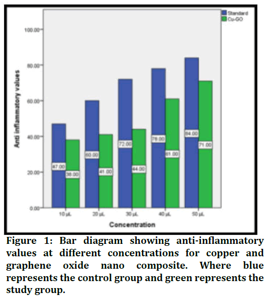
Anti-inflammatory assay showed the following values at the end of the study:
Anti-inflammatory property of nanoparticles was higher than the standard values at 40μL, 50 μL concentrations. Percentage of inhibition was highest at 40 μL (86%) and 50 μL (84.6%) (Figure 1).

Figure 1. Bar diagram showing anti-inflammatory values at different concentrations for copper and graphene oxide nano composite. Where blue represents the control group and green represents the study group.
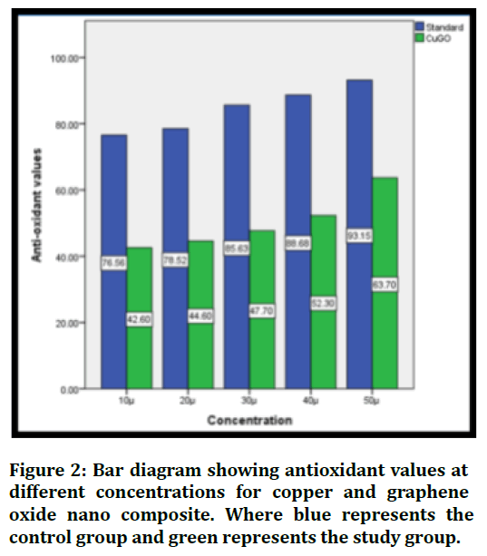
Antioxidant test showed the following values for the nanocomposite: The values for antioxidant property of nanoparticles were found to be higher than the standard values at concentrations except at 40μL, 50 μL. Percentage of inhibition was highest at 20 μL (86.2%) (Figure 2).

Figure 2. Bar diagram showing antioxidant values at different concentrations for copper and graphene oxide nano composite. Where blue represents the control group and green represents the study group.
Discussion
Effective drug delivery systems with the ability to improve the therapeutic profile and efficacy of therapeutic agents is one of the key issues faced by modern medicine.
Advances in nanoscience and nanotechnology, enabling the synthesis of new nanomaterials, have led to the development of several new drug delivery systems.
There has been a rapid evolution of nanoparticle synthesis recently as compared to the early part of the century [17].
Earlier conventional methods were used for the synthesis of the nanoparticles. Even though less time is utilized for synthesizing large quantities of nanoparticles using conventional physical and chemical methods, toxic chemicals are required as capping agents to maintain stability.
These methods resulted in toxicity in the environment due to use of toxic chemicals. Green Synthesis method was proposed and is widely used all over the world to avoid the use of such toxic chemicals. It is an eco-friendly method as well as it is highly cost effective [18].
We therefore undertook this study to evaluate the cytotoxicity of the copper and graphene oxide nanocomposite reinforced with amla extract. Antibacterial properties of the same composite were found to be excellent against oral microbes in the previous studies [16].
Amla or Phyllanthus emblica is known since ancient times for its medicinal value and is commonly used in Ayurvedic medicine.
Amla is a rich source of vitamin C which is a highly essential vitamin in maintenance of epithelial homeostasis. Superoxide radicals (O2–.) have been implicated in several pathological disorders and are responsible for elevated oxidative stress.
Studies have shown that amla extract acts as a very good antioxidant by scavenging the reactive oxygen species and protects the antioxidant enzymes like SOD required for the cellular defence.
Copper has been known for its antibacterial activity. Copper nanoparticles impart the antioxidant effect by inhibition of chain reaction, decomposition of peroxides, binding of transition metal ion catalysts, radical scavenging activity and inhibition of continued hydrogen abstraction.
The prop- erties of absorbing, neutralizing these free radicals or quenching singlet and triplet oxygen are few crucial factors that are responsible for the antioxidant activity. The highest antioxidant activity is attributed due to the presence of various bio-reductive groups of the phyto- chemicals present on the surface of the CuNPs.
Copper not only is an excellent antioxidant but also an effective anti-inflammatory agent [12,19].
Remarkable physical–chemical properties, including a high Young’s modulus, high fracture strength, excellent electrical and thermal conductivity, fast mobility of charge carriers, large specific surface area and biocompatibility are few of the major advantages of graphene nanoparticles.
Graphene oxide is studied in the medicinal field for over 40 years for its potential for being the ideal drug delivery properties.
Hence the combination of copper and graphene oxide with amla was selected to be tested for its antiinflammatory and antioxidant property in the current study.
Conclusion
Within the limits of this study, it can be concluded that copper and graphene oxide nanocomposite has exceptional anti-inflammatory and antioxidant properties. With a possibility to be included in the dental materials to improve the properties.
Conflict of Interest
The author declares that there are no conflicting interests.
References
- Vimbela GV, Ngo SM, Fraze C, et al. Antibacterial properties and toxicity from metallic nanomaterials. Int J Nanomedicine 2017; 12:3941.
- Morens DM, Folkers GK, Fauci AS. Emerging infections: a perpetual challenge. Lancet Infec Dis 2008; 8:710-9.
- Alpaslan E, Geilich BM, Yazici H, et al. pH-controlled cerium oxide nanoparticle inhibition of both gram-positive and gram-negative bacteria growth. Scientific Rep 2017; 7:1-2.
- Beyth N, Houri-Haddad Y, Domb A, et al. Alternative antimicrobial approach: nano-antimicrobial materials. Evidence-based Comple Alter Med 2015; 2015.
- Tran Phong A, Neil O’Brien-Simpson, Eric C. Reynolds, et al. Low cytotoxic trace element selenium nanoparticles and their differential antimicrobial properties against S.aureus and E. Coli. Nanotechnol 2016.
- Gholami A, Ebrahiminezhad A, Abootalebi N, et al. Synergistic evaluation of functionalized magnetic nanoparticles and antibiotics against Staphylococcus aureus and Escherichia coli. Pharmaceut Nanotechnol 2018; 6:276-86.
- Morones JR, Elechiguerra JL, Camacho A, et al. The bactericidal effect of silver nanoparticles. Nanotechnol 2005; 16:2346.
- Asiri A, Mohammad A. Applications of nanocomposite materials in dentistry. Woodhead Publishing 2018.
- Gour JK, Srivastava A, Kumar V, et al. Nanomedicine and leishmaniasis: Future prospects. Digest J Nanomat Biostruct 2009; 4.
- Haris Z, Khan AU. Selenium nanoparticle enhanced photodynamic therapy against biofilm forming Streptococcus mutans. Int J Life Sci Scienti Res 2017; 3:1287-94.
- Bitiren M, Karakilcik AZ, Zerin M, et al. Protective effects of selenium and vitamin E combination on experimental colitis in blood plasma and colon of rats. Biolog Trace Element Res 2010; 136:87-95.
- Mao L, Wang L, Zhang M, et al. In situ synthesized selenium nanoparticles‐decorated bacterial cellulose/gelatin hydrogel with enhanced antibacterial, antioxidant, and anti‐inflammatory capabilities for facilitating skin wound healing. Adv Healthcare Mat 2021: 2100402.
- Marcello E, Maqbool M, Nigmatullin R, et al. Antibacterial composite materials based on the combination of polyhydroxyalkanoates with selenium and strontium co-substituted hydroxyapatite for bone regeneration. Fron Bioengine Biotech 2021; 9.
- Ren G, Hu D, Cheng EW, et al. Characterisation of copper oxide nanoparticles for antimicrobial applications. Int J Antimicrob Agents 2009; 33:587-90.
- Minko T, Rodriguez-Rodriguez L, Pozharov V. Nanotechnology approaches for personalized treatment of multidrug resistant cancers. Adv Drug Deliv Rev 2013; 65:1880-95.
- Cao W, Zhang Y, Wang X, et al. Novel resin-based dental material with anti-biofilm activity and improved mechanical property by incorporating hydrophilic cationic copolymer functionalized nanodiamond. J Mat Sci 2018; 29:1-3.
- Keystone JS. Advantages and disadvantages of antimalarials for chemoprophylaxis. In Travel Medi 1989; 102-112.
- Aronoff BL. Advantages and disadvantages of the laser. In Lasers Med Surg 1982; 357:86-88.
Indexed at , Google Scholar, Cross Ref
Indexed at , Google Scholar, Cross Ref
Indexed at , Google Scholar, Cross Ref
Indexed at , Google Scholar, Cross Ref
Indexed at , Google Scholar, Cross Ref
Indexed at , Google Scholar, Cross Ref
Indexed at , Google Scholar. Cross Ref
Indexed at , Google Scholar, Cross Ref
Indexed at , Google Scholar, Cross Ref
Indexed at , Google Scholar, Cross Ref
Indexed at , Google Scholar, Cross Ref
Author Info
Gaurav N Ketkar1, Sankari Malaiappan1* and Rajeshkumar S2
1Department of Periodontics, Saveetha Dental College and Hospital, Saveetha Institute of Medical and Technical Sciences, Chennai, India2Department of Pharmacology, Saveetha Dental College and Hospital, Saveetha Institute of Medical and Technical Sciences, Chennai, India
Received: 06-Dec-2021, Manuscript No. JRMDS-21-44369; , Pre QC No. JRMDS-21-44369 (PQ); Editor assigned: 08-Dec-2021, Pre QC No. JRMDS-21-44369 (PQ); Reviewed: 22-Dec-2021, QC No. JRMDS-21-44369; Revised: 27-Dec-2021, Manuscript No. JRMDS-21-44369 (R); Published: 03-Jan-2022
