Research - (2021) Volume 9, Issue 4
Newly Proposed Classification of Celiac Trunk Variations Based on Embryology-Correlation with Computed Tomography Angiography
*Correspondence: Prabakaran M, Department of Radio Diagnosis, Sree Balaji Medical College & Hospital,, Bharath Institute of Higher Education and Research, India, Email:
Abstract
To analyze variations of celiac trunk branching pattern using Computed Tomography Angiography(CTA) and proposing new classification on the basis of embryology Statistical analysis to identify incidence of different variations of celiac trunk reiterate the clinical importance of anatomical variants like IPA and celioemesenteric trunk and pathological conditions of celiac artery like median arcuate ligament syndrome.
Keywords
Computed tomography angiography, Hepatic artery, Embryology
Introduction
Celiac trunk (CT) was first described by Haller as Tripus halleri in 1756. CT is the main artery in the upper abdomen supplying major organs like the pancreas, liver, gall bladder, stomach, lower end of esophagus, part of duodenum and the spleen [1] .The CT is the first ventral branch of the abdominal aorta and normally has trifurcation branching pattern (Left gastric artery, Common hepatic artery and Splenic artery), but sometimes various other branching patterns like bifurcation, quadrifurcation, pentafurcation and hexafurcation can be seen. These include additional branches like SMA, inferior phrenic artery, Gastro duodenal artery (GDA), Middle colic artery (MCA), accessory hepatic artery, suprarenal arteries, retroportal artery, aberrant bronchial artery and dorsal pancreatic artery as reported in literature [2].
In this research study I have proposed to reclassify CT variation on the basis embryology correlated with routine CTA studies on 402 patients. The previous studies on same subject are touched upon below. CT variations were first classified by Lipshutz et al. into four types based on origin of common hepatic artery (CHA), Splenic artery (SA) and left gastric artery (LGA), followed by various authors like Adachi et al. and Michel et al. who classified CT into six types by adding origin of superior mesenteric artery (SMA) with CT. Uflacker’s et al. classified CT into eight types by adding origin of SMA, MCA with CT and no CT [3-6]. Song et al. classified CT into 15 types which included the above mentioned arteries [7]. Panagouli et al. classified CT into 10 types in which along with the above mentioned arteries he added IMA. The knowledge of CT branching pattern, origin and its course is not only limited to anatomy, but also it has significant role clinically. It may be the source of pathological conditions like celiac compression syndrome and gastrointestinal bleeding. Patients should undergo diagnostic angiography prior to operative procedures and interventional procedures to identify the anatomical variations, since knowledge of vascular anatomy is obligatory before for planning of treatment [8].
Materials and Methods
We retrospectively assessed celiac trunk variations in patients who had come for diagnostic CT Angiography. The patient data was acquired from Picture Archiving and Communication System of our institution. For this study permission was obtained from the ethical committee of the institution and the study duration was of one and a half year (July 2019 to January 2018). Multiphasic helical CT was done on 128 slice Computed Tomography (Siemens, Software-syngo CT VC 40, SOMATOM, Shanghai, China,) for the study.We reviewed 402 cases which included 254 males and 148 females respectively. The area of CT study extended from just above diaphragm up to iliac vessel bifurcation. The scanning parameters included the data acquired in 5.0 mm slice direction: cranio-caudal, (Rotation time: 0.6 seconds) detector configuration with 64 x 0.6 mm, 150mAs; 130 kVp; pitch: 0.8.The images obtained were later reconstructed in to 1.0 mm slice thickness. Nonionic iodinated contrast material was injected through sterile 18 gauge cannula inserted in to antecubital vein. The dose range was 100-120 ml and rate of injection was 5-6 ml per second. Iodine concentration used was 350 mg/mL. Images were obtained at 3 seconds after AA threshold.
The data from PACS were first transferred to workstation for multiplanar reformation, volume rendering as well as 3D reconstruction with Maximum Intensity Projection (MIP). The images obtained were individually analyzed by 2 experts and one junior resident of Radio- Diagnosis. The experts have an experience of 7 years and 15 years respectively. We analyzed origin and branching pattern of celiac trunk including LGA, SA, CHA, SMA and IPA.
Initially images were reconstructed in axial section at the level of celiac trunk origin, First bilateral IPA were assessed after adjusting the MIP. Later Celiac Trunk and SMA origin and branching pattern were assessed. Overall origin and branching of the above mentioned arteries were analyzed after 3D reconstruction and volume rendering.
Exclusion criteria
• Major vascular pathology.
• Major upper abdominal surgeries.
Inclusion criteria
Patients undergoing routine diagnostic CT angiography of study of upper abdomen.
Results
We have proposed a new classification of celiac trunk and its branching pattern based on embryology and classified it into six main types, which is further divided into 18 subtypes (Tables 1,2 and Figure 1). The study was conducted in 402 patients in whom 254 were males and 148 females (Table 3 and Figure 2). Type I was normal trifurcation pattern seen in 174 patients (40%) (Figure 3). Type II seen in 17 patients (4%) was further subdivided into three subtypes. Type IIa (Hepatosplenic trunk) was seen in 8 patients (2%). Type IIb (Hepatogastric trunk) was not seen in any of our patients. Type IIc (Gastrosplenic trunk) was seen in 9 patients (2%). Type III (No celiac trunk) and was seen in 1 patient (0.002%) (Figure 4). Type IV is further divided into seven subtypes. Type IVa (Celiomesenteric trunk) was seen in one patient (0.002%), Type IVb (Hepatomesenteric trunk) was seen in five patients (1%). Type IVc (Right hepatomesenteric trunk) was seen in thirty patients (7%), Type IVd (Gastromesenteric trunk) was not seen in any patient, Type IVe (Splenomesenteric trunk) was not seen in any patients, Type IVf (Hepatosplenomesenteric trunk) was seen in three patients (1%), Type IVg (Gastrosplenomesenteric trunk) was not seen in any patient. Type V (Celiacocolic trunk) was not seen in any patient. Type VI is subdivided into four subtypes. Type VIa (Celiacophrenic trunk) (CT + CIPA) was seen in thirty six patients (8%), Type VIb (Celiacophrenic trunk) (CT+ RIPA) was seen in twenty six patients (6%), Type VIc (Celiacophrenic trunk) (CT + LIPA) was seen in fifty nine patients (14%). Type VId (Celiacophrenic trunk) (CT + RIPA + LIPA) was seen in sixty two patients (14%)(Figure 5). And finally ‘Others’ which consists of other variants was seen in twenty two patients (5%) (Figure 6).
| S.No | Types | CT Branching pattern |
|---|---|---|
| 1 | I | Normal trifurcation |
| 2 | II(a) | Hepatosplenic trunk |
| 3 | II(b) | Hepatogastric Trunk |
| 4 | II(c) | Gastrosplenic Trunk |
| 5 | III | No celiac Trunk |
| 6 | IV(a) | Celiomesenteric Trunk |
| 7 | IV(b) | Hepatomesenteric Trunk |
| 8 | IV(c) | Right Hepatomesenteric Trunk |
| 9 | IV(d) | Gastro mesenteric Trunk |
| 10 | IV(e) | Splenomesenteric Trunk |
| 11 | IV(f) | Hepatosplenomesenteric Trunk |
| 12 | IV(g) | Gatrosplenomesenteric Trunk |
| 13 | V | Celiaccolic Trunk |
| 14 | VI(a) | Coeliophrenic Trunk (CT+ CIPA) |
| 15 | VI(b) | Coeliophrenic Trunk (CT+ RIPA) |
| 16 | VI(c) | Coeliophrenic Trunk (CT+ LIPA) |
| 17 | VI(d) | Coeliophrenic Trunk (CT+ RIPA + LIPA) |
| 18 | Others | Additional branches |
Table 2: Subtypes of CT variations incidence in 402 cases.
| Types | Incidence in total |
|---|---|
| Type I | 174 |
| Type II a | 8 |
| Type II b | 0 |
| Type II c | 9 |
| Type III | 1 |
| Type IV a | 1 |
| Type IV b | 5 |
| Type IV c | 30 |
| Type IV d | 0 |
| Type IV e | 0 |
| Type IV f | 3 |
| Type IV g | 0 |
| Type V | 0 |
| Type VI a | 36 |
| Type VI b | 26 |
| Type VI c | 59 |
| Type VI d | 62 |
| Others | 22 |
Table 2: Subtypes of CT variations incidence in 402 cases.
| Type | Incidence total | Total |
|---|---|---|
| Type I | 174 | 402 |
| Type II | 17 | 402 |
| Type III | 1 | 402 |
| Type IV | 39 | 402 |
| Type V | 0 | 402 |
| Type VI | 183 | 402 |
| Other | 22 | 402 |
Table 3: Main types of CT variations incidence in 402 cases.
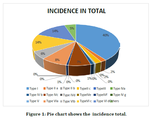
Figure 1. Pie chart shows the incidence total.
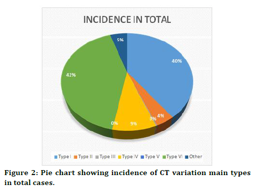
Figure 2. Pie chart showing incidence of CT variation main types in total cases.
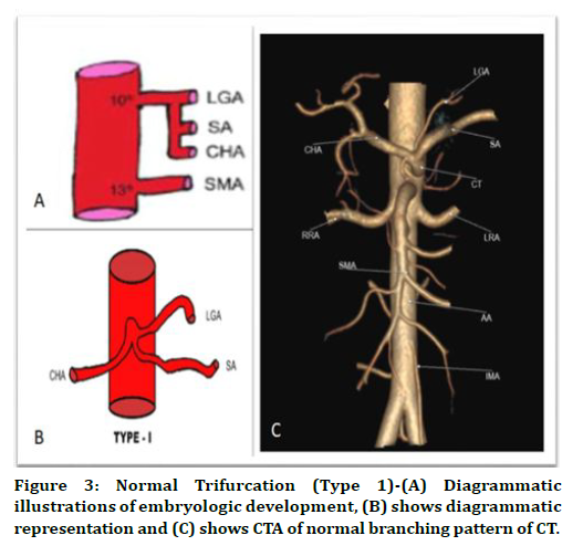
Figure 3. Normal Trifurcation (Type 1)-(A) Diagrammatic illustrations of embryologic development, (B) shows diagrammatic representation and (C) shows CTA of normal branching pattern of CT.
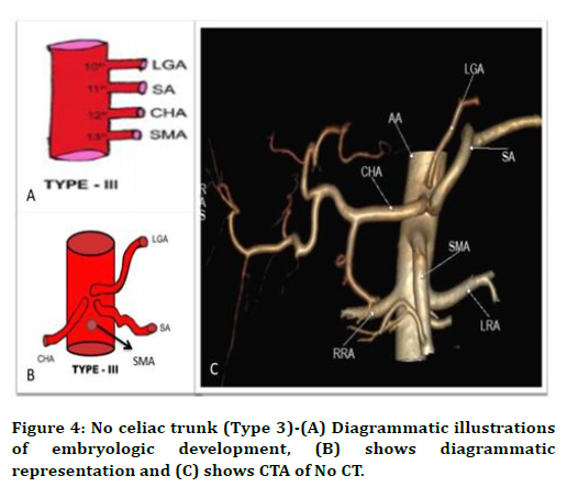
Figure 4. No celiac trunk (Type 3)-(A) Diagrammatic illustrations of embryologic development, (B) shows diagrammatic representation and (C) shows CTA of No CT.
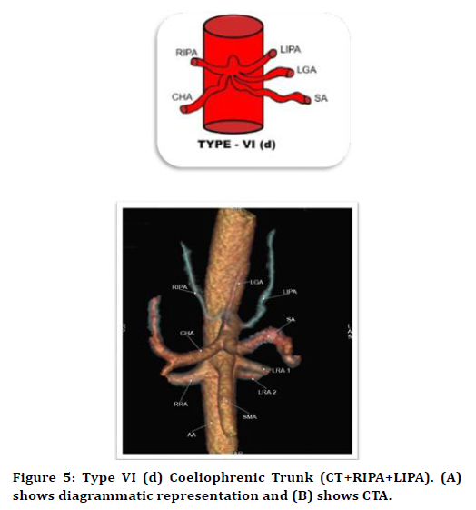
Figure 5. Type VI (d) Coeliophrenic Trunk (CT+RIPA+LIPA). (A) shows diagrammatic representation and (B) shows CTA.
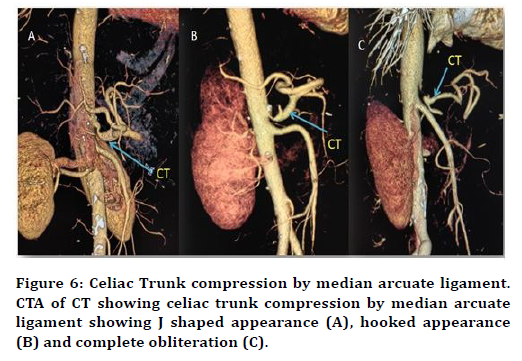
Figure 6. Celiac Trunk compression by median arcuate ligament. CTA of CT showing celiac trunk compression by median arcuate ligament showing J shaped appearance (A), hooked appearance (B) and complete obliteration (C).
Discussion
The celiac trunk is the short and first ventral branch of abdominal aorta arising at the level of 12th thoracic vertebrae [9,10]. The variations of celiac trunk branching pattern are more common than other abdominal vessels. In this study, we propose a new classification of CT branching pattern into six main types and 18 subtypes based on embryology. In this classification, we have included following vessels CT, SMA, IPA and MCA, unlike the previous studies so far. This newly proposed classification of CT is correlated with routine diagnostic CTA studies in 402 cases.
The study was conducted in 402 patients in whom 254 were males and 148 females. We have proposed a new classification of the celiac trunk and its branching pattern based on embryology and classified it into six main types, which is further divided into 18 subtypes [11-13]. The study was conducted in 402 patients in whom 254 were males and 148 females. Type I (normal trifurcation) was seen in 174 patients (40%). Type II was seen in total 17 patients (4%) it is further subdivided into three subtypes; Type IIa (Hepatosplenic trunk) was seen in 8 patients (2%). Type IIb (Hepatogastric trunk) was not seen in any patients, Type IIc (Gastrosplenic trunk) was seen in 9 patients (2%). Type III (No celiac trunk) and was seen in 1 patient (0.002%). Type IV is further divided into seven subtypes, Type IVa (Celiomesenteric trunk) was seen in one patient (0.002%), Type IVb (Hepatomesenteric trunk) was seen in five patients (1%), Type IVc (Right hepatomesenteric trunk) was seen in thirty patients (7%), Type IVd (Gastromesenteric trunk) was not seen in any patient, Type IVe (Splenomesenteric trunk) was not seen in any patient, Type IVf (Hepatosplenomesenteric trunk) was seen in three patients (1%), Type IVg (Gastrosplenomesenteric trunk) was not seen in any patient. Type V (Celiacocolic trunk) was not seen in any patient. Type VI was subdivided into four subtypes, Type VIa - Celiacophrenic trunk (CT + CIPA) was seen in thirty six patients (8%), Type VIb - Celiacophrenic trunk (CT + RIPA) was seen in twenty six patients (6%) Type VIc - Celiacophrenic trunk (CT + LIPA) was seen in fifty nine patients (14%) Type VId -Celiacophrenic trunk (CT + RIPA + LIPA) was seen in sixty two patients (14%). And finally, ‘Others’ which consist of other variants and seen in twenty-two patients (5%) [14-17].
In this present study we could not find some of the variants like Hepatogastric Trunk (Type II b), Gastro mesenteric Trunk (Type IVd), Splenomesenteric Trunk (Type IVe), Gatrosplenomesenteric Trunk (Type IVg) and Celiaccolic Trunk (Type V). These variants were found in literature with CTA correlation and cadaveric studies were Wang et al. [17] song et al.[7]. We compared the present study with a previously proposed classification and major CTA studies of CT [18].
Most of endovascular treatment for hemoptysis failed due to anomalous origin of bronchial artery and it accounts approximately to 30%. Aberrant BA may arise from the internal mammary artery, aortic arch, thyrocervicl trunk, costocervical artery, subclavian artery, brachiocephalic artery, inferior phrenic artery, pericardiophrenic artery, abdominal aorta and celiac trunk [19]. Bronchial artery is considered abnormal if vessel diameter is more than 2mm. In case of hemoptysis, MDCT aids in identify the Non bronchial systemic arteries as a source of bleeding. MDCT criteria for Non Bronchial Systemic Artery (NBSA) arepleural thickness greater than 3mm and it is due to systemic vessels passing through extra pleural fat layer [20,21]. Bhalla et al. had done a case series of 3 patients on aberrant systemic artery to a lungs and cause for hemoptysis. In one case they done a selective celiac trunk angiography to identify the source for hemoptysis ,angiography showed an aberrant systemic artery from celiac trunk which supplies to right middle lobe and it was a source of hemoptysis for that they done a embolization as a treatment [22-25].
Conclusion
In the present study we have tried to classify CT branching pattern into six main types with their subtypes and proposed embryological basis for such variations in CT. We analyzed previous studies from the literature Including Cadaveric, Postmortem, CTA and Digital Subtraction Angiography studies.
Variations of CT classifications reported till date were analyzed and found that it could not encompass all variants of CT branching pattern. So we have proposed this new classification which incorporates most of the variants. We reiterate the clinical importance of anatomical variants like IPA and celioemesenteric trunk and pathological conditions of celiac artery like median arcuate ligament syndrome
Today in the era of minimal invasive procedures, interventional and robotic surgeries it is essential to understand the normal anatomy and variations of CT. Knowledge of these variations is important for accurate diagnostic approach, which will help surgeons and interventional radiologists to avoid any iatrogenic complications while performing any abdominal and thoracic procedures.
Funding
No funding sources.
Ethical Approval
The study was approved by the Institutional Ethics Committee.
Conflict of Interest
The authors declare no conflict of interest.
Acknowledgments
The encouragement and support from Bharath University, Chennai is gratefully acknowledged. For provided the laboratory facilities to carry out the research work.
References
- Haller A. The anatomical images of the human body in which some parts are delineated set forth above and arterial history continetur. Göttingen: Vandenhoeck 1756.
- Toro JS, Prada G, Takeuchi SY, et al. Prevalence of anatomical celiac trunk variations using 3D angiography computed tomography images in a reference hospital. J Clin Exp Res Cardiol 2017; 3:201.
- Lipshutz B. A composite study of the celiac axisartery. Ann Surg 1917; 65:159-169.
- Das AB. Arterial system of the Japanese. Published by the Imperial Japanese University of Kyoto 1928; 2:18-71.
- Micahels NA. The hepatic, cystic and retro duodenal arteries and their relations to the biliary duct. Ann Surg 1951; 133:503-524.
- Uflacker R. Atlas of vascular anatomy: an angiographic approach. Br J Anatomy 1997; 83:661-667.
- Young SS, Chung JW, Yin YH, et al. Celiac axis and common hepatic artery variations in 5002 patients: Systematic analysis with spiral CT and DSA. Radiology 2010; 255.
- Panagouli E, Venieratos D, Lolis E, et al. Variations in the anatomy of the celiac trunk: a systematic review and clinical implications. Annals Anatomy 2013; 195:501-511.
- Susan S. Gray’s anatomy. The anatomical basis of clinical practice. 40th Edn 2008; 494-495.
- Dilli Babu E, Khrab P. Celiac trunk variations: Review with proposed new classification. Int J Anat Res 2013; 3:165-170.
- White RD, Weir-McCall JR, Sullivan CM, et al. The celiac axis revisited: Anatomic variants, pathologic features, and implications for modern endovascular management. Radiographics 2015; 35:879-98.
- Tandler J. About the varieties of the Arteia celiaca and their development. Anat Hefte 1094; 25:473-500.
- Kalthur SG, Sarda R, Bankar M. Multiple vascular variations of abdominal vessels in a male cadaver: Embryological perspective and clinical importance. J Morpholog Sci 2011; 28:152-156.
- Felix, W. Mesonephric arteries. In Keibel F, Mall FP. Edn. Manual of human embryology. 2th Edn London: Lippincott 1912; 820-825.
- Isogai S, Horiguchi M, Hitomi J. The para-aortic ridge plays a key role in the formation of renal, adrenal and gonadal vascular systems. J Anat 2010; 216:656–670.
- Lopamodra M, Subhasis C, Indrajit G. Embryological basis of variation of origin of inferior phrenic artery-a cadaveric study in South Bengal. India J Basic Appl Med Res 2017; 6:45-51.
- Wang Y, Cheng C, Wang L, et al. Anatomical variations in the origins of the celiac axis and the superior mesenteric artery: MDCT angiographic findings and their probable embryological mechanisms. Eur Radiol 2014; 24:1777-1784.
- Aslaner R, Pekcevik Y, Sahin H, et al. Variations in the origin of inferior phrenic arteries and their relationship to celiac axis variations on CT angiography. Korean J Radiol 2017; 18:336.
- Gwon DI, Ko GY, Yoon HK, et al. Inferior phrenic artery: anatomy, variations, pathologic conditions, and interventional management. Radiographics 2007; 27:687-705.
- Ashalata K. Variations of the branches of the celiac trunk: A case report. Asian J Med Sci 2011; 2:148-150.
- Yalcin B, Kocabiyik N, Yazar F, et al. Variations of the branches of the celiac trunk. Gulhane Tip Dergisi 2004; 46:163-165.
- Astik B. Rajesh, Urvi H. Dave. Uncommon branching pattern of the celiac trunk: Origin of seven branches. Int J Anatomical Variations 2011; 4:83–85.
- Samarawickrama MB. Hepatosplenomesenteric trunk: A rare variation of the celiac trunk. Galle Med J 2010; 15:1.
- Rajendran SS, Anbumani SK, Subramaniam A, et al. Variable branching pattern of the common hepatic artery and the celiac artery. J Clin Diagnostic Res 2011; 1433-6.
- Sankar D, Bhanu S, Susan PJ. Variant anatomy of the celiac trunk and its branches. Int J Morphol 2011; 29:581-584.
Author Info
Department of Radio Diagnosis, Sree Balaji Medical College & Hospital,, Bharath Institute of Higher Education and Research, Chennai, Tamil Nadu, IndiaCitation: Dillibabu E, Prabakaran M, Development, Newly Proposed Classification of Celiac Trunk Variations Based on Embryology-Correlation with Computed Tomography Angiography, J Res Med Dent Sci, 2021, 9 (4): 325-330.
Received: 20-Mar-2021 Accepted: 07-Apr-2021
