Research - (2021) Volume 9, Issue 1
Morphometric Analysis of Femoral Condyles in Dry Human Femur Bones and its Clinical Implications
Akifa Begum and Karthik Ganesh Mohanraj*
*Correspondence: Karthik Ganesh Mohanraj, Department of Anatomy, Saveetha Dental College, Saveetha Institute of Medical and Technical Sciences (SIMATS), Saveetha University Tamilnadu, India, Email:
Abstract
The femur bone is the thigh bone which extends from the hip to the knee. It serves as an attachment point for all the muscles that exert their force over the hip and knee joints. The femur bone has 2 joints, the medial condyle and the distal condyle which has its specific length and breadth. The medial condyle is larger than the lateral condyle due to more weight bearing caused by the centre of mass being medial to the knee. The aim of the present study is to analyze the morphology and morphometry of condyles of femur and its clinical implication. The digital vernier calliper is used for the measurement of the condyles of femur bone. For this study, 36 femur bones were used from the department of anatomy of Saveetha dental college. The results revealed that there was a difference in dimensions of the right and the left femoral condyles. The present study thus concluded that prior knowledge of this morphological data might be useful in performing surgical procedures and many other studies.
Keywords
Femur bone, Morphometry, Femoral condyles, Knee joint, Knee arthroplasty
Introduction
The femur bone has two condyles which are the medial condyle and lateral condyle. The medial condyle is one of the 2 projections on the lower extremity of femur, the other being the lateral condyle [1]. The medial condyle is larger than the lateral condyle due to more weight bearing caused by the centre of mass being medial to the knee [2,3]. The femur articulates proximally with the acetabulum of the pelvis for in the hip joint [4]. According to the previous research or literature, the length of the condyles was taken, and the breadth of the condyles were taken and even the diameter of the femoral condyles were taken [5]. The femur customized anatomical coordinate system was constructed according to the X-Y, Y-Z and X-Z planes [6]. The other study has researched the length of the medial condyle, but in this study, both the length and breadth of the medial and lateral condyle has been taken. In this study there is also comparison done between the right and left femoral condyles.
Generally, the bicondylar width as well as the medial and lateral condylar depths of the femur are important parameters for the design of total knee prostheses. And in this study, both the morphology and morphometry of the condyles of femur is also studied. The lateral condyle depth of the femur has been associated with osteoarthritis, but it remains unclear whether the increased depth of the lateral condyle is a predisposing factor or the effect of knee osteoarthritis.
Recent studies have shown that gender differences of distal femur morphometry depend on other morphometric measurements of femur, such as the femur length and width. The knee joint is a complex synovial joint consisting of the tibiofemoral and patellofemoral articulations [7]. It functions to control the centre of body mass and posture in the activities of daily living [8]. Most of the morphometric large sample size studies of the distal femur include measurements on radiographs, computerized tomography, or magnetic resonance imaging. The femoral condyles have many dimensions which can be measured, which are the bicondylar width, the diameter of the intercondylar notch, etc. [9].
Previously our team had conducted numerous survey studies [10-15], in vivo animal studies [16], bioinformatic studies [17], genetic studies [18], anthropometric studies [19] and morphometric studies [20-24] over the past 5 years. Now we are focusing on epidemiological surveys and related research. The idea for this morphometric study stemmed from the current interest in our community and to analyse the morphometry of the condyles of femur. The aim of this study was to analyze the morphology and morphometry of the condyles of femur bone and to correlate it with its clinical implications.
Materials and Methods
This study was conducted in the department of anatomy, saveetha dental college, Chennai. A total of 36 femur bones were used for the research study, among which 18 were right femur bones and 18 were left femur bone. The damaged femur bone was removed from evaluation. The measurements of the condyles of the femur bone such as the length and the breadth were measured using the instrument 'vernier caliper’. The observed data was analysed statistically.
Results and Discussion
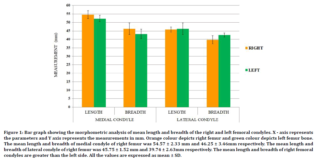
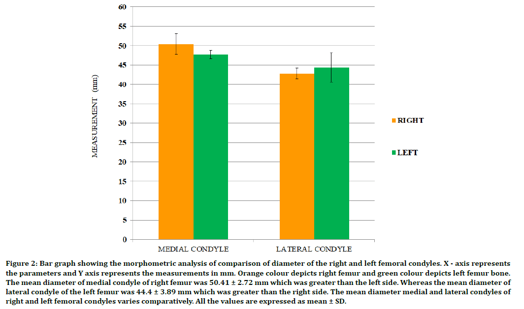
The mean length of the right medial condyle is 54.57 ± 2.3mm and the breadth is 46.25 ± 3.4mm. The mean length of the left medial condyle is 52.25 ± 1.9mm and the breadth was 43.15 ± 2.9mm. The mean length of the right lateral condyle is 45.75 ± 1.5mm and the breadth is 39.75 ± 2.5mm. The mean length left lateral condyle is 46.25 ± 3.5mm and the breadth is 42.55 ± 1.2mm. Diameter of the right medial condyle is 50.41 ± 2.7mm, diameter of the left medial condyle is 47.70 ± 1.1mm, diameter of the right lateral condyle is 42.75 ± 1.4mm and diameter of the left lateral condyle is 44.40 ± 3.8mm (Figure 1 and Figure 2).

Figure 1. Bar graph showing the morphometric analysis of mean length and breadth of the right and left femoral condyles. X - axis represents the parameters and Y axis represents the measurements in mm. Orange colour depicts right femur and green colour depicts left femur bone. The mean length and breadth of medial condyle of right femur was 54.57 ± 2.33 mm and 46.25 ± 3.46mm respectively. The mean length and breadth of lateral condyle of right femur was 45.75 ± 1.52 mm and 39.74 ± 2.63mm respectively. The mean length and breadth of right femoral condyles are greater than the left side. All the values are expressed as mean ± SD.

Figure 2. Bar graph showing the morphometric analysis of comparison of diameter of the right and left femoral condyle s. X - axis represents the parameters and Y axis represents the measurements in mm. Orange colour depicts right femur and green colour depicts left femur bone. The mean diameter of medial condyle of right femur was 50.41 ± 2.72 mm which was greater than the left side. Whereas the mean diameter of lateral condyle of the left femur was 44.4 ± 3.89 mm which was greater than the right side. The mean diameter medial and lateral condyles of right and left femoral condyles varies comparatively. All the values are expressed as mean ± SD.
This study elucidated the anatomy of the condyles of the femur bone. The clinical implication is that the condyle with fracture as a result of severe impaction from activities such as downhill skiing and parachuting. According to the results obtained statistically, the morphometric analysis of the condyles of femur, in case of the medial condyle, the left medial condyle is much more prominent than the right medial condyle. In case of the lateral condyle, the right lateral condyle is less prominent compared to the left lateral condyle. In case of the comparison of the diameter of the femoral condyles, the medial condyle has a larger diameter as compared to the lateral condyle.
In a study done by, Hu et al. [25], the study was mainly based on obtaining the morphological differences between the condyles of femur of male and female. The result of their study revealed that, there weren't many differences in the morphology of the condyles of the femur except for the few small changes which occur due to the age changes in male and female. In a study done by, Streitbuerger et al. [26], the study revealed that there were si de to side differences in the condyles of femur in case of the male and female when the measurement was taken and evaluated.
In a study done by Matthesen et al. [27], the study conveyed the result that with the help of a computed tomography various morphological features of the lower end of the condyles of femur of the right and left side were evaluated and studied. In a study done by, Yan et al. [28], the study was done mainly on the measurements of length and breadth of the condyles of femur bone and the results revealed that, there is a difference with the length and breadth of the condyles of femur bone between a male and female condyle. In a study done by, Jonkers et al. [29], the study revealed that the maximum damage to the condyles of femur occurs due to the major accidents occurring.
If the age of the person is more than 60 years, the chances of the femoral displacement may also occur due to the minor accidents or injuries as well. Since the demur is a bone, a normal level of calcium is also required in a person’s body for the strong bone development [30]. Even the type of the diet we follow also plays a major role in the maintenance of a healthy bone. And the femur bone, it is a place where the main weight of the body resides as the age increases.
Conclusion
The knowledge about the condyles of femur provides the age differences observed in the condyles and also it differs in the gender of the person. The major clinical implication is seen or observed when a person is facing an accident while sky diving or jumping from a long distance. There can be clinical issue when the person is very obese and the whole weight of the person may fall on the person’s knee and may damage the femur bone.
Acknowledgment
Nil.
Conflict of Interest
The authors declare that there are no conflicts of interest in the present study.
References
- Pujol A, Rissech C, Ventura J, et al. Ontogeny of the female femur: geometric morphometric analysis applied on current living individuals of a Spanish population. J Anatomy 2014; 225:346-357.
- Eckhoff DG, Bach JM, Spitzer VM, et al. Three-dimensional morphology and kinematics of the distal part of the femur viewed in virtual reality: Part II. JBJS 2003; 85:97-104.
- Marangalou JH, Ito K, Taddei F, et al. Inter-individual variability of bone density and morphology distribution in the proximal femur and T12 vertebra. Bone 2014; 60:213-220.
- Terzidis I, Totlis T, Papathanasiou E, et al. Gender and side-to-side differences of femoral condyles morphology: Osteometric data from 360 Caucasian dried femori. Anatomy Res Int 2012; 2012:679658.
- Vasta S, Andrade R, Pereira R, et al. Bone morphology and morphometry of the lateral femoral condyle is a risk factor for ACL injury. Knee Surg Sports Traumatol Arthroscopy 2018; 26:2817-2825.
- Lewis PM, Waddell JP. Fractured neck of femur: A review of three seminal papers and their implications to clinical management. Bone Joint Open 2020; 1:198-202.
- Van der Wiel HE, Lips P, Nauta J, et al. Bone loss in the upper femur after immobilization following unstable fractures of the lower leg. Bone Mineral 1992; 17:144.
- Wyss UP, Doerig M, Frey O, et al. Dimensions of the femur condyles. J Biomech 1982; 15:807-808.
- Caldwell CB, Rosson J, Surowiak J, et al. Use of the fractal dimension to characterize the structure of cancellous bone in radiographs of the proximal femur. InFractals Biol Med 1994; 300-306.
- Thejeswar EP, Thenmozhi MS. Educational research-iPad system vs textbook system. Res J Pharm Technol 2015; 8:1158-1160.
- Sriram N, Yuvaraj S. Effects of mobile phone radiation on brain: a questionnaire-based study. Res J Pharm Technol 2015; 8:867-870.
- Samuel AR, Thenmozhi MS. Study of impaired vision due to amblyopia. Res J Pharm Technol 2015; 8:912–914.
- Hafeez N, Thenmozhi. Accessory foramen in the middle cranial fossa. Res J Pharm Technol 2016; 1880.
- Choudhari S, Thenmozhi MS. Occurrence and importance of posterior condylar foramen. Res J Pharm Technol 2016; 9:1083–1085.
- Kannan R, Thenmozhi MS. Morphometric study of styloid process and its clinical importance on eagle’s syndrome. Res J Pharm Technol 2016; 9:1137–1139.
- Seppan P, Muhammed I, Mohanraj KG, et al. Therapeutic potential of Mucuna pruriens (Linn.) on ageing induced damage in dorsal nerve of the penis and its implication on erectile function: An experimental study using albino rats. Aging Male 2018; 13:1-4.
- Johnson J, Lakshmanan G, Biruntha M, et al. Computational identification of MiRNA-7110 from pulmonary arterial hypertension (PAH) ESTs: a new microRNA that links diabetes and PAH. Hypertension Res 2020; 43:360-362.
- Sekar D, Lakshmanan G, Mani P, et al. Methylation-dependent circulating microRNA 510 in preeclampsia patients. Hypertension Res 2019; 42:1647-1648.
- Krishna RN, Babu KY. Estimation of stature from physiognomic facial length and morphological facial length. Res J Pharm Technol 2016; 9:2071–2073.
- Nandhini JT, Babu KY, Mohanraj KG. Size, shape, prominence and localization of gerdy's tubercle in dry human tibial bones. Res J Pharm Technol 2018; 11:3604-3608.
- Subashri A, Thenmozhi MS. Occipital emissary foramina in human adult skull and their clinical implications. Res J Pharm Technol 2016; 9:716-718.
- Keerthana B, Thenmozhi MS. Occurrence of foramen of huschke and its clinical significance. Res J Pharm Technol 2016; 9:1835–1836.
- Pratha AA, Thenmozhi MS. A study of occurrence and morphometric analysis on meningo orbital foramen. Res J Pharm Techno 2016; 9:880-882.
- Menon A, Thenmozhi MS. Correlation between thyroid function and obesity. Res J Pharm Techno 2016; 9:1568–1570.
- Hu J, Yu D, Wu Y. Primary non-hodgkin lymphoma of the right femur and subsequent metastasis to the left femur: A case report and literature review. Oncology Letters 2018; 15:4427-4431.
- Streitbuerger A, Hardes J, Gosheger G, et al. Knee salvage in revision arthroplasty after massive bone loss of the femur condyles (≥ Engh III) with a single-modular-hinged knee revision implant. Archives Orthop Trauma Surg 2016; 136:1077-1083.
- Matthesen G, Teifke JP. Multiple subchondral cystic lesions in condyles of femur and tibia plateau of a horse. Pferdeheilkunde 1994; 10:153–159.
- Yan M, Wang J, Wang Y, et al. Gender-based differences in the dimensions of the femoral trochlea and condyles in the Chinese population: correlation to the risk of femoral component overhang. Knee 2014; 21:252-256.
- Jonkers I, Sauwen N, Lenaerts G, et al. Relation between subject-specific hip joint loading, stress distribution in the proximal femur and bone mineral density changes after total hip replacement. J Biomec 2008; 41:3405-3413.
- Chapman MW. Closed intramedullary bone-grafting and nailing of segmental defects of the femur. A report of three cases. JBJS 1980; 62:1004-1008.
Author Info
Akifa Begum and Karthik Ganesh Mohanraj*
Department of Anatomy, Saveetha Dental College, Saveetha Institute of Medical and Technical Sciences (SIMATS), Saveetha University Tamilnadu, Chennai, IndiaCitation: Akifa Begum, Karthik Ganesh Mohanraj, Morphometric Analysis of Femoral Condyles in Dry Human Femur Bones and its Clinical Implications, J Res Med Dent Sci, 2021, 9 (1): 283-286.
Received: 23-Sep-2020 Accepted: 04-Jan-2021
