Research - (2021) Volume 9, Issue 7
Morphological and Radiological Study of Liver and Portal Vein
*Correspondence: S Kalaivani, Department of Anatomy, Sree Balaji Medical College & Hospital Affiliated to Bharath Institute of Higher Education and Research, India, Email:
Abstract
Morphology of liver in 50 cadavers shows Mean maximum transverse diameter was 19.94 ± 2.45 cm, maximum vertical diameter was 14.95 ± 1.87 cm, and volume of liver 1140.15 ± 244.68 ml. mean transverse diameter was 19.67 ± 2.19 cm, mean vertical diameter was 14.37 ± 2.0 cm and volume of the liver 122 7.40±127.28 ml, mean weight of the liver was l .59 ± 0.25kg. In this study, mean transverse diameter was 19 .67 ± 2.19 cm, mean vertical diameter was 14 .3 7 ± 2.0 cm and mean antero posterior was8.70 ± 1.16 cm and volume of the liver 12 27.40 ± 127.2 8 ml. Mean weight of the liver was l .59 ± 0.2 5 kg. Ultrasonic measurement of portal vein diameter in correlation with parameters like age, sex and height of the individual and stated that in males the PVD does not vary with the age, but height had correlation with PVD. In females there is no correlation between them. Correlation study between maximum diameter of the portal vein with various physical parameters like age, sex, height, weight and BSA (body surface area) of subjects. Mean diameter of the portal vein was 1.2 with the standard deviation of 0.2. In current study mean diameter of the portal vein was 0.9.
Keywords
Cadavers, Portal vein, UltrasoundIntroduction
The morphological sign of the embryonic liver is the formation of the hepatic diverticulum, an out-pocket of thickened ventral foregut epithelium adjacent to the developing heart. The anterior portion of the hepatic diverticulum gives rise to the liver and intra hepatic biliary tree, while the posterior portion forms the gall bladder and extra hepatic biliary apparatus Liver is synthesizing clotting factors, producing immune factors, and removing bacteria. Hepatic tissue made from stem cells is the unlimited source of material for transplantation. The portal vein contributes a proximately 75 percent of hepatic blood flow.
The main functions of portal vein are to supply metabolic substrates to the liver and to maintain that ingested substances are first processed by the liver before entering normal circulation.
All the nutrient rich blood from the GIT flows into the portal vein which determines the anatomical division of the hepatic lobes. Several studies have been performed to establish the normal upper limits of the portal vein diameter (PVD) but these values vary according to the sonographer, mode of technique and the population being studied upon.
The portal vein contributes almost 75 % of hepatic blood flow. All the nutrient rich blood from the GIT flows into the portal vein which determines the anatomical division of the hepatic lobes. Normal blood flow in portal vein disturbed in diseased person [1-4]. Hence this study aims to examine the morphology and radiological aspect of adult liver and the portal vein and to measure the maximum transverse, anteroposterior diameter, and vertical diameter of the cadaveric liver by using Vernier caliper, thread, measuring tape and scale. To measure the weight of the liver by weighing machine and volume of the cadaveric liver after dissection by volume displacement method and to measure the length and diameter of the portal vein (PVD) by Vernier calliper, thread, and scale. To correlate the length of the portal vein with the liver volume and finding out its significance and also to compare the present study with previous studies.
Methodology
Study design
The morphological study was carried out in Sree Balaji Medical College & Hos pit al, Chrompet, Chennai and SRM Medical College , Hospital & Research centre, Potheri, Chennai in 2014& 2015.Apparently normal liver specimen obtained from 50 cadavers, in Anatomy department of two colleges, were utilized for this study. Photographs of various dimensions of liver and portal vein were taken.
Various dimensions of the liver and the length and diameter of portal vein were measured by vernier caliper. Radiological studies were performed by measuring the maximum transverse diameter and vertical diameter of liver by doing ultrasound abdomen and correlated with the vertical diameter of liver with BMI of same, and diameter of portal vein with height of the same individual.
Results
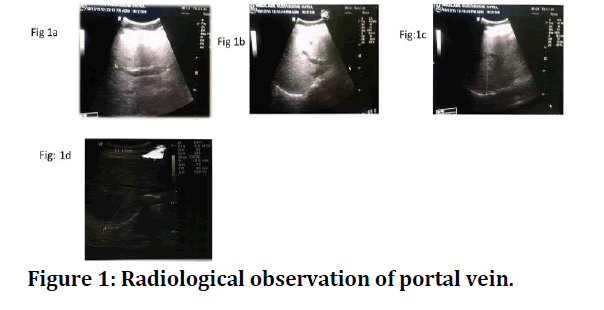
Radiological study of liver and portal vein was done in 100 individuals which includes both male and female. Maximum number of cases had the portal vein diameter of o.65-0.85 &0.85-1.06 cm equally. In mean age and above age group, maximum number of cases had the portal vein diameter of 0.65- o.85 cm. maximum number of cases had the portal vein length of 7.6-8.2 cm. The maximum cases had the transverse diameter of 19-23 cm. the vertical diameter of 12.55 -13.56cm. Diameter of portal vein of .06-1.25 cm. The maximum cases had the diameter of portal vein 0.65-0.85 cm (Figure 1 A to 1D). There was no significant correlation between ultrasound measurement of vertical diameter of liver and BMI of same individual and the p value is >0.05. Correlation between diameter of portal vein and height was significant. Other correlations like portal vein diameter with BMI and weight was not significant and the parametrs are tabulated in Table 1.

Figure 1: Radiological observation of portal vein.
Table 1: Parameters.
S.no |
Parameters | Mean | Standard deviation |
|---|---|---|---|
| 1 | Transverse diameter of liver | 19.67 | 2.194 |
| 2 | Vertical diameter of liver | 14.37 | 2.073 |
| 3 | Antero posterior diameter of liver | 8.7 | 1.162 |
| 4 | Volume of liver | 1227.4 | 127.288 |
| 5 | Weight of liver | 1.59 | 0.251 |
| 6 | Portal vein diameter | 0.95 | 0.164 |
| 7 | Length of portal vein | 7.68 | 0.669 |
Discussion and Conclusion
The correlation study between maximum diameter of the portal vein with various physical parameters like age, sex, height, weight and BSA (body surface area) of subjects. Mean diameter of the portal vein was 1.2 with the standard deviation of 0.2. In current study mean diameter of the portal vein was 0.9 with the standard deviation of 0.27. In this study there was no correlation with liver size and BMI. Like the study results carried out in Nigerian population. A better knowledge of the expected normal portal vein diameter at a given anatomical location is the first step towards developing a quantitative estimate of the severity of the portal vein abnormalities [5-8].
References
- Dan YY, Yeoh GC. Liver stem cells: A scientific and clinical perspective. J Gastroenterol Hepatol 2008; 23:687-98.
- Lavon N, Benvenisty N. Study of hepatocyte differentiation using embryonic stem cells. J Cellular Biochem 2005; 96:1193-202.
- Fausto N, Campbell JS, Riehle KJ. Liver regeneration. Hepatology 2006; 43:S45-53.
- Koç Z, Oguzkurt L, Ulusan S. Portal vein variations: clinical implications and frequencies in routine abdominal multidetector CT. Diagnostic Intervent Radiol 2007; 13:75.
- Phad VV, Syed SA, Joshi RA. Morphological variations of liver. Int J Health Sci Res 2014; 4:119-124.
- Saritha S, Ramani N, Nagajyothi D, et al. Cadaveric study of morphological variations in the human liver and its clinical importance. Int J Med Sci Clin Invent 2015; 2:1020-31.
- Khedekar DN, Hattangdi SS. Some interesting morphological features of liver lobes in Mumbai population. Int J Med Res Health Sci 2014; 3:656-659.
- Mamatha Y, Murthy CK, Prakash BA. Study on morphological surface variations in human liver. Int J Health Sci Res 2014; 4:97-102.
Author Info
Department of Anatomy, Sree Balaji Medical College & Hospital Affiliated to Bharath Institute of Higher Education and Research, Chennai, Tamil Nadu, IndiaCitation: S Kalaivani, Morphological and Radiological Study of Liver and Portal Vein, J Res Med Dent Sci, 2021, 9(7): 449-450
Received: 07-Jul-2021 Accepted: 26-Jul-2021
