Research - (2021) Volume 9, Issue 2
Knowledge and Awareness of Microbial Flora in Patients with Potentially Malignant Disorders among Dental Students: A Survey
Varusha Sharon Christopher and Gheena S*
*Correspondence: Gheena S, Department of Oral and Maxillofacial Pathology, Saveetha Dental College and Hospitals, Saveetha Institute of Medical and Technical Sciences, Saveetha University Tamilnadu, India, Email:
Abstract
Oral cancer is a serious and growing problem in many parts of the globe. Oral and pharyngeal cancer, grouped together, is the sixth most common cancer in the world (World Health Organization, 2006). Oral cancer is sometimes preceded by clinically visible lesions which are noncancerous to begin with and which have therefore been termed precancerous. Certain microbial species have been found to be cancer associated. For instance, Prevotella melaninogenica, Capnocytophaga gingivalis and Streptococcus mitis appear to be significantly elevated in tumour tissues compared to normal counterparts from the same patient. The aim of the study is to assess the awareness and knowledge about the microbial flora in oral potentially malignant disorder patients among dental students. The survey was conducted on an online platform called Google Forms. The survey was answered by 100 participants belonging to Saveetha Dental College, Chennai. I BDS was the least knowledgeable about the oral microbial flora in oral potentially malignant disorders when compared to II BDS, III BDS, IV BDS and interns. Moreover, this association between the different study years and their knowledge in PMDs were significant (p<0.05, chi-square test). Within the limits of study the awareness about oral microflora in oral potentially malignant disorders among dental students is less. I BDS and II BDS students have less awareness when compared to the other groups. III BDS, IV BDS and Interns are aware of the shift in Oral microbial flora in patients affected by PMDs.
Keywords
Oral potentially malignant disorders, Oral microflora, H. pylori, Precancerous
Introduction
Oral cancer is a serious and growing problem in many parts of the globe. Oral and pharyngeal cancer, grouped together, is the sixth most common cancer in the world [1]. Oral cancer is sometimes preceded by clinically visible lesions which are noncancerous to begin with and which have therefore been termed precancerous. The common term oral potentially malignant disorders’ (OPMD) has been suggested for oral precancers, including both oral precancerous lesions (e.g. leukoplakia, erythroplakia, and oral proliferative verrucous leukoplakia) and oral precancerous conditions (e.g. lichen planus and submucous fibrosis) [2,3]. All oral mucosal lesions that carry a risk of malignant transformation are included under this term [4].
It has been well established by researchers that virtually all oral cancers are preceded by visible clinical changes in the oral mucosa usually in the form of white or red patch (two-step process of cancer development) [5]. Prevention and early detection of such potentially malignant disorders have the potential of not only decreasing the incidence but also in improving the survival of those who develop oral cancer. Lack of public awareness about the signs, symptoms and risk factors, along with the absence of knowledge for early detection by health-care providers are believed to be responsible for the diagnostic delay in identifying the OPMDs [6-8].
Many etiological factors have been put forward by various authors, some etiological factors are Tobacco, Alcohol – synergistic action along with tobacco, Virus infection (HSV,HPV,EBV,HIV) , Bacterial infection (Treponema pallidum), Fungal infection (Candida), Electro-galvanic restorative metals, Ultraviolet radiation from sunlight -associated with lip lesions (actinic chelitis), Chronic inflammation or irritation from sharp teeth or chronic cheek-bite (tissue modifiers rather than true carcinogens), Genetic (5% are hereditary), Immunosuppression–organ transplant, HIV, Malnutrition – iron (anemia), vitamin A, B, C deficiency [2,9].
Certain microbial species have been found to be cancer associated. For instance, Prevotella melaninogenica, Capnocytophaga gingivalis and Streptococcus mitis appear to be significantly elevated in tumour tissues compared to normal counterparts from the same patient. Besides that, the salivary microbiome of oral cancer patients formed a distinct cluster separated from the normal group in a study using the Denaturing Gradient Gel Electrophoresis (DGGE) fingerprint technique. It has been suggested that this association between oral cancer and microbiome can be exploited for the development of diagnostic tools since bacteria is able to selectively bind to and colonize mucosal surfaces similar to a “lock and key” mechanism [10,11].
OPMD with subtle abnormalities present diagnostic challenges while the potential for multifocal cancer development not only complicates prognosis but also reduces treatment effectiveness. The associated oral microbiome was not addressed in previous studies which otherwise could uncover OPMD associated oral microbes that have the potential to be biomarkers for the detection of genetically abnormal oral mucosa prior to the early oral cancer stage [12-14].
No previous studies were presented based on the surveys on oral microbial flora in patients with potentially malignant disorders. Various studies on etiology, diagnosis, treatment and management of these conditions were reported in literature. The aim of the study is to assess the awareness and knowledge about the microbial flora in oral potentially malignant disorder patients among dental students. Our extensive slide collection has enabled us to publish numerous articles in the past 3 years [15-22]. Based on these inspirations, we have planned to study the awareness of oral microbial flora in oral potentially malignant disorder patients among dental students.
Materials and Methodology
Study setting
The study was conducted with the approval of the Institutional Ethics Committee [SDC/ SIHEC/2020/DIASDATA/0619-0320]. The study consisted of one reviewer, one assessor and one guide.
Study design
The study was designed to include all students belonging to the year I BDS, II BDS, III BDS, IV BDS and interns.
Sampling technique
The study was based on a non-probability consecutive sampling method. To minimize sampling bias, all responses were reviewed and included.
Data collection and tabulation
The survey was conducted on an online platform called Google Forms. To minimize sampling bias all data were included. Data was downloaded from google forms and imported to Excel, Tabulation was done. The values were tabulated and analysed.
Statistical analysis
Descriptive statistics were performed using SPSS by IBM on the tabulated values. Chi-Square test was performed and the p value was determined to evaluate the significance of the variables it was used to evaluate the association between the year of study and the responses obtained for various questions. The results were obtained in the form of graphs.
Results and Discussion
The survey was conducted among 100 students and the results were collected and analyzed. Of 100 participants, 20% were I BDS, 20% were II BDS, 20% were III BDS, 20% were IV BDS and 20% were clinical practitioners (Figure 1).
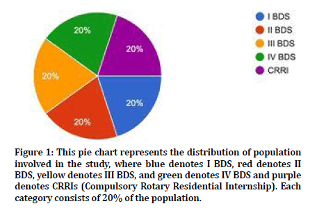
Figure 1. This pie chart represents the distribution of population involved in the study, where blue denotes I BDS, red denotes II BDS, yellow denotes III BDS, and green denotes IV BDS and purple denotes CRRIs (Compulsory Rotary Residential Internship). Each category consists of 20% of the population.
All the figures in response with the questions such as regarding whether they knew the term potentially malignant disorders (Figure 2), the risk factors in causing potentially malignant disorders (Figure 3), knowledge regarding few of the potentially malignant disorders (Figure 4), about the oral microbial flora (Figure 5), about how important the oral microbial flora is (Figure 6), knowledge regarding some of the normal oral microbial flora (Figure 7), knowledge regarding some of the microbial flora present in with potentially malignant disorders (Figure 8), if there is any difference between the normal oral microbial flora and microbial flora in precancerous lesions (Figure 9) and if oral microbial flora can help in diagnosing these precancerous conditions (Figure 10). Comparison among the different study years and their responses id represented in graph (Figures 11-14).
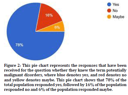
Figure 2. This pie chart represents the responses that have been received for the question whether they knew the term potentially malignant disorders, where blue denotes yes, and red denotes no and yellow denotes maybe. This pie chart shows that 78% of the total population responded yes, followed by 16% of the population responded no and 6% of the population responded maybe.
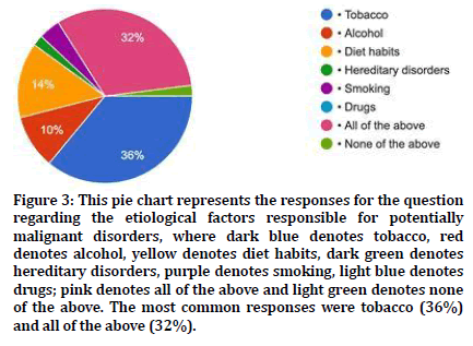
Figure 3. This pie chart represents the responses for the question regarding the etiological factors responsible for potentially malignant disorders, where dark blue denotes tobacco, red denotes alcohol, yellow denotes diet habits, dark green denotes hereditary disorders, purple denotes smoking, light blue denotes drugs; pink denotes all of the above and light green denotes none of the above. The most common responses were tobacco (36%) and all of the above (32%).
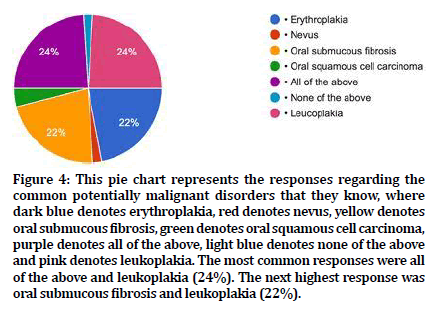
Figure 4. This pie chart represents the responses regarding the common potentially malignant disorders that they know, where dark blue denotes erythroplakia, red denotes nevus, yellow denotes oral submucous fibrosis, green denotes oral squamous cell carcinoma, purple denotes all of the above, light blue denotes none of the above and pink denotes leukoplakia. The most common responses were all of the above and leukoplakia (24%). The next highest response was oral submucous fibrosis and leukoplakia (22%).
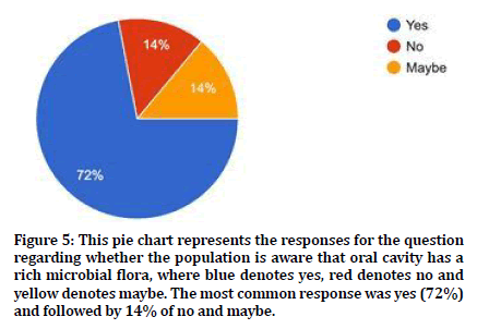
Figure 5. This pie chart represents the responses for the question regarding whether the population is aware that oral cavity has a rich microbial flora, where blue denotes yes, red denotes no and yellow denotes maybe. The most common response was yes (72%) and followed by 14% of no and maybe.
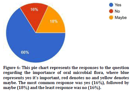
Figure 6. This pie chart represents the responses to the question regarding the importance of oral microbial flora, where blue represents yes it's important, red denotes no and yellow denotes maybe. The most common response was yes (16%), followed by maybe (18%) and the least response was no (16%).
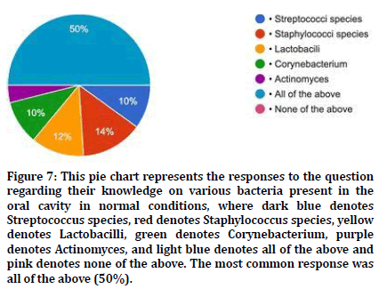
Figure 7. This pie chart represents the responses to the question regarding their knowledge on various bacteria present in the oral cavity in normal conditions, where dark blue denotes Streptococcus species, red denotes Staphylococcus species, yellow denotes Lactobacilli, green denotes Corynebacterium, purple denotes Actinomyces, and light blue denotes all of the above and pink denotes none of the above. The most common response was all of the above (50%).
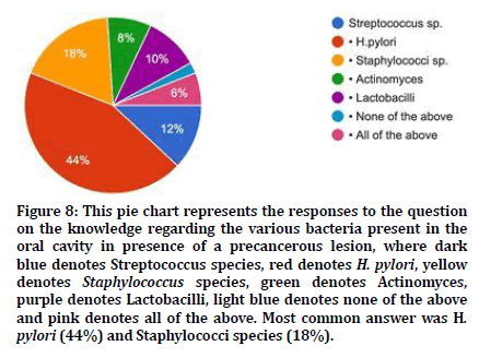
Figure 8. This pie chart represents the responses to the question on the knowledge regarding the various bacteria present in the oral cavity in presence of a precancerous lesion, where dark blue denotes Streptococcus species, red denotes H. pylori, yellow denotes Staphylococcus species, green denotes Actinomyces, purple denotes Lactobacilli, light blue denotes none of the above and pink denotes all of the above. Most common answer was H. pylori (44%) and Staphylococci species (18%).
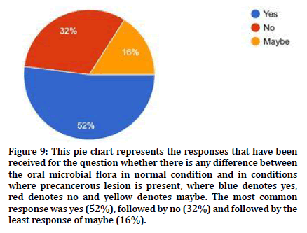
Figure 9. This pie chart represents the responses that have been received for the question whether there is any difference between the oral microbial flora in normal condition and in conditions where precancerous lesion is present, where blue denotes yes, red denotes no and yellow denotes maybe. The most common response was yes (52%), followed by no (32%) and followed by the least response of maybe (16%).
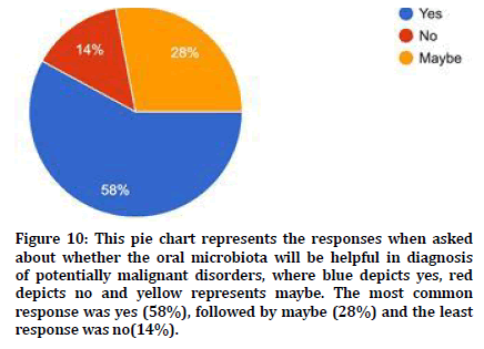
Figure 10. This pie chart represents the responses when asked about whether the oral microbiota will be helpful in diagnosis of potentially malignant disorders, where blue depicts yes, red depicts no and yellow represents maybe. The most common response was yes (58%), followed by maybe (28%) and the least response was no(14%).
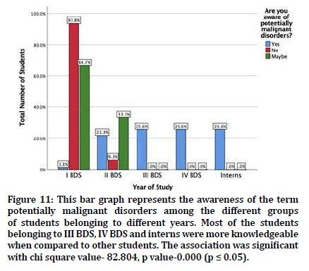
Figure 11. This bar graph represents the awareness of the term potentially malignant disorders among the different groups of students belonging to different years. Most of the students belonging to III BDS, IV BDS and interns were more knowledgeable when compared to other students. The association was significant with chi square value- 82.804, p value-0.000 (p ≤ 0.05).
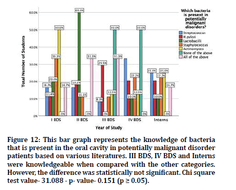
Figure 12. This bar graph represents the knowledge of bacteria that is present in the oral cavity in potentially malignant disorder patients based on various literatures. III BDS, IV BDS and Interns were knowledgeable when compared with the other categories. However, the difference was statistically not significant. Chi square test value- 31.088 - p- value- 0.151 (p ≥ 0.05).
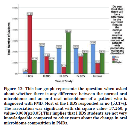
Figure 13. This bar graph represents the question when asked about whether there is any difference between the normal oral microbiome and an oral oral microbiome of a patient who is diagnosed with PMD. Most of the I BDS responded as no (53.1%). The association was significant with chi square value- 37.260, p value-0.000(p≤0.05).This implies that I BDS students are not very knowledgeable compared to other years about the change in oral microbiome composition in PMDs.
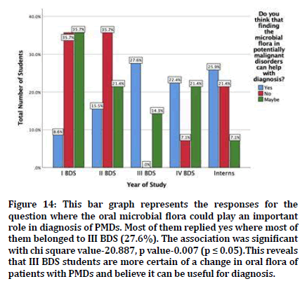
Figure 14. This bar graph represents the responses for the question where the oral microbial flora could play an important role in diagnosis of PMDs. Most of them replied yes where most of them belonged to III BDS (27.6%). The association was significant with chi square value-20.887, p value-0.007 (p ≤ 0.05).This reveals that III BDS students are more certain of a change in oral flora of patients with PMDs and believe it can be useful for diagnosis.
Students belonging to the academic year I BDS, II BDS, III BDS, IV BDS and CRRI were included so that the knowledge about these oral microbiota and potentially malignant disorders could be compared among these groups. I BDS would not have much knowledge since they mainly focus on their basic science. II BDS would have somewhat better knowledge than I BDS since they study about pathology and microbiology in their curriculum. III BDS, IV BDS and CRRI have better awareness as they responded better than the other two groups. Moreover, this association between the different study years and their knowledge in PMDs were significant (p<0.05, chi-square test).
Many studies have been reported about the increasing status of potentially malignant disorders in the Indian subcontinent [23,24], which urges the need to diagnose these conditions faster and with ease so that proper treatment plan could be adopted before it transforms to malignancy. Since our oral cavity is rich in microbes, there could be alterations found in the oral microbiota when precancerous conditions are present. Hence microbial tests and cultures could give us a faster diagnosis to treat such conditions.
It has long been known that oral bacteria preferentially colonize different surfaces in the oral cavity as a result of specific adhesions on the bacterial surface binding to complementary specific receptors on a given oral surface [25]. Indeed, the profiles of 40 cultivable bacterial species differed markedly on different oral soft tissue surfaces, saliva, and supragingival and subgingival plaques from healthy subjects. Normal microflora of the oral cavity consists of organisms such as species of Streptococcus, Gemella, Eubacterium, Selenomonas, Veillonella and Actinomyces [26].
The Helicobacter pylori (H. pylori) is one of the most common and well-known bacterium in the world. It colonizes the human stomach and it is responsible for chronic gastritis, peptic ulcer disease and has been recognized as a risk factor for gastric adenocarcinoma. Apart from its presence in the stomach, the H. pylori has also been found in dental plaque and feces. Hence, it is thought that the route of infection can be oraloral or fecal-oral [27]. The role of dental plaque as a reservoir of H. pylori and a possible source of infection or reinfection of gastric mucosa has been discussed for a long time. Nevertheless, it is also suggested that H. pylori may be present in the oral environment as normal bacterial flora [28]. H. pylori has been detected both in supragingival and subgingival plaques, and in saliva with and without a concomitant stomach infection [29].
Helicobacter pylori are one of the most important bacteria associated with oral potentially malignant disorders. Helicobacter pylori (H. pylori), a Gram-negative microaerophilic bacteria, usually found in the stomach, is frequently associated with gastric and duodenal ulcers. Kazanowska-Dygdala et al. [30] compared the H. pylori prevalence with the oral health status between OPMDs and healthy individuals through PCR. The OPMDs included oral lichen planus (OLP) and oral leukoplakia (OL). A total of 23.6% of OLP and 20% of OL were positive for H. pylori, while the control subjects were all negative.
Hulimavu et al. [31] investigated the presence of H. pylori in OLP, normal buccal mucosa. Biopsies of peptic ulcers were taken as positive controls. Immunohistochemistry was used to identify the presence of H. pylori. While the control samples (peptic ulcer tissues) were positive, none of the OLP or normal buccal mucosa samples were positive for H. pylori. This study was in contradiction with the study conducted by Kazanowska-Dygdala et al. [30]
Gupta et al. [32] assessed the prevalence of H. pylori in OPMDs and Oral Squamous Cell Carcinoma. Unlike previous studies, Chaudhary et al. [33] used salivary samples for estimating the presence of H. pylori. The OPMDs included oral submucous fibrosis (OSMF) and Oral Leukoplakia. The study used a specialized Campylobacter Supplement medium (Skirrow’s) to detect H. pylori. Overall, OSCC showed a higher prevalence than OPMDs and healthy subjects. Within OPMDs, OSMF showed higher H. pylori prevalence than Oral Leukoplakia.
The importance of oral microbial flora and its importance in diagnosis of oral potentially malignant disorders and oral squamous cell carcinoma could help in faster diagnosis and adopt a proper treatment plan so that all patients could be benefitted so they could lead a proper quality of life.
Conclusion
Within the limits of study the awareness about oral microflora in oral potentially malignant disorders among dental students is less. I BDS and II BDS students have less awareness when compared to the other groups. III BDS, IV BDS and Interns are aware of the shift in Oral microbial flora in patients affected by PMDs. The subject is important since the ultimate goal in any management of disease is to have a proper diagnosis and adopt a proper treatment plan so that the patient has an improved quality of life. Awareness of the oral microbiota and its shift in various pathologies should be added in the curriculum since it could be a thrust area of research for diagnostic tests and also because oral potentially malignant disorders are one of the most common conditions seen in the Indian subcontinent because of their cultural and culinary habits.
Acknowledgements
The authors would like to acknowledge the help and support rendered by the department of oral pathology and also the management of Saveetha Dental College and hospitals for their constant assistance with the research.
Conflict of Interest
The authors declare no potential conflict of interest.
References
- World Health Organization (2006) Constitution of the World Health Organization. Create Space Independent Publishing Platform.
- George A, Sreenivasan BS, Sunil S, et al. Potentially malignant disorders of oral cavity. Oral Maxillofac Pathol J 2011; 2:95-100.
- Van der Waal I. Oral potentially malignant disorders: Is malignant transformation predictable and preventable? Medi Oral Patol Oral 2014; 19:e386.
- Lodi G, Porter S. Management of potentially malignant disorders: Evidence and critique. J Oral Pathol Med 2008; 37:63–69.
- Ord RA, Blanchaert RH. Oral cancer: The dentist’s role in diagnosis, management, rehabilitation, and prevention. Chicago; 2000.
- Warnakulasuriya KA, Harris CK, Scarrott DM, et al. An alarming lack of public awareness towards oral cancer. Br Dent J 1999; 187:319-322.
- Amarasinghe HK, Usgodaarachchi US, Johnson NW, et al. Public awareness of oral cancer, of oral potentially malignant disorders and of their risk factors in some rural populations in Sri Lanka. Community Dent Oral Epidemiol 2010; 38:540-548.
- Satgé D, Nishi M, Culine S, et al. Awareness on oral cancer in people with intellectual disability. Oral Oncol 2012; 11:e44-e45.
- Bugshan A, Farooq I. Oral squamous cell carcinoma: Metastasis potentially associated malignant disorders, etiology and recent advancements in diagnosis. F1000Research. 2020; 9:229.
- Meurman JH. Oral microbiota and cancer. J Oral Microbiol 2010; 2.
- Subramaniam N, Muthukrishnan A. Oral mucositis and microbial colonization in oral cancer patients undergoing radiotherapy and chemotherapy: A prospective analysis in a tertiary care dental hospital. J Investigative Clin Dent 2019; 10:e12454.
- McGurk M, Scott SE. The reality of identifying early oral cancer in the general dental practice. Br Dent J 2010; 208:347–351.
- Streckfus CF. Advances in salivary diagnostics. Springer 2015.
- Chattopadhyay I, Verma M, Panda M. Role of oral microbiome signatures in diagnosis and prognosis of oral cancer. Technol Cancer Res Treatment 2019; 18:1533033819867354.
- Krishnan RP, Ramani P, Sherlin HJ, et al. Surgical specimen handover from operation theater to laboratory: A survey. Annals Maxillofac Surg 2018; 8:234-238.
- Padavala S, Sukumaran G. Molar incisor hypomineralization and its prevalence. Contemporary Clin Dent 2018; 9:S246–S250.
- Sujatha G, Muruganandan J, Priya VV. Knowledge and attitude among senior dental students on forensic dentistry: A survey. World J Dent 2018; 9:187-191.
- Sujatha G, Muruganandhan J, Priya VV, et al. Determination of reliability and practicality of saliva as a genetic source in forensic investigation by analyzing DNA yield and success rates: A systematic review. J Oral Maxillofac Surg Med Pathol 2019; 31:218-227.
- Abitha T, Santhanam A. Correlation between bizygomatic and maxillary central incisor width for gender identification. Br Dent Sci 2019; 22:458–466.
- Alexander AJ, Ramani P, Sherlin HJ, et al. Quantitative analysis of copper levels in areca nut plantation area–A role in increasing prevalence of oral submucous fibrosis: An in vitro study. Indian J Dent Res 2019; 30:261-266.
- Jayaraj G, Sherlin HJ, Ramani P, et al. Malignant glomus tumour of the head and neck–A review. J Oral Maxillofac Surg Med Pathol 2019; 31:228-230.
- Sridharan G, Ramani P, Patankar S, et al. Evaluation of salivary metabolomics in oral leukoplakia and oral squamous cell carcinoma. J Oral Pathol Med 2019; 48:299-306.
- Kumar S, Debnath N, Ismail MB, et al. Prevalence and risk factors for oral potentially malignant disorders in Indian population. Adv Preventive Med 2015; 2015.
- Gopinath D, Thannikunnath BV, Neermunda SF. Prevalence of carcinomatous foci in oral leukoplakia: A clinicopathologic study of 546 Indian samples. J Clin Diagnostic Res 2016; 10:ZC78–ZC83.
- Marsh PD. Role of the oral microflora in health. Microbial ecology in health and disease. Taylor & Francis, 2000; 12:130–137.
- Aas JA, Paster BJ, Stokes LN, et al. Defining the normal bacterial flora of the oral cavity. J Clin Microbiol 2005; 43):5721-5732.
- De Francesco V, Giorgio F, Hassan C, et al. Worldwide H. pylori antibiotic resistance: A systematic. J Gastrointestin Liver Dis 2010; 19:409-14.
- Nguyen AM, El-Zaatari FA, Graham DY. Helicobacter pylori in the oral cavity: a critical review of the literature. Oral Surg Oral Med Oral Pathol Oral Radiol Endodontol 1995; 79:705-709.
- Gebara EC, Faria CM, Pannuti C, et al. Persistence of Helicobacter pylori in the oral cavity after systemic eradication therapy. J Clin Periodontol 2006; 33:329-333.
- Kazanowska-Dygdała M, Duś I, Radwan-Oczko M. The presence of Helicobacter pylori in oral cavities of patients with leukoplakia and oral lichen planus. J Applied Oral Science 2016; 24:18–23.
- Hulimavu SR, Mohanty L, Tondikulam N, et al. No evidence for Helicobacter pylori in oral lichen planus. J Oral Pathol Med 2014; 43:576-578.
- Gupta AA, Kheur S, Raj AT, et al. Association of Helicobacter pylori with oral potentially malignant disorders and oral squamous cell carcinoma-A systematic review and meta-analysis. Clin Oral Investigations 2020; 24:13-23.
- Chaudhary M, Gawande M, Sharma P. Evaluation of prevalence of bacteria helicobacter pylori in potentially malignant disorders and oral squamous cell carcinoma. World J Dent 2015; 82–86.
Author Info
Varusha Sharon Christopher and Gheena S*
Department of Oral and Maxillofacial Pathology, Saveetha Dental College and Hospitals, Saveetha Institute of Medical and Technical Sciences, Saveetha University Tamilnadu, Chennai, IndiaCitation: Varusha Sharon Christopher, Gheena S, Knowledge and Awareness of Microbial Flora in Patients with Potentially Malignant Disorders among Dental Students: A Survey, J Res Med Dent Sci, 2021, 9 (2): 117-123.
Received: 23-Sep-2020 Accepted: 29-Jan-2021
