Research - (2020) Volume 8, Issue 1
Investigation in to the Efficacy of Ultrasonic Activation of the Solvent in Removal of Residual Obturation Materials in Endodontic Retreatment Procedures
Sarah Muayad Mahmood* and Saif Alarab A Mohmmed
*Correspondence: Sarah Muayad Mahmood, Department of Conservative Dentistry, College of Dentistry, University of Baghdad, Iraq, Email:
Abstract
Complete removal of endodontic obturation materials and remnant from the root canal is the key to Endodontic retreatment success however no single method has shown to achieve this purpose so far. The aim of this study is to evaluate the efficacy of ultrasonic activation solvent as final irrigant in removal of residual obturation materials during endodontic retreatment procedure using protaper universal retreatment system. Using digital microscope and Scanning electron microscope SEM. forty lower premolars were selected and divided into five groups of 8 one control group without solvent the other groups were divided according to the solvent and the ultrasonic activation G1: without solvent, G2 orange oil without ultrasonic activation, G3 orange oil with ultrasonic activation, G4 chloroform without ultrasonic activation, G5 chloroform with ultrasonic activation. After retreatment the roots were split and examined under digital microscope for residual obturation materials then 3 samples of each group is examined under SEM for observation. The result showed significant difference between G4 and G5 compared to G1 and G2. Conclusion no method show complete removal of obturation materials however the use of solvent and ultrasonic activation enhance the removal of residual obturation materials.
Keywords
Endodontic obturation, Endodontic retreatment, Ultrasonic activation, Distilled water
Introduction
The endodontic retreatment procedures are challenging with high level difficulty and require much time [1]. When initial root canal treatment fail retreatment procedure are performed, retreatment procedures have lower success rate than initial endodontic treatment, the factors having the most effect on the prognosis of the retreatment are perforation and under restoration [2]. When planning endodontic retreatment removal of the entire obturation material should be considered, however the complete removal or the filling material is not possible [3]. The aim of this study is to evaluate the effectiveness of ultrasonic activation of the solvent in removing residual obturation materials from the root canal.
Materials and Methods
40 extracted lower premolars were collected the collection criteria were single canal with fully formed apices with no internal or external resorption. Radiographs were taken for all the teeth to confirm these criteria. The teeth were standardized by cutting part of the crown setting the working length to 19 mm. After initial negotiation of the root canal with manual files size 10 and 15 k files., the teeth were prepared using protaper universal system up to size F3 then obturated using corresponding gutta percha with resin based sealer in single cone technique. The teeth were placed in an incubator at 37°C for two weeks. After the period of incubation the 40 teeth were divided into 5 Groups N=8.
G1 retreated using protaper universal retreatment system D1, D2, D3 in sequence mounted on eighteeth endomotor without solvent speed 600 rpm and torque 3N. To remove the obturation materials using slight apical pressure and brushing motion for each file all teeth were finally irrigated using 5.25% sodium hypochlorite 1 ml per canal and flushed with distilled water.
G2 retreated using protaper universal retreatment system D1, D2, D3 in sequence mounted on eighteeth endomotor speed 600 rpm and torque 3N with orange oil as solvent. The solvent is placed in the pulp chamber and left for 1 minute and D1 file was used to remove the obturation materials from the coronal third more solvent then was place into the canal and left for 1 minute then D2 file was used for middle third, the same is repeated for the apical third using D3 file total of 2 ml orange oil was used. All the canals were finally irrigated using 5.25% sodium hypochlorite 1 ml per canal and flushed with distilled water.
G3 retreated using protaper universal retreatment system D1, D2, D3 in sequence mounted on eighteeth endomotor speed 600 rpm and torque 3N with orange oil as solvent. The solvent was placed in the pulp chamber and left for 1 minute and D1 file was used to remove the obturation materials from the coronal third more solvent then was place into the canal and left for 1 minute then D2 file was used for middle third, the same was repeated for the apical third using D3 file. then for each canal orange oil solvent was injected inside the canal and ultrasonic activation was used using woodpecker ultrasonic scaler and tip size 20 for one minute at power 7 in 3 periods each last for 20 seconds with replacing the solvent between periods, all canals then were irrigated with 5.25% NaOCl 1 ml per canal and finally flushed with distilled water.
G4 retreated using protaper universal retreatment system D1, D2, D3 in sequence mounted on eighteeth endomotor speed 600 rpm and torque 3N with chloroform as solvent. The solvent was placed in the pulp chamber and left for 1 minute and D1 file was used to remove the obturation materials from the coronal third more solvent then was place into the canal and left for 1 minute then D2 file was used for middle third, the same was repeated for the apical third using D3 file total of 2 ml chloroform was used. All the canals were finally irrigated using 5.25% sodium hypochlorite 1 ml per canal and flushed with distilled water.
G5 retreated using protaper universal retreatment system D1, D2, D3 in sequence mounted on eighteeth endomotor speed 600 rpm and torque 3N with chloroform as solvent. The solvent was placed in the pulp chamber and left for 1 minute and D1 file was used to remove the obturation materials from the coronal third more solvent then is place into the canal and left for 1 minute then D2 file was used for middle third, the same was repeated for the apical third using D3 file. then for each canal chloroform was injected inside the canal and ultrasonic activation was performed using woodpecker ultrasonic scaler and tip size 20 for one minute at power 7 in 3 periods each last for 20 seconds with replacing the solvent between periods, all canals then was irrigated with 5.25% NaOCl 1 ml per canal and finally flushed with distilled water.
All the teeth then were longitudinally sectioned by making a groove on the buccal and lingual sides without reaching the canal and the two parts are separated using chisel, the samples are examined under digital microscope 10X to evaluate the amount of residual obturation materials left on the walls of the root canal. Images obtained and analyzed using image analysis software to measure the pre area covered with residual obturation materials. After completion of the measuring 3 samples of each group are examined using scanning electron microscope under 500X and 3000X for observational analysis.
Results
The results showed that there was a decrease in the amount of residual obturation materials when solvents were used. The chloroform showed better results than orange oil. Furthermore, the ultrasonic activation of the solvent showed even better results.
Statistical analysis
Kruskal-Wallis test (Table 1) shows that the value of median difference of the of residual obturation materials in Group G4 (chloroform) was 26.875% less than Group G1 (no solvent), and 17.875% less than group P2 (orange oil). These were statistically significant p value=(0.000) and (0.022) respectively.
| Experimental Groups | Median Difference (%) ± SE | P-value |
|---|---|---|
| G2- G1 | 9.000 (± 5.845) | 1 |
| G3 – G1 | 13.250 (± 5.845) | 0.234 |
| G4-G1 | 26.875 (± 5.845) | 0 |
| G5-G1 | 26.500 (± 5.845) | 0 |
| G3-G2 | 4.250 (± 5.845) | 1 |
| G4-G2 | 17.875 (± 5.845) | 0.022 |
| G5-G2 | 17.500(± 5.845) | 0.028 |
| G4- G3 | 13.625 (± 5.845) | 0.198 |
| G5-G3 | 13.250 (± 5.845) | 0.234 |
Table 1: Median difference, standard error.
The value of median difference of the residual obturation materials in Group G5 (chloroform with activation) was 26.500% less than Group G1 and 17.500% less than Group G2. These were statistically significant p value=(0.000) and (0.0.028) respectively.
Observational SEM images were taking for each group the images confirmed the results obtained by the digital microscope images of the coronal, middle and apical thirds of the root canal for each group are shown in Figures 1-5.
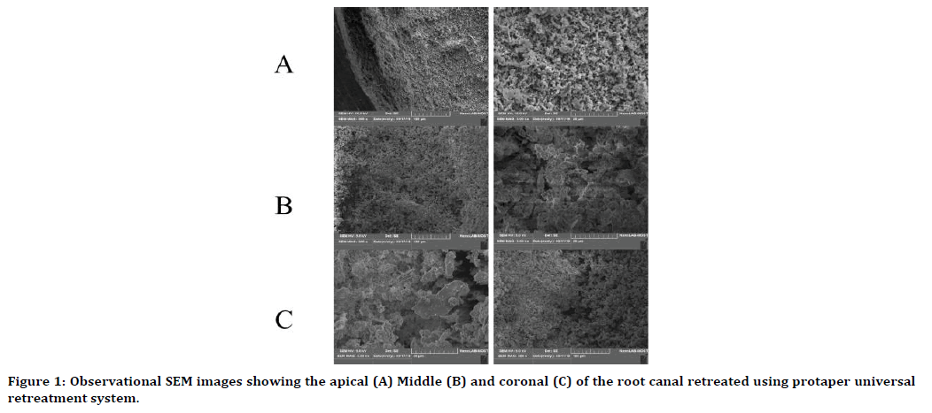
Figure 1. Observational SEM images showing the apical (A) Middle (B) and coronal (C) of the root canal retreated using protaper universal retreatment system.
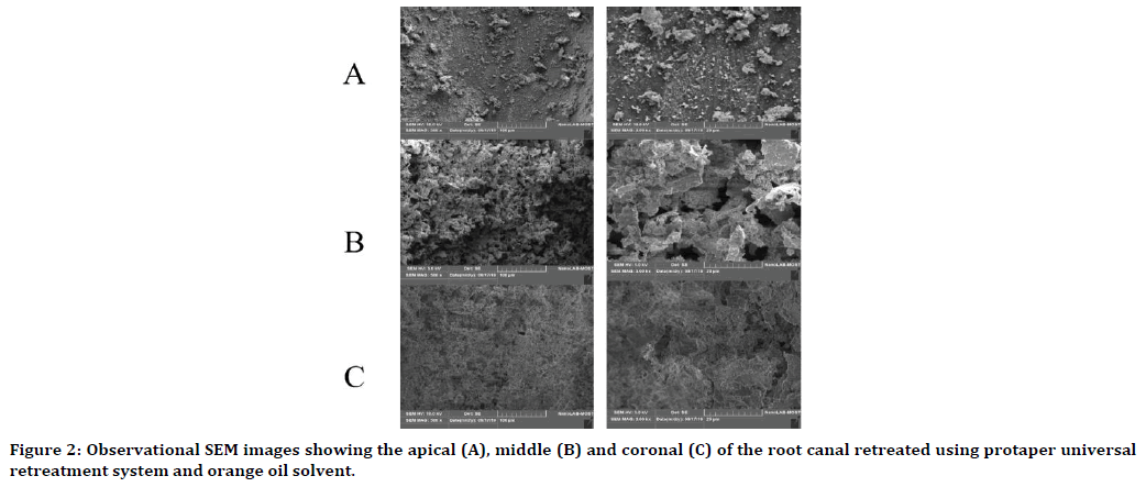
Figure 2. Observational SEM images showing the apical (A), middle (B) and coronal (C) of the root canal retreated using protaper universal retreatment system and orange oil solvent.
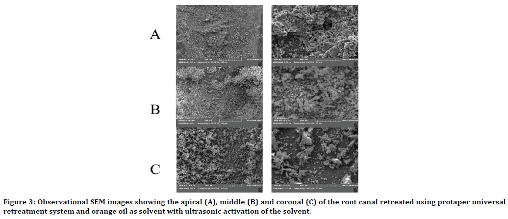
Figure 3. Observational SEM images showing the apical (A), middle (B) and coronal (C) of the root canal retreated using protaper universal retreatment system and orange oil as solvent with ultrasonic activation of the solvent.
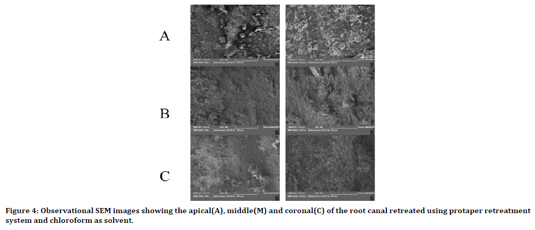
Figure 4. Observational SEM images showing the apical(A), middle(M) and coronal(C) of the root canal retreated using protaper retreatment system and chloroform as solvent.
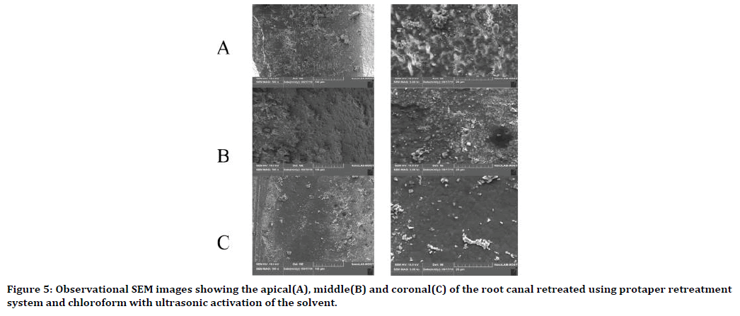
Figure 5. Observational SEM images showing the apical(A), middle(B) and coronal(C) of the root canal retreated using protaper retreatment system and chloroform with ultrasonic activation of the solvent.
Discussion
Complete removal of obturation materials from the root canal system is the key to success of the retreatment procedure [4]. Many retreatment methods were used in order to remove the obturation materials from the root canal; however, none of them showed complete removal of the obturation materials [3, 5]. The aim of this study was to investigate the effectiveness of ultrasonic agitation of the solvent on the removal of residual obturation materials. For this experimental study extracted lower premolars with Single straight canal with fully formed apices and no internal or external resorption were used in the experimental work as they are easily collected ( extracted for orthodontic work) and they have single root [6]. The collected teeth were prepared and obturated using resin based sealer and size F3 gutta percha using single cone technique the gutta perch in single cone obturation act as key and lock with the canal as it has the same taper of the final shaping instrument used [7]. Activation of the irrigation was shown to improve the removal of obturation materials during retreatment [8]. For that one of two groups for each solvent was ultrasonically activated.
Evaluation of the percentage of the residual obturation materials left on the canal walls after retreatment was done using digital microscope under 10x magnification, after longitudinal sectioning of the root, as the digital microscope provides clear and colored images of the residual materials on the root canal walls [9], the obtained images was evaluated using image analysis software ( image pro-plus) this software has been used in other studies to analyze images [10,11].
Observational scanning electron microscope images were taking to verify the results obtained in the digital microscope, SEM provide images at high magnification (in this study 500x and 3000x) and allow viewing the dentinal tubules [12]. Residual obturation materials were observed in all the retreated teeth regardless of the method used. There was statistically significant difference in the amount of residual obturation materials left on the canal wall when using chloroform as solvent compared to the (no solvent and orange oil) groups. Chloroform resulted in less residual obturation materials; this may be due to the higher solubility of gutta percha in chloroform as compared with orange oil [13]. This result was supported by SEM images as it showed more areas free of residual materials compared to no solvent and orange oil groups and few open dentinal tubules.
There was significant difference between chloroform with ultrasonic activation group and (no solvent and orange oil) groups besides the effectiveness of the chloroform as solvent ultrasonic activation has shown to decrease the amount of residual obturation materials, other than the role of ultrasonic agitation of the fluid inside the canal to reach more inaccessible areas, the file may have come in contact with the canal walls during ultrasonic activation which may led to further cleaning of the canal [14]. The observational SEM images confirmed this result; few areas of residual materials and more open dentinal tubules were seen.
Conclusion
The use of orange oil as solvent showed more residual materials removal than the control group and the ultrasonic activation of orange oil showed further decrease in the residual obturation materials as compared to no solvent and orange oil without activation Groups. However, these results were statically insignificant.
Within the limitation of this study it can be concluded that the use of solvent result in less residual obturation materials. Chloroform showed better results than orange oil. More over the ultrasonic activation of the solvent showed better result in the term of removal of residual obturation materials.
References
- Wilcox LR. Endodontic retreatment: ultrasonics and chloroform as the final step in reinstrumentation. J Endod 1989; 15:125-128.
- De Chevigny C, DAO TT, Basrani BR, et al. Treatment outcome in endodontics: The Toronto study—phases 3 and 4: Orthograde retreatment. J Endod 2008; 34:131-137.
- Hammad M, Qualtrough A, Silikas N. Three-dimensional evaluation of effectiveness of hand and rotary instrumentation for retreatment of canals filled with different materials. J Endod 2008; 34:1370-1373.
- Poggio C. Gutta percha solvents alternative to chloroform: an in vitro comparative evaluation. EC Dental Science 2017; 15:51-56.
- De Campos Fruchi L, Ordinola-zapata R, Cavenago BC, et al. Efficacy of reciprocating instruments for removing filling material in curved canals obturated with a single-cone technique: a micro–computed tomographic analysis. J Endod 2014; 40:1000-1004.
- Samran A, Al-afandi M, Kadour JA, et al. Effect of ferrule location on the fracture resistance of crowned mandibular premolars: An in vitro study. J Prost Dent 2015; 114:86-91.
- Iglecias EF, Freire LG, De Miranda Candeiro GT, et al. Presence of voids after continuous wave of condensation and single-cone obturation in mandibular molars: A micro-computed tomography analysis. J Endod 2017; 43:638-642.
- Bernardes R, Duarte M, Vivan R, et al. Comparison of three retreatment techniques with ultrasonic activation in flattened canals using micro‐computed tomography and scanning electron microscopy. Int Endod J 2016; 49:890-897.
- Adl A, Sedigh-shams M, Majd M. The effect of using RC prep during root canal preparation on the incidence of dentinal defects. J Endod 2015; 41:376-379.
- Huang TY, Gulabivala K, NG YL. A bio‐molecular film ex‐vivo model to evaluate the influence of canal dimensions and irrigation variables on the efficacy of irrigation. Int Endod J 2008; 41:60-71.
- Mcgill S, Gulabivala K, Mordan N, et al. The efficacy of dynamic irrigation using a commercially available system (RinsEndo®) determined by removal of a collagen ‘bio‐molecular film’from an ex vivo model. Int Endod J 2008; 41:602-608.
- Vidal F, Nunes E, Horta M, et al. Evaluation of three different rotary systems during endodontic retreatment-Analysis by scanning electron microscopy. J Clin Exp Den 2016; 8:125-129.
- Rubino GA, Akisue E, Nunes BG, et al. Solvency capacity of gutta-percha and resilon using chloroform, eucalyptol, orange oil or xylene. J Health Sci Inst 2012; 30:22-25.
- Grischke J, Müller-Heine A, Hülsmann M. The effect of four different irrigation systems in the removal of a root canal sealer. Clin Oral Inves 2014; 18:1845-1851.
Author Info
Sarah Muayad Mahmood* and Saif Alarab A Mohmmed
Department of Conservative Dentistry, College of Dentistry, University of Baghdad, IraqCitation: Sarah Muayad Mahmood, Saif Alarab A Mohmmed, Investigation in to the Efficacy of Ultrasonic Activation of the Solvent in Removal of Residual Obturation Materials in Endodontic Retreatment Procedures, J Res Med Dent Sci, 2020, 8(1): 10-15.
Received: 17-Dec-2019 Accepted: 30-Dec-2019
