Research - (2021) Volume 9, Issue 1
Intracanal Adaptation, Intratubular Penetration, and Push-Out Bond Strength of Different Root Canal Sealers: A Comparative In vitro Study
Mohammed S Khalil1* and Anas F Mahdee2
*Correspondence: Mohammed S Khalil, Department of Conservative Dentistry, College of Dentistry, University of Baghdad, Iraq, Email:
Abstract
Aim: The purpose of this study was to evaluate the intracanal adaptation, intratubular penetration, and push-out bond strength of Total fill Bioceramic, AH Plus, Gutta-flow BIOSEAL and MTAF sealers.
Material and method: Sixty freshly extracted human lower 1st premolars were collected, drowned, and endodontically filled using different types of sealers. Specimens were randomly divided into four groups (A, B, C, and D) (n=15) depending on the sealer type (Total Full Bioceramic, AH Plus, Gutta-flow BIOSEAL and MTAF sealers respectively). The used sealer in five samples from each group was mixed with 0.1% fluorescein die before obturating the canals with Thermafil. These samples then were embedded in clear acrylic before sectioning into 0.5 mm disks at 3, 7 and 11 mm from the root apex. Disks were examined by fluorescent microscope to identify the intratubular penetration of the root canal sealers. The rest samples (n=10 in each group) were sectioned into 2mm thickness at the same positions from root apex. These samples were examined by stereoscope to measure the root filling adaptation before testing the push-out bond strength.
Results: the results of the adaptation and push out bond strength tests have shown higher values for the Total Fill Bioceramic sealer in comparison to the other types of sealers within all regions of the tested roots, especially within the apical sections. However, no statistically significant differences have been detected in the sealer penetration test among all sealer groups which may suggested further future analysis using more sensitive testing procedure.
Conclusions: The Total fill Bioceamic sealer has better sealing ability in comparison to other types of sealers. The use of this sealer may improve the success rate with better prognosis for endodontic treatment outcomes.
Keywords
Intracanal adaptation, Intratubular penetration, Push-out bond strength, Bioceramic, AH plus, Gutta-flow, MTA
Introduction
Successful root canal relies on thorough cleaning of the root canal system, the elimination of microorganisms and finally, complete sealing of the canal to block the entry of the bacteria from the oral environment and its spreading to the apical tissue [1]. The excellent sealing and filling of the perfectly debrided and shaped root canal system are important steps that can affect the success of root canal treatment [2,3]. Due to of the complexity of root canal system, sealers must be used to occupy the irregularities and to penetrate dentinal tubules to obtain a hermetic seal of the root canal system. Meanwhile, root canal sealers should provide adhesion between gutta-percha and dentinal walls to avoid gap formation at the sealer-dentine interface [4]. Grossman stated the requirements of an ideal sealer, including the following: Provides perfect adhesion between it and the canal wall when set; provides a hermetic seal; no gaps upon setting; have no solubility in tissue fluids; tolerated by tissues; and others. The current available commercial sealers can be broadly categorized into the following groups: zinc oxide eugenol-based, calcium hydroxide-based, glass ionomer-based, resin-based, silicone-based, and the recently introduced, calcium silicate-based sealers. However, at present not one of the existing sealers satisfies all the criteria [5,6]. Zinc oxide eugenol-based [7], calcium hydroxide-based and glass ionomer-based sealers have the common problem of solubility during contact with peri radicular tissue. In addition, zinc oxide eugenolbased sealer has shrinkage settings [5,8]. From the above the need for a study to compare the different types of sealer is important to assess these sealers and identify their sealing ability to suggest recommendations depending on their properties.
Aim
To evaluate the intracanal adaptation, intratubular penetration, and push-out bond strength of endodontically treated teeth using Total fill Bioceramic, AH Plus, Gutta-flow BIOSEAL and MTAF root canal sealers.
Materials and Method
Sixty freshly extracted human mandibular first premolar was collected. Immediately after extraction, all attached soft tissues on the tooth surface were removed manually with periodontal curette [9]. The teeth were stored at room temperature in plastic containers contain distilled water and 0.1% concentration of thymol to prevent dehydration and bacterial growth [10]. All root surfaces were verified for absence of any obvious cracks or fractures using Stereomicroscope at 20X magnification. For standardization of root lengths, 15 mm from the anatomical apex was determined using digital caliper and marked on the root using permanent marker. Teeth were sectioned perpendicular to their long axis using diamond disc mounted on a straight handpiece with water coolant to provide straight line access during root canal preparations, with flat coronal surface that served as a stable reference point for root length measurements. This would prevent the production of extra variables that might contribute to variations in the canal instrumentation procedure [11].
The Pulp tissue was removed using barbed broach and the patency of the canals was examined by using of size10 K-file into the root canal until it was visualized at the apical foramen. The correct working length (WL) was measured by subtracting 1mm from root length. Also size 10 K-file was used to determine the initial size of the canal, only roots with initial file size 10 were included in the study.
For proper handling of the samples during instrumentation and obturation procedures, each sample was fixed in vinyl polysiloxane impression material (putty consistency) that placed inside a silicon mold (20 mm in height and 10. mm in diameter). The impression material was mixed (base and catalyst) according to manufacture instructions, and then each root was centered inside the putty material with the aid of dental surveyor to make the long axis of the root parallel to that of the mold [10].
Samples then were randomly divided into FOUR main groups (n=15) according to the types of sealer material used for root canals:
AH group for AH PLUS sealer.
GF group for Gutta Flow Bioseal sealer.
MTA group for MTA Fillapex sealer.
BC group for Total Fill bioceramic sealer.
Then each group was further divided into two unequal subgroups: subgroup A (n=10) and subgroup B (n=5). These subgroups were used in different experiments i.e., subgroup A samples for the root filling adaptation and push-out bond strength tests, while subgroup B for sealer penetration test. All samples were processed similarly during obscuration procedure except the subgroup B where the used sealers were mixed with 0.1% fluoresce in die for better identification during examination of sealer penetration by using fluorescent microscopy. The sealers were mixed according to the manufacturer instructions and introduced within the prepared root samples using paper points before obstructing the roots with Thermafil obturators (CMS dental Denmark).
After complete root obscurations samples were sectioned horizontally at three points using sectioning discs mounted on a surveyor under constant water cooling at 3, 7 and 11mm from the root species. The thickness of samples was different according to the divided subgroups. In subgroup A 3 discs of 2mm thickness were performed, while in subgroup B the thickness of the 3 discs was only 0.5mm.
Intra canal adaptation test
Samples of subgroup A from all groups were tested. By using a stereomicroscope at 40x, the samples were examined to detect the adhesive interface integrity between dentine and root canal sealer. This was measured on the four quadrants within each root sections by identifying the presence of gaps, and/or continuous and homogenous dentine-sealer interface. A score system, as proposed was used to classify the findings (0–4): 0: absence of a continuous and homogeneous interface, with gaps in all areas; 1: continuous and homogenous interface in one area; 2: continuous and homogeneous interface in two areas; 3: continuous and homogeneous interface in three areas; and finally, 4: Continuous and homogeneous interface in four areas.
Push out bond strength test
After finishing the adaptation test, same samples were used for push-out bond strength test. At the beginning, samples were examined using Nikon camera with macro lens 105 mm and pictures of both sides of each section were taken and circumference measurements calculated using Image J software analysis program. The circumference of both apical and coronal side of the section at each level was calculated. The area under load was calculated by ½ * (circumference of coronal aspect + circumference of apical aspect) *thickness)
Three different sizes of pins were used, 0.7 mm, 0.5mm, and 0.4mm diameter for the coronal, middle and apical slices respectively to complete coverage over the main core without interfering the canal walls and sealer. Push-out test was done through applying a compressive load from apical to coronal direction using Universal Testing Machine with loading speed of 0.5 mm / min until the first dislodgment of obturating material.
The maximum failure load was recorded and was used to calculate the push-out value using the following formula [12,13]:
Push-out bond strength (MPa)= failure load (N)/ the area under load (mm2)
Measuring the intradentinal tubular pentration
Samples in subgroup B were used to measure the intradentinal tubular penetration of the sealers into dentin tested under fluorescent microscope. This was done to evaluate the formation of the sealer tags and to measure the penetration depth of these tags into dentinal tubules. Images were taken and processed by using image J software. The percentage of resin tag penetration was calculated according to the following formula:
Surface area of sealer penetration/ total surface area of the specimen* 100%
Results
Adaptation
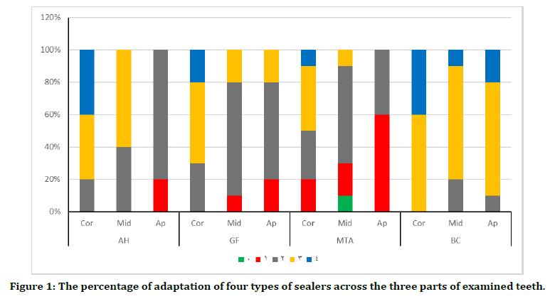
BC sealer shows the highest adaptation (Table 1 and Figure 1) at all regions of the canal in comparison to the other sealers with mean values (3.10 ± .568, 2.90 ± .568 and 3.40 ± .516) at the apical, middle, and coronal regions, respectively. The MTA sealer has the lowest adaptation across all regions with the lowest measurable mean at the apical region (1.40 ± .516). The adaptation values for AH and GF appear in between to the former sealers. During microscopical examination of the sections, almost all the voids were found between the sealer and dentin surface, no voids were found between sealer and gutta percha.
| Sealer | Site | Median | IQR | Min. | Max. |
|---|---|---|---|---|---|
| AH | Coronal | 3 | 1 | 2 | 4 |
| Middle | 3 | 1 | 2 | 3 | |
| Apical | 2 | 0 | 1 | 2 | |
| GF | Coronal | 3 | 1 | 2 | 4 |
| Middle | 2 | 0 | 1 | 3 | |
| Apical | 2 | 1 | 1 | 3 | |
| MTA | Coronal | 2.5 | 1 | 2 | 4 |
| Middle | 2 | 1 | 0 | 3 | |
| Apical | 1 | 1 | 1 | 2 | |
| BC | Coronal | 3 | 1 | 3 | 4 |
| Middle | 3 | 0 | 2 | 4 | |
| Apical | 3 | 0 | 2 | 4 |
Table 1: Descriptive statistics of root filling adaptation at three levels of root canals for the four sealers.

Figure 1. The percentage of adaptation of four types of sealers across the three parts of examined teeth.
A Kruskal-Wallis test was performed to explore the statistical differences in the adaptation scores between different regions of sealers which were nonparametric data. Statistically significant differences (p ≤ 0.05) are identified between the middle and apical regions of the sealers, whist no statistical difference (p>0.05) is detected in the coronal region (Table 2) Pairwise comparison with Bonferroni corrections show statistically significant differences between BC and MTA (p< .05) in the middle region only (Table 3). In the apical region, only BC sealer shows statistically significant difference (p< .05) with other types of sealers (MTA, AH and GF).
| Site | Sealer | Kruskal-Wallis | Pairwise comparisons | Adjusted P-value |
|---|---|---|---|---|
| P-value | ||||
| Coronal | AH | 0.063 | ||
| GF | ||||
| MTA | ||||
| BC | ||||
| Middle | AH | 0.002 | BC vs. MTA | 0.003 |
| GF | ||||
| MTA | ||||
| BC | ||||
| Apical | AH | 0 | BC vs. MTA | 0.000 |
| GF | BC vs. AH | 0.004 | ||
| MTA | BC vs. GF | 0.027 | ||
| BC | ||||
Statistical non-significant result at p value >0.05
Table 2: Kruskal-Wallis and pairwise comparisons (Bonferroni correction) between sealer adaptation for different types of sealers.
| Sealer | Site | Kruskal-Walis | Pairwise comparisons | Adjusted P-value |
|---|---|---|---|---|
| P-value | ||||
| AH | Coronal | 0.001 | Api>Cor | 0.000 |
| Middle | Api>Mid | 0.048 | ||
| Apical | ||||
| GF | Coronal | 0.017 | Api>Cor | 0.029 |
| Middle | ||||
| Apical | ||||
| MTA | Coronal | 0.037 | Api>Cor | 0.032 |
| Middle | ||||
| Apical | ||||
| BC | Coronal | 0.148 | ||
| Middle | ||||
| Apical | ||||
Statistical non-significant result at p value >0.05
Table 3 Kruskal-Walis with pairwise (Bonferroni correction) tests for sealer adaptation test between different regions (coronal, middle and apical) for each sealer.
A Kruskal-Wallis test with Bonferroni Pairwise corrections was also performed to explore the statistically significant differences between sealer adaptations within different regions of root canals for each sealer. Three of sealers used in this study (AH, GF and MTA) show statistically significant differences at different regions of the root canal. Most of these statistically significant differences are notice between the apical and coronal regions of these three sealers. In addition, the AH sealer also shows statistical significance (p<0.048) between apical and middle regions. On the other hand, not statistically significant (p=0.148) appears within different root canal regions of BC sealer subgroups (Table 3).
Penetration
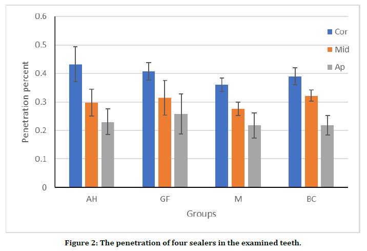
All coronal region for the sealers used show the highest penetration in comparison to other regions of the root canal (see table 4 and Figure 2). MTA sealer shows the lowest penetration values (Table 3) at all regions of the canal in comparison to other sealers. The highest penetration results varied between AH sealer in the coronal region (0.432 ± 0.062), BC sealer in the middle region (0.322 ± 0.109), and GF sealer in the apical region (0.258 ± 0.070).
| Sealer | Site | Mean | ± SD | Min. | Max. |
|---|---|---|---|---|---|
| AH | Coronal | 0.432 | 0.004 | 0.36 | 0.51 |
| Middle | 0.298 | 0.047 | 0.23 | 0.36 | |
| Apical | 0.23 | 0.045 | 0.18 | 0.28 | |
| GF | Coronal | 0.408 | 0.031 | 0.38 | 0.46 |
| Middle | 0.314 | 0.061 | 0.22 | 0.37 | |
| Apical | 0.258 | 0.07 | 0.19 | 0.37 | |
| MTA | Coronal | 0.36 | 0.023 | 0.33 | 0.38 |
| Middle | 0.276 | 0.024 | 0.25 | 0.31 | |
| Apical | 0.218 | 0.044 | 0.17 | 0.27 | |
| BC | Coronal | 0.39 | 0.029 | 0.35 | 0.43 |
| Middle | 0.322 | 0.019 | 0.3 | 0.35 | |
| Apical | 0.218 | 0.034 | 0.17 | 0.25 |
Table 4: Descriptive statistics of root filling penetration of four sealers at three levels of root canal filling.

Figure 2. The penetration of four sealers in the examined teeth.
One-way ANOVA test between different types of sealers indicates no statistically significant difference along all regions (p>0.05) across all tested regions. One-way ANOVA test (Table 4) was also performed to assess the statistical significance between sealer penetration percentages at different root regions for all tested sealers. All regions show statistically significant level at P<0.01. The regions differed for the AH sealer group (p< .001). Pairwise comparison with Bonferroni test showed almost all tested root regions have statistically significant differences between each other’s, except for GF which showed statistically significant differences (p<0.05) only between coronal and apical regions (Table 5).
| Sealer | Site | ANOVA | Pairwise comparisons | Adjusted P-value |
|---|---|---|---|---|
| P-value | ||||
| AH | Coronal | 0 | Cor vs. Mid | <0.05 |
| Middle | Cor vs. Api | <0.001 | ||
| Apical | ||||
| GF | Coronal | 0.004 | Cor vs. Api | <0.05 |
| Middle | ||||
| Apical | ||||
| MTA | Coronal | 0 | Cor vs. Mid | <0.05 |
| Middle | Cor vs. Api | <0.001 | ||
| Apical | Mid vs. Api | <0.05 | ||
| BC | Coronal | 0 | Cor vs. Mid | <0.05 |
| Middle | Cor vs. Api | <0.001 | ||
| Apical | Mid vs. Api | <0.05 | ||
Statistical non-significant result at p value >0.05
Table 5: One-way ANOVA and Bonferroni comparisons for penetration test between different regions of root canal.
Push out bond strength
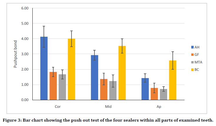
BC sealer shows the highest push-out at all regions of the canal in comparison to the other sealers with mean values (Table 5). The AH sealer comes after with comparable values in the coronal and middle thirds (4.13 ± 0.690, 2.93 ± 0.326 respectively) but much lower in the apical region (1.42 ± 0.276). The third less push-out bond strength values are for GF sealer, while the least values are for the MTA sealer with the lowest measurable mean at the apical region (0.712 ± 0.158) (Figure 3). Bonferroni test was also showed to identify the statistical significance between different sealers at tested regions. All details about statistically significant values are illustrated in table 6.

Figure 3. Bar chart showing the push out test of the four sealers within all parts of examined teeth.
| Sealer | Site | Mean | ± SD | Min. | Max. |
|---|---|---|---|---|---|
| AH | Coronal | 4.13 | 0.69 | 2.98 | 5.34 |
| Middle | 2.93 | 0.326 | 2.34 | 3.5 | |
| Apical | 1.42 | 0.276 | 1.02 | 1.84 | |
| GF | Coronal | 1.83 | 0.324 | 1.14 | 2.15 |
| Middle | 1.37 | 0.381 | 0.872 | 1.93 | |
| Apical | 0.783 | 0.321 | 0.06 | 1.08 | |
| MTA | Coronal | 1.67 | 0.298 | 1 | 2.05 |
| Middle | 1.23 | 0.402 | 0.762 | 1.78 | |
| Apical | 0.712 | 0.158 | 0.497 | 0.963 | |
| BC | Coronal | 4.01 | 0.544 | 3.05 | 4.72 |
| Middle | 3.53 | 0.47 | 2.76 | 4 | |
| Apical | 2.57 | 0.574 | 1.66 | 3.55 |
Table 6: Descriptive statistics of root filling push-out of four sealers at three levels of root canal filling.
The One-way ANOVA test for pushout bond strength of different types of sealers across all regions reveals statistically significant differences at p<0.05 (Table 7).
| Site | Sealer | Anova | Pairwise comparisons | Adjusted P-value |
|---|---|---|---|---|
| P-value | ||||
| Coronal | AH | 0 | AH vs. GF | <0.000 |
| AH vs. MTA | <0.000 | |||
| AH vs. BC | >0.05 | |||
| GF | GF vs. MTA | >0.05 | ||
| MTA | BC vs. GF | <0.000 | ||
| BC | BC vs. MTA | <0.000 | ||
| Middle | AH | 0 | AH vs. GF | <0.000 |
| AH vs. MTA | <0.000 | |||
| AH vs. BC | <0.000 | |||
| GF | GF vs. MTA | >0.05 | ||
| MTA | BC vs. GF | <0.000 | ||
| BC | BC vs. MTA | <0.000 | ||
| Apical | AH | 0 | AH vs. GF | <0.000 |
| AH vs. MTA | <0.000 | |||
| AH vs. BC | <0.000 | |||
| GF | GF vs. MTA | >0.05 | ||
| MTA | BC vs. GF | <0.000 | ||
| BC | BC vs. MTA | < 0.000 |
Statistical non-significant result at p value >0.05
Table 7: One-way ANOVA test of push-out test and pairwise comparisons (Bonferroni correction) between different types of sealers.
One-way ANOVA test was also used to point the statistically significant difference among regions along the four sealers (Table 8). Pairwise comparison with Bonferroni test showed that all regions have statistically significant differences among each other within each type of sealers except between the middle and coronal thirds of BC sealers.
| Sealer | Site | ANOVA | Pairwise comparisons | Adjusted P-value |
|---|---|---|---|---|
| P-value | ||||
| AH | Coronal | 0 | Cor vs. Mid | <0.001 |
| Middle | Cor vs. Api | |||
| Apical | Mid vs. Api | |||
| GF | Coronal | 0 | Cor vs. Mid | <0.001 |
| Middle | Cor vs. Api | |||
| Apical | Mid vs. Api | |||
| MTA | Coronal | 0 | Cor vs. Mid | <0.05 |
| Middle | Cor vs. Api | |||
| Apical | Mid vs. Api | |||
| BC | Coronal | 0 | Cor vs. Api | <0.05 |
| Middle | Mid vs. Api | |||
| Apical |
Statistical non-significant result at p value >0.05
Table 8: One-way ANOVA test for push-out test with pairwise (Bonferroni correction) tests among different regions for each sealer.
Discussion and Conclusion
This study was performed in in_vitro on an extracted tooth, with a standardized instrumentation and irrigation protocol during samples preparation. Only one type rotary file, Micro Mega 25/.06 was used during instrumentation of all samples to ensure similar Root canal debridement and tapering within all portions of the prepared canals. At the same time maintaining the apical region at 0.25mm size with good remaining dentin thickness. On the same way, proper tapering of the canals 0.06 can improve irrigation, filling adaptation with minimum sealer amount and better push out bond strength result [14].
The irrigation protocol used in this study containing 17% EDTA solution after completion of instrumentation to ensure removal of the smear layer for greater sealer adhesion in addition; distilled water was used as final rinse to wash out all the remaining irrigation chemicals that may interfere with the setting reaction of the used sealers especially the resin-based ones.
According to the results of this study, the BC sealer demonstrated greater adaptation and push out bond strength in comparison to the other types of sealer used at different regions of the examined root. This may be attributable to the alkaline effect of hydration products of the calcium silicate sealer with high pH (11.16) [15]. This could lead to degrade the interfacial dentin collagen portion which may facilitate sealer penetration into the dentinal tubules [16]. The fine particle scale (less than 2 micron) and the proper premixed consistency introduced with a capillary tip delivery system could have increased its penetration into the entire cannel walls [14].
Another possible cause is the prolonged working time of BC sealer in compared to the other types of sealers used in this study. According to manufacturer's instructions the working time for these sealers can be arranged as follow:
Gutta flow Sealer < MTA Fillapex < AH Plus < BC [17].
The results of present study are compatible with other studies [13] who explained the higher bond strength for Total Fill BC sealer could be attributed to a process known as alkaline etching. This process may allow ion exchange at the sealer dentine inter-surface creating a mineral infiltration zone, which possibly decrease the gap and increase the adhesion between sealer and dentine surface [18] on the other hand, MTA sealer had the lowest adaptation and poor bond strength comparing to AH plus and BC sealer, like the findings of other studies [19]. This could be due to poor micro tags formed on setting for this sealer [20-24]. There are some controversies in research that disagree with the present findings [25]. This could be due to using different brands for the same sealer type that may affect their results. This further suggests future investigations on different brands for the same sealer type.
The findings of the current study also showed different in sealer adaptation and bond strength within different regions of root canals for each sealer. All used sealers All used sealers (BC, AH, GF and MTA) have less apical sealer adaptation and bond strength in comparison to other regions. The apical region can be considered as the most problematic region during endodontic treatment [26-28]. The limited access, small cross section, anatomical complexity, and communication with the periradicular tissue make this region difficult in debridement, disinfection, smear layer removal and even dryness. All these may impair proper sealing and adhesion of the root filling material within this region.
In addition, The higher percentages of gaps at the middle thirds of the prepared canals have also been identified in compression to coronal thirds this could be due to the cross section of the canals for premolar teeth that were used in this study these teeth have an oval cross section which may interfere with proper instrumentation and adequate obturation of root canal [29-31]. While the lower density and scale of dental tubules observed at apical level may results in lower penetration of sealers [32-35] Moreover, the smear layer removal is difficult Apical third that could Act as a physical barrier that interfered with root canal dentin sealer adaptation [36-38].
In the penetration test, there was no statistically significant difference between the different types of the used sealers. The current study employed the fluorescent microscope to examine sealers mixed with fluorescein Dye. However, most of the previous investigations were using either laser confocal [39-42] or electron microscopy to evaluate sealer penetration within dentinal tubules, and these two testing methods were not under the scope of the current study [43-45] found that iRoot SP as a type of bioceramic had much larger zone of penetration in comparison to AH plus, MTA Fillapex and Gutta Flow Bioseal.
Also, El [46] reported that BC sealer has better penetration into the dentinal tubules compared to AH Plus.
Furthermore, also found that Total Fill BC showed superior tubular penetration than AH Plus.
However, the current study showed higher penetration of the used sealers within the coronal thirds in comparison to the middle and apical thirds, and in the middle more than the apical thirds. Many studies of the sealer penetration into dentinal tubules have also shown reduction in the penetration values from the coronal to apical sections. The weakness in sealer penetration within the apical regions of root canals can be explained due to several reasons including: inadequate delivery of irrigant into this region, the smaller diameter and reduced number of dentinal tubules in the apical root portion, and the presence of greater tubular sclerosis that may close the dentinal tubules.
In conclusion, sealer adaptation within root canal, bond strength and penetration into the dentinal tubules are influenced by the type of sealer and the level within root canal of the endodontically treated teeth. The Total Fill BC sealer showed the best properties followed by AH Plus, Guttaflow BIOSEAL and MTAF sealers. However, all of the tested sealers failed to obtain consistent properties through the entire circumference of the root canal wall.
References
- Jainaen A, Palamara JEA, Messer HH. Push‐out bond strengths of the dentine–sealer interface with and without a main cone. Intern Endodo J 2007; 40:882-890.
- André T, De Gramont A, Vernerey D, et al. Adjuvant fluorouracil, leucovorin, and oxaliplatin in stage II to III colon cancer: Updated 10-year survival and outcomes according to BRAF mutation and mismatch repair status of the MOSAIC study. J Clinil Oncol 2015; 33: 4176-4187.
- Song BK, Kim JS. U.S. Patent Application No. 29/369,300. 2011.
- Bolacha E, Moita de Deus H, Fonseca P E, et al. The concept of analogue modelling in geology: An approach to mountain building. In Proceedings of the 9th ESERA Conference.
- Wennberg A, Ørstavik D. Evaluation of alternatives to chloroform in endodontic practice. Dental Traumatol 1989; 5:234-237.
- Ramirez ME, Müller DG, Peters AF, et al. Life history and taxonomy of two populations of ligulate desmarestia (Phaeophyceae) from Chile. Canad J botany 1986; 64:2948-2954.
- Peters RH, Peters RH. The ecological implications of body size. Cambridge University Press 1986; 2.
- Kazemi RB, Safavi KE, Spångberg LS, et al. Dimensional changes of endodontic sealers. Oral Surg Oral Med Oral Pathol 1993; 76:766-771.
- Malur MH, Goud M. Comparative analysis of morphology of lateral canals by modified tooth clearing technique-An in vitro study. Endodo 2011; 23:35-41.
- Soares DJ, Tsallis C, Mariz AM, et al. Preferential attachment growth model and nonextensive statistical mechanics. EPL (Europhysics Letters), 2005; 70:70.
- Bürklein S, Tsotsis P, Schäfer. Incidence of dentinal defects after root canal preparation: reciprocating versus rotary instrumentation. J Endod 2013; 39:501-504.
- Jainaen A, Palamara JEA, Messer HH, et al. Push‐out bond strengths of the dentine–sealer interface with and without a main cone. Intern Endod J 2007; 40:882-890.
- Nagas E, Uyanik MO, Eymirli A, et al. Dentin moisture conditions affect the adhesion of root canal sealers. J Endod 2012; 38:240-244.
- McMichen FRS, Pearson G, Rahbaran S, et al. A comparative study of selected physical properties of five root‐canal sealers. Int Endod J 2003; 36:629-635.
- de Miranda Candeiro, Correia GT, Duarte FC, et al. Evaluation of radiopacity, pH, release of calcium ions, and flow of a bioceramic root canal sealer. J Endod 2012; 38: 842-845.
- Atmeh AR, Chong E Z, Richard, Festy G, et al. Dentin-cement interfacial interaction: calcium silicates and polyalkenoates. J Dental Res 2012; 91:454-459.
- Zhou J, Zhi X, Wang L, et al. Linc00152 promotes proliferation in gastric cancer through the EGFR-dependent pathway. J Exper Clini Cancer Res 2015; 34:135.
- Al-Hiyasat AS, Alfirjani SA. The effect of obscuration techniques on the push-out bond strength of a premixed bioceramic root canal sealer. J Dentistry 2019; 89:103169.
- Polineni S, Bolla N, Mandava P, et al. Marginal adaptation of newer root canal sealers to dentin: A SEM study. J Conservative Dent 2016; 19:360.
- Leonardo C. Gendered roles and stress (Doctoral dissertation, Adelphi University) 2000.
- Ricucci D, Rôças IN, Alves FR, et al. Apically extruded sealers: Fate and influence on treatment outcome. J Endod 2016; 42:243-249.
- Vogelstein B, Papadopoulos N, Velculescu VE, et al. Cancer genome landscapes. Science 2013; 339:1546-1558.
- Chang WY. A literature review of wind forecasting methods. J Power Energy Eng 2014; 2:161.
- Du E, Gan L, Li Z, et al. In vitro antibacterial activity of thymol and carvacrol and their effects on broiler chickens challenged with Clostridium perfringens. J Ani Sci Biotech 2015; 6:58.
- Ersahan S, Aydin C. Solubility and apical sealing characteristics of a new calcium silicate-based root canal sealer in comparison to calcium hydroxide-, methacrylate resin-and epoxy resin-based sealers. Acta Odontologica Scandinavica 2013; 71:857-862.
- Kolemen S, Ozdemir T, Lee D, Ki, et al. Remote‐controlled release of singlet oxygen by the plasmonic heating of endoperoxide‐modified gold nanorods: Towards a paradigm change in photodynamic therapy. Angewandte Chemie Inter Edition 2016; 55:3606-3610.
- Cola BA, Xu J, Cheng C, et al. Photoacoustic characterization of carbon nanotube array thermal interfaces. J Applied Physics 2007; 101:054313.
- Plotino G, Grande NM, Isufi A, et al. Fracture strength of endodontically treated teeth with different access cavity designs. J Endod 2017; 43:995-1000.
- Pascual J, Berger SP, Witzke O, et al. Everolimus with reduced calcineurin inhibitor exposure in renal transplantation. J Amer Society Nephrol 2018; 29:1979-1991.
- Eltair M, Pitchika V, Hickel R, et al. Evaluation of the interface between gutta-percha and two types of sealers using scanning electron microscopy (SEM). Clini oral invest 2018; 22:1631-1639.
- de Miranda Candeiro GT, Correia FC, Duarte MAH, et al. Evaluation of radiopacity, pH, release of calcium ions, and flow of a bioceramic root canal sealer. J Endod 2012; 38:842-845.
- Atmeh AR, Chong EZ, Richard G, et al. Dentin-cement interfacial interaction: Calcium silicates and polyalkenoates. J Dental Res 2012; 91:454-459.
- Bahammam M, Black SA, Sume SS, et al. Requirement for active glycogen synthase kinase-3β in TGF-β1 upregulation of connective tissue growth factor (CCN2/CTGF) levels in human gingival fibroblasts. Amer J Physiology-Cell Physiol 2013; 305:C581-C590.
- Vogelstein B, Papadopoulos N, Velculescu VE, et al. Cancer genome landscapes. Science 2013; 339:1546-1558.
- Wong K, Ren XR, Huang YZ, et al. Signal transduction in neuronal migration: Roles of GTPase activating proteins and the small GTPase Cdc42 in the Slit-Robo pathway. Cell 2001; 107:209-221.
- Corrigan OI, Crean AM. Comparative physicochemical properties of hydrocortisone–PVP composites prepared using supercritical carbon dioxide by the GAS anti-solvent recrystallization process, by co precipitation and by spray drying. Inter J pharmaceutics 2002; 245:75-82.
- Violich DR, Chandler NP. The smear layer in endodontics–a review. Intern Endod J 2005; 43:2-15.
- Weis S, Cui J, Barnes, et al. Endothelial barrier disruption by VEGF-mediated SRC activity potentiates tumor cell extravasation and metastasis. J Cell biol 2004; 167:223-229.
- Mamootil K, Messer HH. Penetration of dentinal tubules by endodontic sealer cements in extracted teeth and in vivo. Inter Endod J 2007; 40:873-881.
- Kocaman S, Ural S, Karakas G, et al. 3D Processing of Gokturk-2 imagery. In 38th Asian Conference on Remote Sensing, New Delhi, India 2017; 23-27.
- Eltair M, Pitchika V, Hickel, et al. Evaluation of the interface between gutta-percha and two types of sealers using scanning electron microscopy (SEM). Clini Oral Invest 2018; 22:1631-1639.
- Nejad HS, Ghiasi, AR. A new adaptive sliding-based congestion control strategy for a large-scale TCP/IP network with differentiated services. IEEE 2013; 28-35.
- Ghoneim MM, El-Desoky HS, Zidan NM, et al. Electro fenton oxidation of sunset yellow FCF azo-dye in aqueous solutions. Desalination 2011; 274:22-30.
- Carneiro P. LME in the provision of health care at home: Risk assessment and construction of statistical forecasting models 2012.
- Saleh AM, Vijayasarathy C, Masoud L, et al. Paraoxon induces apoptosis in EL4 cells via activation of mitochondrial pathways. Toxicol Applied Pharma 2013; 190:47-57.
- Evrengül H, Dursunoglu D, Cobankara V, et al. Heart rate variability in patients with rheumatoid arthritis. Rheumatol Intern 2004; 24:198-202.
Author Info
Mohammed S Khalil1* and Anas F Mahdee2
1Department of Conservative Dentistry, College of Dentistry, University of Baghdad, Iraq2Department of Restorative and Aesthetic Dentistry, College of Dentistry, University of Baghdad, Iraq
Citation: Mohammed S Khalil, Anas F Mahdee, Intracanal Adaptation, Intratubular Penetration, and Push-Out Bond Strength of Different Root Canal Sealers: A Comparative In vitro Study, J Res Med Dent Sci, 2021, 9 (1): 242-250.
Received: 01-Dec-2020 Accepted: 24-Dec-2020
