Research - (2022) Volume 10, Issue 8
Institution Based Assessment of Incidence and Association of Abrasion between Different Age Groups-A Retrospective Study
Nivethitha R and K Anjaneyulu*
*Correspondence: K Anjaneyulu, Department of Conservative Dentistry, Saveetha Dental College and Hospitals, Saveetha Institute of Medical and Technical Sciences, Saveetha University, Chennai, India, Email:
Abstract
Introduction: Defects in the gingival third of a tooth crown, which may be on the facial or lingual surface, are prominent in cervical lesions. Non-carious cervical lesions are pathological losses of tooth structure produced by reasons other than dental caries, such as cervical abrasion (NCCL). Cervical abrasion is an example of NCCL, in which exposing the teeth to mechanical pressures on a regular basis causes pathological wear of the hard tissues. Cervical tooth lesions are more common as people get older in the majority of situations. The prevalence of cervical abrasion differs between males and females, according to several studies. To evaluate the incidence and association of cervical abrasion between age groups. Materials and methods: A total of 1000 patients were included in the present study. Demographic details like age, gender of patients with cervical abrasion were recorded. All the data was entered on the excel sheet. Data was analysed by the SPSS software version. Results: Cervical abrasion was observed in individuals of this study. High prevalence of cervical abrasions was seen in males (30.80%) compared to females (28.60%). Most of the cases were observed in individuals within the 41-50 years of age group (30.80%), and the least observed within 71-80 years of age groups (3.70%). Conclusion: Within the limitations of the study, most of the cervical abrasions cases are recorded within 41-50 years of age groups with high predilection in males. There is a statistically non-significant association between age groups and gender of patients with cervical abrasion.
Keywords
Age, Cervical abrasion, Sensitivity, Non carious lesion, Gender
Introduction
Defects in the gingival third of a tooth crown, which may be in the facial or lingual surface, are common in cervical lesions [1]. Carious cervical lesions (CCL) and non- carious cervical lesions (NCCL) are the two types [2]. The loss of tooth structure at the Cement Enamel Junction (CEJ) level that is unrelated to caries is referred to as a Non Carious Cervical Lesion (NCCL). Tooth sensitivity, plaque retention, caries incidence, structural integrity, and pulpal vitality can all be affected by these lesions [3]. Other than caries, the causes that cause tooth structural loss in the cervical portions of the teeth are complex and poorly understood. Teeth discomfort and aesthetic issues may be caused by loss of tooth structure in the cervical area of the teeth [4]. Early research revealed that tooth loss was associated with abrasion (brushing) and erosion (acidic drinks). This theory did not fully explain why single teeth, subgingival tooth loss, and wedge-shaped lesions in the cervical area were losing tooth structure [5].
The NCCL is becoming more common, and it poses particular obstacles for its restoration. In diverse study populations, the prevalence of cervical lesions has been found to range from 5% to 85% [6,7]. It is critical to consider the origin of such a lesion in order to adequately treat it. The cement enamel junction is a vulnerable spot in the structure because the enamel layer is at its thinnest. The production of NCCLs in this sensitive area of enamel is thought to be caused by erosion, abrasion, and abfraction. When the cervical fulcrum area of a tooth is subjected to unique stress torque and moments as a result of occlusal function, bruxing, and parafunctional activity, abfraction occurs [8]. By causing cyclic fatigue, these flexural stresses can disrupt the typical ordered crystalline structure of the thin enamel and underlying dentin, resulting in fractures, chips, and ruptures [9,10].
Shallow depression, disk-shaped lesions with flat, sharp, or indentation doors are common features of NCCLs. After a lengthy period of time, NCCLs may cause dental caries and plaque, tooth sensitivity, and loss of structural integrity and pulp vitality [11,12]. According to various articles, cervical abrasion is a multifactorial process whose etiologies include poor teeth brushing, the use of abrasive dentifrices, or other abrasive materials like coal [13]. According to many studies, improper brushing behaviours, such as brushing technique, duration, and pressures utilised when brushing, are the main causes of cervical abrasion [14]. Individuals, who brush their teeth horizontally and at least twice daily, as opposed to those with poor brushing practices, are thought to apply more force on the cervical area of the teeth [15].
NCCLs must be detected early in order to prevent further progression into a severe illness and to implement suitable preventive measures. Similar to other ailments, correct diagnosis is an important element of treatment that allows a dentist to provide the best possible care. Our team has extensive knowledge and research experience that has translate into high quality publications [16–35].
This study is done to evaluate the incidence and association of abrasion between different age groups.
Materials and Methods
Study designs and study setting
The present study was conducted in a university setting [Saveetha dental college and hospitals, Chennai, India]. Thus the data available is of patients from the same geographic location and have similar ethnicity. The retrospective study was carried out with the help of digital case records of 250 patients who reported to the hospital. Ethical clearance to conduct this study was obtained from the Scientific Review Board of the hospital.
Sampling
Data of 1000 patients [71.4% males and 28.6% females] were reviewed and then extracted. All patients with age, gender. Only relevant data was included to minimize sampling bias. Simple random sampling method was carried out. Cross verification of data for error was carried out. Cross verification of data for error was done by presence of additional reviewer and by photographic evaluation. Incomplete data collection was excluded from the study.
Data collection
A single calibrated examiner evaluated the digital case records of patients who reported to Saveetha Dental College from June 2020 to March 2021. For the present study, inclusion criteria were data of patients who had cervical abrasion. Data obtained were age, gender. All obtained data were tabulated into Microsoft excel documents.
Statistical analysis
The collected data was tabulated and analysed with Statistical Package for Social Sciences for Windows, version 20.0 [SPSS Inc., Vancouver style] and results were obtained. Categorical variables were expressed in frequency and percentage. Chi square test was used to test association between categorical variables. Chi square tests were carried out using age, gender and as independent variables and dependent variables. The statistical analysis was done by pearson chi square test. P value < 0.05 was considered statistically significant.
Results
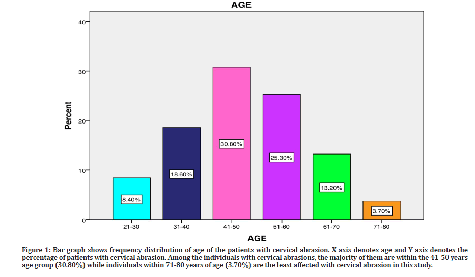
Figure 1 shows frequency distribution of age of the patients with cervical abrasion. X axis denotes age and Y axis denotes the percentage of patients with cervical abrasion. Among the individuals with cervical abrasions, majority of them are within the 41-50 years age group (30.80%) while individuals within 71-80 years of age (3.70%) are the least affected with cervical abrasion in this study.

Figure 1: Bar graph shows frequency distribution of age of the patients with cervical abrasion. X axis denotes age and Y axis denotes the percentage of patients with cervical abrasion. Among the individuals with cervical abrasions, the majority of them are within the 41-50 years age group (30.80%) while individuals within 71-80 years of age (3.70%) are the least affected with cervical abrasion in this study.
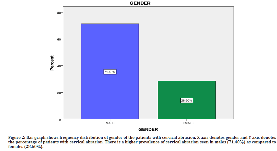
Figure 2 shows frequency distribution of gender of the patients with cervical abrasion. X axis denotes gender and Y axis denotes the percentage of patients with cervical abrasion. From this study we observed that 30.80% were males, 28.60% were females.

Figure 2: Bar graph shows frequency distribution of gender of the patients with cervical abrasion. X axis denotes gender and Y axis denotes the percentage of patients with cervical abrasion. There is a higher prevalence of cervical abrasion seen in males (71.40%) as compared to females (28.60%).
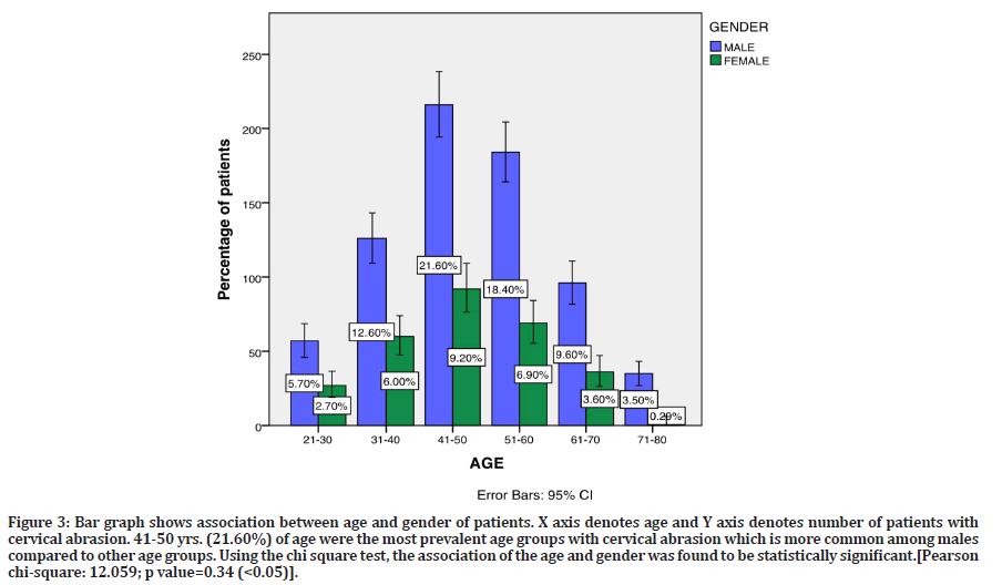
Figure 3 shows association between age and gender of patients. X axis denotes age and Y axis denotes number of patients with cervical abrasion. 41-50 yrs (21.60%) of age were the most prevalent age groups with cervical abrasion which is more common among males compared to females. Using the chi square test, the association of the age and gender was found to be statistically significant.[Pearson chi-square: 12.059 ; p value = 0.34 (>0.05)].

Figure 3: Bar graph shows association between age and gender of patients. X axis denotes age and Y axis denotes number of patients with cervical abrasion. 41-50 yrs. (21.60%) of age were the most prevalent age groups with cervical abrasion which is more common among males compared to other age groups. Using the chi square test, the association of the age and gender was found to be statistically significant.[Pearson chi-square: 12.059; p value=0.34 (<0.05)].
Discussion
A total of 1000 patients were evaluated in this study. In this present study, we observed that the majority of them are within the 41-50 years age group (30.80%) while individuals within 71-80 years of age (3.70%) are the least affected with cervical abrasion. Cervical lesions occur more frequently and are more severe in older people, according to several studies [36]. According to a study by Vaghasiya, et al. people between the ages of 31 and 40 had a higher incidence of cervical abrasion (32.5%), while those between the ages of 21 and 30 have the lowest prevalence (17%). Previous research has shown that the incidence of cervical abrasion rises with age, and that this is linked to poor brushing technique and a lack of understanding about basic oral hygiene in older age groups.
Males (71.40%) have a higher prevalence of cervical abrasions than females (28.60%), according to a study. In both urban and rural locations, males had more cervical abrasion cases than females, according to the findings of a prior study [37]. However, Atalay, et al. discovered that females (54%) have more cervical abrasions than males (46%) in a research [38]. Males brush their teeth with more force and for longer periods of time than females, which contributes to a higher prevalence of cervical abrasions. The way of teeth brushing with various strokes such as horizontal, vertical, and complex motion also influences the development of cervical abrasion. Patients brushing with horizontal strokes have a higher incidence of cervical abrasion than those brushing with vertical or complex strokes, according to a prior study [39]. Individuals who change their toothbrush after a month due to fraying of the bristles have a higher likelihood to acquire cervical abrasion than those who change their toothbrush after six months or more [40].
Conclusion
Within the limits of the study, most of the cervical abrasion cases are recorded in individuals within the 41-50 years age group with higher predilection in males. Excessive brushing and use of hard toothbrushes have strong associations with cervical tooth wear. There is a need to educate the patients about proper brushing techniques.
Authors Contribution
Nivethitha R: Literature search, data collection, analysis, manuscript drafting.
Anjaneyulu: Data verification, manuscript drafting.
Acknowledgement
The authors would thank all the participants for their valuable support and authors thank the dental institution for the support to conduct.
Conflict of Interest
All the authors declare that there was no conflict of interest in present study.
Source of Funding
The present project is supported/ funding/sponsored by the
✔ Saveetha Institute of Medical and Technical Sciences, Saveetha Dental College and Hospitals, Saveetha University, India
✔ Pavithra Harvesters and Earth Movers.
References
- Amin AH, Yaakob MH, Nasir WZ, et al. Evaluation the correlation between age, gender, and the incidence of cervical lesions. J Clin Res Dent 2018; 1:1-5.
- Terry DA, McGuire MK, McLaren E, et al. Perio esthetic approach to the diagnosis and treatment of carious and noncarious cervical lesions: Part II. J Esthet Restor Dent 2003; 15:284â??296.
- MiLoseviC A. Abrasion: A common dental problem revisited. Primary Dent J 2017; 6:32-36.
- Teixeira DN, Thomas RZ, Soares PV, et al. Prevalence of noncarious cervical lesions among adults: A systematic review. J Dent 2020; 95:103285.
- Wood I, Jawad Z, Paisley C, et al. Non-carious cervical tooth surface loss: A literature review. J Dent 2008; 36:759â??766.
- Van Meerbeek B, Kanumilli P, De Munck J, et al. A randomized, controlled trial evaluating the three-year clinical effectiveness of two etch & rinse adhesives in cervical lesions. Oper Dent 2004; 29:376-385.
- Jarvinen VK, Rytomaa II, Heinonen OP. Risk factors in dental erosion. J Dent Res 1991; 70:942-947.
- Lee WC, Eakle WS. Stress-induced cervical lesions: Review of advances in the past 10 years. J Prosthet Dent 1996; 75:487-494.
- Heymann HO, Sturdevant JR, Bayne S, et al. Examining tooth flexure effects on cervical restorations: A two-year clinical study. J Am Dent Assoc 1991; 122:41â??47.
- Mayhew RB, Jessee SA, Martin RE. Association of occlusal, periodontal, and dietary factors with the presence of non-carious cervical dental lesions. Am J Dent 1998; 11:29â??32.
- Kumar D, Antony SD. Calcified canal and negotiation-A review. J Adv Pharm Technol Res 2018; 11:3727.
- Teja KV, Ramesh S, Priya V. Regulation of matrix metalloproteinase-3 gene expression in inflammation: A molecular study. J Conserv Dent 2018; 21:592.
- Rajendran R, Kunjusankaran RN, Sandhya R, et al. Comparative evaluation of remineralizing potential of a paste containing bioactive glass and a topical cream containing casein phosphopeptide-amorphous calcium phosphate: An in vitro study. Pesquisa Br Odont Clin Integrada 2019; 19.
- Kelleher MG, Bomfim DI, Austin RS. Biologically based restorative management of tooth wear. Int J Dent 2012; 2012.
- Noor SS. Chlorhexidine: Its properties and effects. Res J Pharm Technol 2016; 9):1755-1760.
- Muthukrishnan L. Imminent antimicrobial bioink deploying cellulose, alginate, EPS and synthetic polymers for 3D bioprinting of tissue constructs. Carbohydr Polym 2021; 260:117774.
- PradeepKumar AR, Shemesh H, Nivedhitha MS, et al. Diagnosis of vertical root fractures by cone-beam computed tomography in root-filled teeth with confirmation by direct visualization: A systematic review and meta-analysis. J Endod 2021; 47:1198â??1214.
- Chakraborty T, Jamal RF, Battineni G, et al. A review of prolonged post-COVID-19 symptoms and their implications on dental management. Int J Environ Res Public Health 2021; 18.
- Muthukrishnan L. Nanotechnology for cleaner leather production: A review. Environ Chem Lett 2021; 19:2527â??2549.
- Teja KV, Ramesh S. Is a filled lateral canal: A sign of superiority? J Dent Sci 2020; 15:562â??3.
- Narendran K, Ms N, Sarvanan A. Synthesis, characterization, free radical scavenging and cytotoxic activities of phenylvilangin, a substituted dimer of embelin. Indian J Pharm Sci 2020; 82:909-912.
- Reddy P, Krithika Datta J, et al. Dental caries profile and associated risk factors among adolescent school children in an urban south-Indian city. Oral Health Prev Dent 2020; 18:379â??386.
- Sawant K, Pawar AM, Banga KS, et al. Dentinal microcracks after root canal instrumentation using instruments manufactured with different NiTi alloys and the SAF system: A systematic review. Adv Sci Inst Ser E Appl Sci 2021; 11:4984.
- Bhavikatti SK, Karobari MI, Zainuddin SLA, et al. Investigating the antioxidant and cytocompatibility of Mimusops elengi linn extract over human gingival fibroblast cells. Int J Environ Res Public Health 2021; 18.
- Karobari MI, Basheer SN, Sayed FR, et al. An In vitro stereomicroscopic evaluation of bioactivity between Neo MTA plus, pro root MTA, biodentine & glass ionomer cement using dye penetration method. Material 2021; 14:3159.
- Rohit Singh T, Ezhilarasan D. Ethanolic extract of Lagerstroemia speciosa (L.) Pers., induces apoptosis and cell cycle arrest in HepG2 cells. Nutr Cancer 2020; 72:146-156.
- Ezhilarasan D. MicroRNA interplay between hepatic stellate cell quiescence and activation. Eur J Pharmacol 2020; 885:173507.
- Romera A, Peredpaya S, Spark Y, et al. Bevacizumab biosimilar BEVZ92 versus reference bevacizumab in combination with FOLFOX or FOLFIRI as first-line treatment for metastatic colorectal cancer: A multicentre, open-label, randomised controlled trial. Lancet Gastroenterol Hepatol 2018; 3:845â??855.
- Raj RK. Ã?-Sitosterol-assisted silver nanoparticles activates Nrf2 and triggers mitochondrial apoptosis via oxidative stress in human hepatocellular cancer cell line. J Biomed Material Res 2020; 108:1899-908.
- Vijayashree Priyadharsini J. In silico validation of the non-antibiotic drugs acetaminophen and ibuprofen as antibacterial agents against red complex pathogens. J Periodontol 2019; 90:1441â??1448.
- Priyadharsini JV, Girija AS, Paramasivam A. In silico analysis of virulence genes in an emerging dental pathogen A. baumannii and related species. Arch Oral Biol 2018; 94:93-98.
- Uma Maheswari TN, Nivedhitha MS, Ramani P. Expression profile of salivary microRNA-21 and 31 in oral potentially malignant disorders. Braz Oral Res 2020; 34:e002.
- Gudipaneni RK, Alam MK, Patil SR, et al. Measurement of the maximum occlusal bite force and its relation to the caries spectrum of first permanent molars in early permanent dentition. J Clin Pediatr Dent 2020; 44:423â??428.
- Chaturvedula BB, Muthukrishnan A, Bhuvaraghan A, et al. Dens invaginatus: A review and orthodontic implications. Br Dent J 2021; 230:345â??350.
- Kanniah P, Radhamani J, Chelliah P, et al. Green synthesis of multifaceted silver nanoparticles using the flower extract of Aerva lanata and evaluation of its biological and environmental applications. Chem Select 2020; 5:2322â??2331.
- Rajakeerthi R, Nivedhitha MS. Natural product as the storage medium for an avulsed toothâ??A systematic review. Cumhuriyet Dent J 2019; 22:249-256.
- Yadav NS, Saxena V, Reddy R, et al. Alliance of oral hygiene practices and abrasion among urban and rural residents of Central India. J Contemp Dent Pract 2012; 13:55-60.
- Atalay C, Ozgunaltay G. Evaluation of tooth wear and associated risk factors: A matched case-Control study. Niger J Clin Pract 2018; 21:1607â??1614.
- Litonjua LA, Bush PJ, Andreana S, et al. Effects of occlusal load on cervical lesions. J Oral Rehabil 2004; 31:225â??332.
- Attin T, Siegel S, Buchalla W, et al. Brushing abrasion of softened and remineralised dentin: An in situ study. Caries Res 2004; 38:62-66.
Indexed at, Google Scholar, Cross Ref
Indexed at, Google Scholar, Cross Ref
Indexed at, Google Scholar, Cross Ref
Indexed at, Google Scholar, Cross Ref
Indexed at, Google Scholar, Cross Ref
Indexed at, Google Scholar, Cross Ref
Indexed at, Google Scholar, Cross Ref
Indexed at, Google Scholar, Cross Ref
Indexed at, Google Scholar, Cross Ref
Indexed at, Google Scholar, Cross Ref
Indexed at, Google Scholar, Cross Ref
Indexed at, Google Scholar, Cross Ref
Indexed at, Google Scholar, Cross Ref
Indexed at, Google Scholar, Cross Ref
Indexed at, Google Scholar, Cross Ref
Indexed at, Google Scholar, Cross Ref
Indexed at, Google Scholar, Cross Ref
Indexed at, Google Scholar, Cross Ref
Indexed at, Google Scholar, Cross Ref
Indexed at, Google Scholar, Cross Ref
Indexed at, Google Scholar, Cross Ref
Indexed at, Google Scholar, Cross Ref
Indexed at, Google Scholar, Cross Ref
Indexed at, Google Scholar, Cross Ref
Indexed at, Google Scholar, Cross Ref
Indexed at, Google Scholar, Cross Ref
Indexed at, Google Scholar, Cross Ref
Indexed at, Google Scholar, Cross Ref
Indexed at, Google Scholar, Cross Ref
Indexed at, Google Scholar, Cross Ref
Indexed at, Google Scholar, Cross Ref
Indexed at, Google Scholar, Cross Ref
Indexed at, Google Scholar, Cross Ref
Indexed at, Google Scholar, Cross Ref
Indexed at, Google Scholar, Cross Ref
Author Info
Nivethitha R and K Anjaneyulu*
Department of Conservative Dentistry, Saveetha Dental College and Hospitals, Saveetha Institute of Medical and Technical Sciences, Saveetha University, Chennai, IndiaCitation: Nivethitha R, K Anjaneyulu, Institution Based Assessment of Incidence and Association of Abrasion between Different Age Groups-A Retrospective Study, J Res Med Dent Sci, 2022, 10 (8):16-21.
Received: 18-Jul-2022, Manuscript No. JRMDS-22-69303; , Pre QC No. JRMDS-22-69303 (PQ); Editor assigned: 11-Jul-2022, Pre QC No. JRMDS-22-69303 (PQ); Reviewed: 25-Jul-2022, QC No. JRMDS-22-69303; Revised: 29-Jul-2022, Manuscript No. JRMDS-22-69303 (R); Published: 05-Aug-2022
