Case Report - (2023) Volume 11, Issue 2
Infected Dermoid Cyst at the Nasion Extending on to the Right Upper Eye Lid: A Case Report
Ashish Shukla*, Chandrashekhar C Mahakalkar, Meenakshi Yeola Pate, Shivani Kshirsagar and Abhishek Chaudhari
*Correspondence: Dr. Ashish Shukla, Department of General Surgery, Datta Meghe Institute of Medical Sciences, Wardha, India, Email:
Abstract
Objective: Dermoid cyst are are benign neoplasia having a mixed origin, i.e., ectodermal as well as mesodermal. These neoplasia are often observed during birth. However, it has a varying age of presentation. The most frequently reported locations are nasal, periorbita, submental, and supra-sternal section.
Case report: Our article presents a case of a 2 years old female patient who reported to the OPD with a sinus over the nose below nasion and swelling in right upper eye lid since 1 month. The history was obtained from the patient's mother who described that there was a swelling over the nose below the nasion, which had a bursting 5 days back leading to pus discharge. Later, there was a formation of another swelling in the right upper eye lid.
Conclusion: Neoplasms known as dermoid cysts are regularly encountered in children. The most often damaged part is the orbit, along the head and neck region. Nasofrontal dermoid sinus cysts develop when an initial embryological link amid an outpouching of dura and nasal tissue is not obliterated. The major goal of surgery should be total resection.
Keywords
Dermoid cyst, Supraorbital cystic extension, Front nasal suture, Pultaceous content
Introduction
Dermoid cysts are atypical neoplasms that are commonly seen in childhood. They are benign neoplasia having a mixed origin, i.e., ectodermal as well as mesodermal. The cyst has a lining of keratinized squamous epithelium associated with dermatic derivatives like hair follicle, sweat glands, sebaceous glands, and fibrous and adipose tissues. These neoplasia are often observed during birth. However, it has a varying age of presentation. An abrupt change in size are frequently found to cause difficulty in diagnosing a dermoid cyst. Around 7% of the total dermoid cysts are found to be occurring in the head and neck region. The most frequently reported locations are nasal, periorbita, submental and supra-sternal section [1].
Case Presentation
Our article presents a case of a 2 years old female patient who reported to the OPD with a sinus over the nose below nasion and swelling in right upper eye lid since 1 month. The history was obtained from the patient's mother who described that there was a swelling over the nose below the nasion, which had a bursting 5 days back leading to pus discharge. Later, there was a formation of another swelling in the right upper eye lid. On examination, a small sinus tract opening in dorsum area of nose was seen (Figure 1). On probing this sinus, it was found that this tract was reaching up to the nasion. There was another inflammatory swelling over the right upper eyelid of size 1 cm × 1 cm, which was soft in consistency suggestive of an abscess. These two lesions were located at the two different sites. So, investigations were performed to obtain data on whether the two has a common pathology. An MRI of face was performed which suggested the presence of a sinus leading to a diffuse swelling at the frontonasal junction. And there was thin tract from this diffuse swelling going across to the right upper eyelid but with no intracranial extension. An exploratory surgery was performed under general anaesthesia (Figures 2 and 3). It was found that there was a whole sinus tract with an attached diffused swelling having a cystic wall with pultaceous contents, and few hairs in it. The cyst was adherent to the frontonasal suture (Figure 4). It was removed meticulously, taking care that no residual cystic wall is left behind. The specimen was sent for histopathological examination. Similarly the infected sinus tract with abscess over the upper eyelid was excised, avoiding any injury to the lacrimal duct and the levator palpebrae superioris. After completion of excision, haemostasis was achieved and wound was closed in layers (Figure 5) [2].
After suture removal, there was a thin scar, that was left behind. This was taken care post-operatively to prevent it from hypertrophy and hyper-pigmentation. There was no loss of function in the right upper eye lid. Histopathological reports confirmed the excised specimen to be a "dermoid cyst". A follow-up was taken after 3 months which showed hyper granulation over suture line which was debrided and de-bulked (Figure 6). The patient was advised topical application anti-bacterial ointment to prevent infection at the site. Telephonic follow-up was carried out later on where the patient reported healing of the suture line [3].
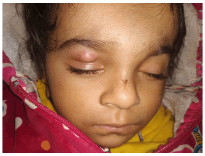
Figure 1: Pre-operative clinical photograph.
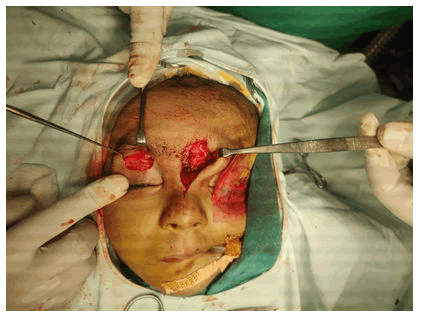
Figure 2: Placement of incision.
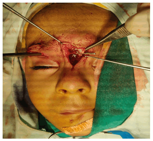
Figure 3: Exploration of dermoid cyst.
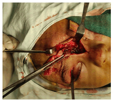
Figure 4: Tracing of the cystic extension over supraorbital region.
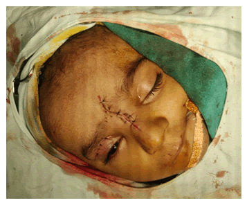
Figure 5: Post operative clinical photograph.
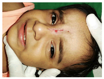
Figure 6: Follow-up after 3 months.
Results and Discussion
The cystic remnants of epidermal components through embryonic fusion lines through embryologic development causes dermoid cyst formation [4]. In the head and neck epidermal and dermal cysts have been documented to affect amongst 1.6 percent and 6.9 percent of people [5]. Despite the fact that over 200 incidents have occurred, of dermoid cysts underlying the anterior fontanelle since birth only four other occurrences of a dermoid cyst outside of the anterior fontanelle have been documented [6].
New and Erich in 1937 dermoid cysts were categorized as acquired implantation, congenital teratoma, or congenital inclusion After the ectopic growth of a dermal cyst lined with squamous epithelium, a component of the skin is traumatically implanted in the deeper layers, resulting in acquired dermoid cysts. They look like epidermal cysts. Congenital teratomas originate in the ovaries and testes and arise from embryonic germinal epithelium of all three types. They contain epithelial, bone, and cartilage components. Congenital inclusion dermoid cysts comprises of dermal and epidermal derivatives and arise along embryologic fusion lines. The congenital inclusion type is believed to cause dermoid cysts of the head and neck. Epidermal inclusion cysts, glioma, meningomyelocele, meningoencephalocele, teratoma, teratoid cyst, thyroglossal duct cyst, branchial cleft cyst, and benign lymphadenopathy are amongst the diagnostic options depending upon position. Because dermoid cysts constitute a pathologic diagnosis, the workup and evaluation of these neoplasms necessitate a medical history and physical examination [7].
In 1965, Pratt developed the most widely accepted theory about the embryological genesis of nasofrontal dermoid sinus cysts. Nasal and cranial cartilage ossification starts towards termination of the second month of pregnancy [8]. The frontal bone and cranial base have not yet connected by the second month of pregnancy, and the nasal and frontal bones are separated by a prospective area known as fonticulus frontalis. The dura ventures in through the nasal area in an opening in the cranial base along this period. Between the nasal bone and the deep cartilaginous capsule in the mesoderm, the dural pouch enters the prenasal region. Between the nasal bone and the deep cartilaginous capsule of the mesoderm, the dural pouch enters the prenasal region. Nasofrontal dermoid sinus tracts, nasal encephaloceles, and nasal gliomas are all the result of ossification failure to remove this transcranial connection [9]. A nontender swelling on the dorsum of the nose could indicate an extracranial nasofrontal dermoid sinus cyst. The bulk is generally mobile beneath the skin, while it is tightly connected to below structures. A nasal dermoid cyst and sinus tract are characteristic for a dimple on the nasal surface with or without projecting hair and sebaceous discharge glabellar edema, nasal broadening, and hypertelorism are all likely indicators. The dura or falx cerebri could be implicated in the lesion's intracranial part, which is often extra-axial. It is quite uncommon to find intraparenchymal extension; it’s solely in the frontal lobes if it's present. Intracranial outcomes include Molaret type chemical meningitis, infectious meningitis, abscess, osteomyelitis, convulsions, or personality disorders [10].
Conclusion
p>Neoplasms known as dermoid cysts are regularly encountered in children. The most often damaged part is the orbit, along the head and neck region. Nasofrontal dermoid sinus cysts develop when an initial embryological link amid an outpouching of dura and nasal tissue is not obliterated. The major goal of surgery should be total resection.Conflict of Interest
The authors have no conflict of interest.
Source of Interest
Nil.
References
- Pryor SG, Lewis JE, Weaver AL, et al. Pediatric dermoid cysts of the head and neck. Otolaryngol Head Neck Surg 2005; 132:938-942.
[Crossref] [Google Scholar] [PubMed]
- Wood J, Couture D, David LR, et al. Midline dermoid cyst resulting in frontal bone erosion. J Craniof Surg 2012; 23:131-134.
[Crossref] [Google Scholar] [PubMed]
- Papadogeorgakis N, Kalfarentzos EF, Vourlakou C, et al. Surgical management of a large median dermoid cyst of the neck causing airway obstruction: A case report. Oral Max Surg 2009; 13:181-184.
[Crossref] [Google Scholar] [PubMed]
- Tateshima S, Numoto RT, Abe S, et al. Rapidly enlarging dermoid cyst over the anterior fontanel: A case report and review of the literature. Child Ner System 2000; 16:875-878.
[Crossref] [Google Scholar] [PubMed]
- Fujimaki T, Miyazaki S, Fukushima T, et al. Dermoid cyst of the frontal bone away from the anterior fontanel. Childs Nerv Syst 1995; 11:424.
[Crossref] [Google Scholar] [PubMed]
- Maniglia AJ, Goodwin WJ, Arnold JE, et al. intracranial abscesses secondary to nasal, sinus, and orbital infections in adults and children. Arch Otolaryngol Head Neck Surg 1989; 115:1424-1429.
[Crossref] [Google Scholar] [PubMed]
- Vanhorebeek I, Latronico N, van den Berghe G. ICU-acquired weakness. Intens Care Med 2020; 46:637-653.
[Crossref] [Google Scholar] [PubMed]
- Zhou C, Wu L, Ni F, et al. Critical illness polyneuropathy and myopathy: A systematic review. Neu Regen Res 2014; 9:101.
[Crossref] [Google Scholar] [PubMed]
- Hugon J, Msika EF, Queneau M, et al. Long COVID: Cognitive complaints (brain fog) and dysfunction of the cingulate cortex. J Neurol 2022; 269:44-46.
[Crossref] [Google Scholar] [PubMed]
- Caronna E, Alpuente A, Torres‐Ferrus M, et al. Toward a better understanding of persistent headache after mild COVID‐19: Three migraine‐like yet distinct scenarios. Headache J Head Face Pain 2021; 61:1277-1280.
[Crossref] [Google Scholar] [PubMed]
Author Info
Ashish Shukla*, Chandrashekhar C Mahakalkar, Meenakshi Yeola Pate, Shivani Kshirsagar and Abhishek Chaudhari
Department of General Surgery, Datta Meghe Institute of Medical Sciences, Wardha, IndiaCitation: Ashish Shukla, Chandrashekhar C Mahakalkar, Meenakshi Yeola Pate, Shivani Kshirsagar, Abhishek Chaudhari,
Infected Dermoid Cyst at the Nasion Extending on to the Right Upper Eye Lid: A Case Report, J Res Med Dent Sci, 2023, 11 (02): 007-010.
Received: 04-Oct-2021, Manuscript No. JRMDS-21-43646; , Pre QC No. JRMDS-21-43646(PQ); Editor assigned: 07-Oct-2021, Pre QC No. JRMDS-21-43646(PQ); Reviewed: 21-Oct-2021, QC No. JRMDS-21-43646; Revised: 31-Jan-2023, Manuscript No. JRMDS-21-43646(R); Published: 28-Feb-2023
