Research - (2020) Volume 8, Issue 1
Importance of Success Criteria in the Validity of Panoramic Radiograph for Measurement of Gonial Angle
*Correspondence: Orthodontics and Preventive Dentistry. Ruba J Mohammad, Department of Pedodontics, College of Dentistry, University of Mosul, Iraq, Email:
Abstract
Introduction: The size of the gonial angle depends on the method of measurement being used. It can be measured on both panoramic and cephalometric radiographs. The study aims to find the effect of the success criteria of a panoramic radiograph on its validity to evaluate the gonial angle.
Materials and methods: Lateral cephalometric and dental panoramic radiographs of 76 individuals of both genders with age range from 12 to 25 years were included in this study. The samples divided into two groups: The first group included 36 samples with non-evaluated panoramic radiographs having good quality and sharpness. The second group included 40 samples of evaluated panoramic radiographs, which were selected according to the specific criteria of success. A paired t-test was used for evaluating the difference between the gonial angle in the lateral cephalometric and panoramic radiographs of both sides. A p-value ≥ 0.05 was considered to be statistically insignificant.
Results: The findings of the study showed a significant difference in the values of the gonial angle between the non-evaluated panoramic radiographs of right and left sides (p=0.00, 0.001) respectively as compared to the cephalometric radiograph. No significant differences observed in the value of gonial angle measurements. They were obtained from cephalometric and evaluated panoramic radiographs of both sides (p=0.888, 0.938) respectively.
Conclusion: The success criteria could serve as an easy and simple method to assess the quality of panoramic radiographs for accurately measuring the gonial angle on both sides and without superimposition as compared to cephalometric radiographs.
Keywords
Gonial angle, Panoramic radiograph, Cephalometry
Introduction
The lateral cephalometric radiography is considered as an essential diagnostic tool for orthodontics, it is used to evaluate the stage of development and craniofacial relationships [1,2]. The value of the gonial angle usually is used to determine the stage of growth and besides the determination of sex and gender [3,4]. The gonial angle is defined as an angle formed by the intersection of the ramus border tangential line and the mandibular lower border tangential line [5,6]. Measurement of the gonial angle is essential in orthodontic treatment and orthodontic surgery. It is important to assess the symmetry of the facial skeleton. Therefore, accurate determination of the gonial angle is essential for assessing the orthodontic cases. Ordinarily, the cephalometric radiograph is used to evaluate the gonial angle, as an intermediate value of the right and left sides angles due to the superimposed of both sides of the mandible [2,7].
The panoramic technique was first described by Paatero in 1952 and since then it has been used in a variety of dental specialties. The main advantages of the panoramic radiography include less radiation dose, fast technique, good patient co-operation, and covering the broad anatomic structures. The ability of dental panoramic radiographs to view both maxilla and mandible in a single image makes it a respected radiological technique [8-10]. Although it can provide a single image of the entire stomatognathic system-teeth, jaws, temporomandibular joints, sinuses. It suffers from the limitation of methodological errors. Many authors considered the panoramic radiograph as the choice to assess the right and left gonial angles accurately without superimposition in a single radiograph [2,11]. Other authors compared the angular measurements on both panoramic and lateral cephalometric radiographs; they found that the panoramic radiography can provide an acceptable measurement of the gonial angle, but that it is not as reliable as a lateral cephalometric radiograph. This study is conducted to find the effect of the success criteria of a panoramic radiograph on its validity to assess the gonial angle [12,13].
Materials and Methods
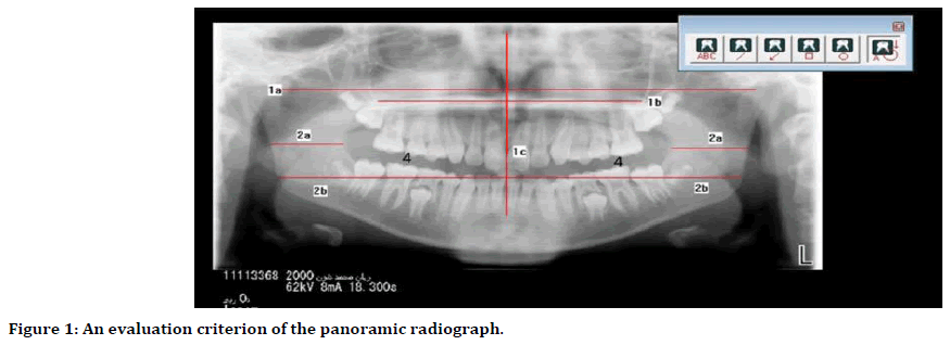
Lateral cephalometric and dental panoramic radiographs of 76 subjects of both genders with skeletal CL-I classification obtained from the database archiving of the digital Dimax Pro system (Helsinki, Finland) in the Dental Radiology Class of Oral and Maxillofacial Surgery Department, College of Dentistry, University of Mosul. The age of patients ranged from 12 to 25 years. The samples of the patients were divided into two groups: the first group included 36 samples (18 female and 18 male) where the panoramic radiographs were of good quality and sharpness (non-evaluated panoramic radiographs). The second group included 40 samples (20 female and 20 male), where the panoramic radiographs were selected according to the specific criteria of success [14] (evaluated panoramic radiographs) as follows Figure 1.

Figure 1. An evaluation criterion of the panoramic radiograph.
1a: The connection line between the two points is drawn through the deepest point of the articular eminence, perpendicular to the median sagittal plane, providing information in normal cases on whether improper positioning has occurred.
1b: The line connecting the deepest part of the innominate line of the facies temporalis of the zygomatic bone and the maxilla perpendicular to the median sagittal plane provide information about the horizontal positioning of the head.
1c: Median sagittal plane in normal cases is perpendicular to 1a and 1b. It demonstrates the vertical positioning of the head.
2a: Bilateral comparison of the width of the ascending mandibular ramus exhibits an asymmetric positioning of the head or nearly lateral displacement of the mandible.
2b: A comparison of the distance between the midline of the mandible to the dorsal border of the ascending ramus provides information about any asymmetric positioning of the skull or the mandible, as evident from the apparent lateral displacement of the mandibular midline.
4: A bilateral comparison of the apparent teeth sizes in the maxilla with asymmetric positioning of the skull and the mandible is also an indication of improper patient positioning.
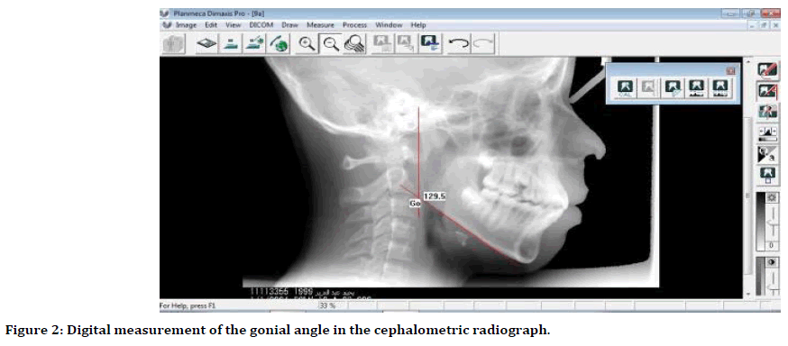
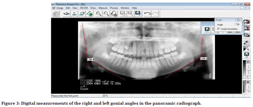
The quality of radiographs was assessed by an expert oral radiologist to compensate for the requirement of each group before inclusion in the study. The radiographs were acquired with the Ceph-Pan Dimax Pro X-ray unit (Helsinki, Finland). The gonial angle formed by the intersection between the tangential lines of the posterior border of the ramus and the lower border of the mandible [2] in cephalometric and panoramic radiographs was used as the standard (Figures 2 and 3). Digital Imaging Software from Dimax Pro (Helsinki, Finland) was used to digitally trace and measure the gonial angle on the lateral cephalometric and on the right and left sides of panoramic radiographs. Measurements were implemented to the nearest 0.5 degrees.

Figure 2. Digital measurement of the gonial angle in the cephalometric radiograph.

Figure 3. Digital measurements of the right and left gonial angles in the panoramic radiograph.
Statistical analysis
Descriptive statistics were carried out to calculate means and standard deviations for all variables (Table 1). The paired t-test was used for evaluating the difference between the measurements of the gonial angle in the lateral cephalometric and panoramic radiographs of both sides. The analyses were performed using the SPSS version 13.0, a p-value of ≥ 0.05 was considered to indicate statistical not significance.
| Group | Samples | Gender | No. | Side | Min. | Max. | Mean | SD |
|---|---|---|---|---|---|---|---|---|
| Non-evaluated | Cephalometric | F | 18 | - | 121.43 | 135 | 129.99 | 4.6889 |
| M | 18 | - | 121.44 | 136 | 126.94 | 4.6801 | ||
| panoramic | F | 18 | R | 117.55 | 132.2 | 126.54 | 6.2442 | |
| L | 117.6 | 131.75 | 126.91 | 6.2031 | ||||
| M | 18 | R | 117.6 | 132.46 | 124.43 | 5.665 | ||
| L | 117.5 | 132.22 | 125.64 | 5.616 | ||||
| Evaluated | Cephalometric | F | 20 | - | 121.43 | 134.96 | 128.98 | 4.448 |
| M | 20 | - | 122.55 | 135.86 | 129.53 | 4.076 | ||
| panoramic | F | 20 | R | 122.6 | 134.56 | 129.82 | 3.3861 | |
| L | 122.33 | 135.78 | 128.18 | 4.3658 | ||||
| M | 20 | R | 122.75 | 134.76 | 129.76 | 4.4432 | ||
| L | 122.7 | 134.56 | 129.36 | 4.0307 |
Table 1: Descriptive statistics of the all variable in both genders.
Results
The results showed a significant difference in the values of the gonial angle in the non-evaluated panoramic radiographs of both right and left sides (p=0.00, 0.001) respectively as compared to its value gained from the cephalometric radiograph. While, no significant differences were seen in the value of gonial angle measurements gained from cephalometric and evaluated panoramic radiographs in both sides (p=0.888, 0.938) respectively (Table 2). Also, no significant differences are seen as comparing the right and left side measurements of the panoramic radiographs either evaluated (p=0.809) or nonevaluated (p=0.888) (Table 3).
| Pairs | Side | Mean | SD | SE mean | t-value | df | p-value |
|---|---|---|---|---|---|---|---|
| Cephalometric-Non-evaluated panoramic measurements | R | 3.889 | 5.467 | 0.911 | 4.268 | 35 | 0 |
| L | 3.514 | 5.656 | 0.942 | 3.728 | 35 | 0.001 | |
| Cephalometric–Evaluated panoramic measurements | R | 0.155 | 6.928 | 1.095 | 0.141 | 39 | 0.888 |
| L | -0.091 | 7.371 | 1.165 | -0.079 | 39 | 0.938 |
Table 2: A comparison between cephalometric and panoramic measurements of gonial angle.
| Pairs | Mean | SD | SE mean | t-value | df | p-value |
|---|---|---|---|---|---|---|
| Right–Left non-evaluated panoramic measurements | -0.375 | 2.25 | 0.375 | -1 | 35 | 0.888 |
| Right–Left evaluated panoramic measurements | -0.246 | 6.425 | 1.015 | -0.243 | 39 | 0.809 |
Table 3: A comparison of gonial angle measurements between the right and left sides of the panoramic radiograph.
Discussion
Lateral cephalometric radiographs are usually used for evaluation of skeletal and dentofacial development through the assessment of different angular and linear measurements [15- 17]. Recently, the dental panoramic radiograph has been used to evaluate different angular and linear measurements to assess the skeletal and dentofacial developments [9,18]. There is a great controversy between authors with respect to the use of two types of radiographs; several authors support the validity of a panoramic radiograph in the measurement of the gonial angle.
The results of this study show a significant difference between the cephalometric radiograph and both sides of non-evaluated panoramic radiographs in measuring the gonial angle, with a mean difference of about 3.5 degrees between the two radiographic techniques. These findings related to that the quality of a panoramic radiograph is greatly affected by the patient head position in vertical and horizontal plains which resulted in angular distortion even this position being standardized using the light-beam positioning marker. In addition to that, the size of the focal trough is very limited, which is very sensitive to the change in the patient head position [19,20]. Therefore, any minor change in the patient`s head position resulted in a low diagnostic quality of the panoramic radiograph. Thus, leading to great changes in the shape and size of the object which affect the linear and angular measurements [14,20]. These results come in agreement with those of other studies; Arkai et al. [21] and Park et al. [22] preferred the use of cephalometric radiograph on the panoramic radiograph in the measurements of gonial angle. They found that the value of the gonial angle from a panoramic radiograph is smaller in 2 to 3.6 degrees than that obtained from a cephalometric radiograph. Similarly, Kundi, et al. [23] concluded that the gonial angle cannot be measured on the panoramic radiograph as accurately as a lateral cephalometric radiograph.
No significant difference is found in the value of the gonial angle between the cephalometric and evaluated panoramic radiographs because the success criteria were considered to select the panoramic radiographs for this group. Which makes the quality of these radiographs in the optimum and free from the effect of patient head positioning errors. Thus, the validity of panoramic radiographs in measuring the gonial angle greatly depends on their quality which should be carefully evaluated to obtain adequate angular and linear measurements by considering the success criteria to display the quality of panoramic radiography [24,25].
A cephalometric radiograph can be used accurately to measure the gonial angle as an intermediate measure between both sides of the mandible, but it is impossible to determine each side separately [3]. The panoramic radiograph could be used to measure the gonial angle on both sides of the mandible in a single radiograph with minimum radiation dose to the patient and minimum cost effect [25,26]. Concerning the amount of radiation exposure for a cephalometric radiograph is equivalent to less than one day of average background radiation. While the panoramic radiographs have a radiation dose equivalent to 1 - 3 days of background radiation [27]. Many authors support the use of cephalometric and panoramic radiographs to measure the gonial angle; Majeed, et al. [9], Park, et al. [22], Trivedi, et al. [28], Katti, et al. [29], Ganeiber, et al. [30], Kundi [31] and Ozkan, et al. [32] they prove that the panoramic radiography can be used to determine the gonial angle as accurately and easily as lateral cephalogram without superimposition of anatomic landmarks, which commonly occurs in a lateral cephalogram.
Also, there is no significant difference in the measurement of the gonial angle between the right and left sides of panoramic radiographs in two groups of samples. This compensates the results of other studies, [3,4,29,33] establish that the panoramic radiograph provides equally reliable information about both left and right side gonial angles.
Conclusion
The panoramic radiograph could be used to assess the gonial angle as accurately as that of a cephalometric radiograph and represents both the right and left gonial angles without superimposition in an easy and simple method with a single radiograph. Respect should be given to the success criteria in observing the quality of panoramic radiographs.
Acknowledgment
I would like to thanks Prof. Dr. Nazar Gh. Jameel, a specialist in oral and dental radiology, Department of Dentistry, Alnoor University College, for his assistance in the selection of the panoramic radiograph and supervision during the application of success criteria. I look forward to cooperate with him in the future.
References
- Okşayan R, Aktan AM, Sokucu O, et al. Does the panoramic radiography have the power to identify the gonial angle in orthodontics? Sci World J 2012; 2012:1-4.
- Thilagarani, Nadkerny PV, Kumar DA, et al. Assessing reliability of mandibular planes in determining gonial angle on lateral cephalogram and panoramic radiograph. J Orthod Res 2015; 3:45-48.
- Radhakrishnan PD, Sapna Varma NK, Ajith VV. Dilemma of gonial angle measurement: Panoramic radiograph or lateral cephalogram. Imaging Sci Dent 2017; 47: 93-97.
- Abbas FK. Analyzing the measurements of gonial angle by panoramic radiographs for forensic estimation in Iraqi population. Mustansiriya Dent J 2018; 15:97-104.
- Upadhyay RB, Upadhyay J, Agrawal P, et al. Analysis of Gonial angle in relation to age, gender, and dentition status by radiological and anthropometric methods. J Forensic Dent Sci 2012; 4:29-33.
- Saikiran CH, Ramaswamy P, Santosh N, et al. Can gonial measurements predict gender? A prospective analysis using digital panoramic radiographs. Forensic Res Criminol Int J 2016; 3:89.
- Abuhijleh E, Warreth A, Qawadi M, et al. Mandibular gonial angle measurement as a predictor of gender-A digital panoramic study. The Open Dent J 2019; 13:399-404.
- Patil SR. Comparative measurement of tooth length: Actual vs. Orthopantomography and CBCT-Based measurements. Pesqui Bras Odontopediatria Clín Integr 2019; 19:4637.
- Majeed M, Ahmed I. Comparison of gonial angle determination from cephalograms and orthopantomogram of patients under orthodontic treatment. JBUMDC 2016; 6:88-91.
- Icoz D, Akgunlu F. Prevalence of detected soft tissue calcifications on digital panoramic radiographs. SRM J Res Dent Sci 2019; 10:21-25.
- Leversha J, McKeough G, Myrteza A, et al. Age and gender correlation of gonial angle, ramus height and bigonial width in dentate subjects in a dental school in far North Queensland. J Clin Exp Dent 2016; 8:49-54.
- Araki M, Kiyosaki T, Sato M, et al. Comparative analysis of the gonial angle on lateral cephalometric radiographs and panoramic radiographs. J Oral Sci 2015; 57:373–378.
- Kumar SS, Thailavathy V, Srinivasan D, et al. Comparison of orthopantomogram and lateral cephalogram for mandibular measurements. J Pharm Bioallied Sci 2017; 9:92–95.
- Pasler FA, Visser H. Panoramic radiography. Pocket atlas of dental radiology. Thieme Com New York 2007; 1:18-19.
- Silva MBG, Sant’Anna EF. The evolution of cephalometric diagnosis in orthodontics. Dental Press J Orthod 2013; 18:63-71.
- Rianti RA, Priaminiarti M, Syahraini SI. Tolerance of image enhancement brightness and contrast in lateral cephalometric digital radiography for Steiner analysis. J Physics Conf Series 2017; 884:12045.
- Moghaddam SF, Sohrabi A, Zokaei M, et al. Investigating validity and reliability of visual inspection of lateral cephalometric radiography (LCR) evaluation in determining dento-skeletal characteristics. Revista Publicando 2018; 16:372-386.
- Rachmadiani DT, Makes BN, Iskandar HHB. The average value of mandible measurements in panoramic radiographs: A comparison of 14–35 and 50–70 year old subjects. J Physics Conf Series 2017; 884:12049.
- Abdinian M, Hashemian A, Sameti AA. The effect of changing focal trough in a panoramic device on the accuracy of distance Measurements. Dent Hypotheses 2018; 9:16-19.
- Chalkoo AH, Maqbool S, Wani BA. Radiographic evaluation of sexual dimorphism in mandibular ramus: A digital orthopantomography study. Inter J Applied Dent Sci 2019; 5:163-166.
- Araki M, Kiyosaki T, Sato M, et al. Comparative analysis of the gonial angle on lateral cephalometric radiographs and panoramic radiographs. J Oral Sci 2015; 57:373-338.
- Park S, Kim Y, Lee S, et al. The simple regression model of gonial angles: Comparison between panoramic radiographs and lateral cephalograms. J Korean Acad Pediatr Dent 2017; 44:129-137.
- Kundi IU, Baig MN. Reliability of panoramic radiography in assessing gonial angle compared to lateral cephalogram. Pakist Oral Dent J 2018; 38:320-323.
- Mayil M, Keser G, Pekiner FN. Clinical image quality assessment in panoramic radiography. MÜSBED 2014; 4:126-132.
- Park JH, Chaea J, Bayd RC, et al. Evaluation of factors influencing the success rate of orthodontic microimplants using panoramic radiographs. Korean J Orthod 2018; 48:30-38.
- Wrzesień M, Olszewski J. Absorbed doses for patients undergoing panoramic radiography, cephalometric radiography and CBCT. Int J Occup Med Environ Health 2017; 30:705–713.
- Mallya SM, Lam EWN. White and pharoah's oral radiology, principles and interpretation. 8th Edn Toronto, Elsevier 2019.
- Trivedi B, Joshipura A, Bhatia AF, et al. Determination of growth pattern using OPG–A new approach in orthodontic diagnosis. BUJOD 2015; 5:63-69.
- Katti G, Katti C, Shahbaz S, et al. Reliability of panoramic radiography in assessing gonial angle compared to lateral cephalogram in adult patients with Class I malocclusion. J Indian Acad Oral Med Radiol 2016; 28:252-255.
- Ganeiber T, Bugaighis I. Assessment of the validity of orthopantomographs in the evaluation of mandibular steepness in Libya. J Orthodont Sci 2018; 7:1-4.
- Kundi I. Accuracy of assessment of gonial angle by both hemispheres of panoramic images and its comparison with lateral cephalometric radiographic measurements. J Dent Health Oral Disord Ther 2016; 4:116.
- Ozkan TH, Arici S, Ozkan E. The better choice for measuring the gonial angle of different skeletal malocclusion types: Orthopantomograms or lateral cephalograms? Inter Med J 2019; 8:93-96.
- Nejad AM, Jamilian A, Meibodi SE, et al. Reliability of panoramic radiographs to determine gonial and frankfurt mandibular horizontal angles in different skeletal patterns. Stoma Edu J 2016; 3:81-85.
