Research - (2021) Volume 9, Issue 1
Histomorphometric Evaluation of the Effect of Local Application of Moringa Oliefera/Marine Collagen on Bone Healing in Rats
Areej Salim Al-Azzawi* and Nada MH Al-Ghaban
*Correspondence: Areej Salim Al-Azzawi, Department of Oral Histology, Dental Teaching Hospital, College of Dentistry, University of Baghdad, Iraq, Email:
Abstract
Backgrounds: Bone repair is a complex multistep process, flavonoids group present in Moringa Oliefera and Marine collagen helps to increase the bone minerals contents density of bone.
Objectives: the aim of study was to evaluation the differences between the healing process when used of Moringa Oleifera (MO), Marine collagen (MC) and their combination (MM) on bone defects (experimental groups) and bone defect that heal normally (CONT) (control group).
Material and Methods: 20 albino rats were anesthetized and both femurs prepared by drilling intrabony defects (2mm diameter, 3mm depth). In 10 rats, right defects consider control while left defects treated with MO, in other 10 rats right defect treated with MC while left defects treated with MM. scarification done in 2 periods: 2 and 4 weeks. Histological and Histomorphometric analysis of bone microarchitectures. Bone cells were counted. Also, trabecular number, trabecular area and bone marrow area per mm2 were measured using image j program.
Results: Histological result revealed acceleration of bone healing in combination group (MO&MC) than that other groups. Highly significant differences were seen between combination of MO with MC and control group in both duration for almost all histomorphometric parameters used in this study.
Conclusions: Combination of MO with MC improve bone healing and increase osteogenic capacity.
Keywords
Moringa oliefera, Marine collagen, Bone healing
Introduction
Bone healing is a greatly complex reformative process. Bone tissue commonly heals spontaneously but in complicated circumstances such as large bone defects [1]. Bone healing can be divided into three phases: Inflammatory Phase, Reparative Phase and Remodeling Phase [2].
Moringa oleifera (MO) contains various flavonoids in leaf, root, flower, and seed coat [3]. MO leaves were evaluated for hypocholesterolemic and anti-obesity activity [4]. Flavonoid in ethanolic extract of moringa olifeira could induce mesenchymal stem cells derived from bone marrow to undergo osteogenic differentiation [5,6]. Soekobagiono revealed that sockets treated with combination of Moringa leaf extract and demineralized freeze-dried bone bovine xenograft DFDBBX had decrease the number of RANKL expressions in Cavia cobaya rats on the day 7 and day 30 after tooth extraction [7].
Marine collagen (MC) derived from marine organisms such as fish, seaweeds, sponges, and jellyfish offer advantages over mammalian [8]. MC has attracted much attention as a mammalian collagen substitute, from biomedical researchers to the cosmetic, food, and nutraceutical industries [9].
Clinical studies proved that Marine collagen promoted cartilage matrix synthesis and reduced pain in osteoarthritic patients, making them a promising candidate for therapeutic agents against human osteoarthritis and osteoporosis [10]. Numerous studies have revealed that MC based biomaterial scaffolds have been used as bone tissue substitutes and reinforcements that promote bone tissue regeneration [11]. MC induced multidirectional differentiation in the primary rat bone marrow mesenchymal stem cells, also it significantly upregulated the expression of osteogenic markers suggesting that MC has the potential to actively promote osteogenic [12].
Materials and Methods
Preparation of Moringa Olifeira extract
Preparation was done in department of chemistry of College of Education-University of Samarra. Dried seeds of MO were collected from local market were grinded into fine powder about 200 gm. extraction process was done by using Soxhlet extractor using 70% ethanol for about 72 hours, then extracted solution was seated in rotary evaporator to get thick jelly solution. Then extract was placed in refrigerator (5-100C) until be used in vivo.
Animal preparation
All experimental procedures were conducted in accordance with the ethical approval of animal experiments of College of Dentistry, University of Baghdad. Supervision and nursing from the stuff of animal house of Collage of Farming/ University of Tikrit. 20males Albino rats, (4-5) months weight (350-450 gm) was used in this experimental study. Animals were anesthetized and both femurs were prepared for surgical operation. Exposure of femur bone (Figure 1 and Figure 2). Scarification of animals was done by giving overdose of anesthesia 2 and 4weeks postoperatively (10 rats for each healing interval). Bone specimen was prepared by cutting the bone about 5 mm away from operation and stored in 10% freshly prepared formalin.
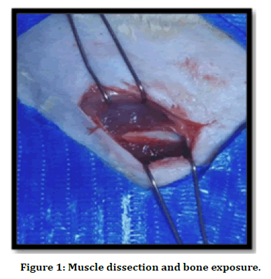
Figure 1. Muscle dissection and bone exposure.
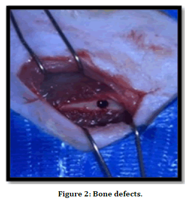
Figure 2. Bone defects.
Histological preparation
Bony specimens were fixed with 10% freshly prepared formalin for 24 hours, then decalcification process started using 10% formic acid for (3-8 days), embedding done using paraffin wax, bone blocks were sectioned by microtome for serial sections of 4μm was taken and placed on slide. Staining was done by using hematoxylin and eosin (H&E) stain.
Histomorphometric analysis of bone microarchitectures. Bone cells number (osteoblast, osteocyte, and osteoclast) cells per mm2 were counted. Also, trabecular number, trabecular area and bone marrow area per mm2 were measured using image j program.
Statistical analysis
The data analyzed using Statistical Package for Social Sciences (SPSS) version 25. Independent t-test and Analysis of Variance (ANOVA) was used to compare the continuous variables accordingly. A level of P–value less than 0.05 was considered significant.
Results
Histological finding observed that all histological sections showed good healing pathway for all experimental and control groups but with difference in rate with their periods consumed. In MO group at 2 weeks irregular trabecular found with irregular distributer osteocytes while at 4 weeks regular mature bone contain obvious osteons observed (Figures 3-6).
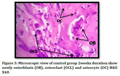
Figure 3. Microscopic view of control group 2weeks duration show newly osteoblasts (OB), osteoclast (OCL) and osteocyte (OC) H&E X40.
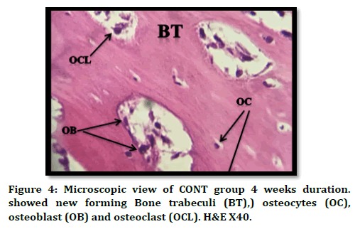
Figure 4. Microscopic view of CONT group 4 weeks duration. showed new forming Bone trabeculi (BT),) osteocytes (OC), osteoblast (OB) and osteoclast (OCL). H&E X40.
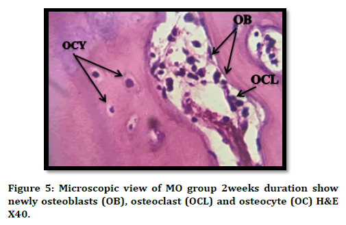
Figure 5. Microscopic view of MO group 2weeks duration show newly osteoblasts (OB), osteoclast (OCL) and osteocyte (OC) H&E X40.
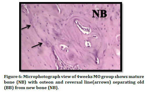
Figure 6. Microphotograph view of 4weeks MO group shows mature bone (NB) with osteon and reversal line(arrows) separating old (BB) from new bone (NB).
In MC group irregular osteoid seen in the defect area at 2weeks duration rimmed with osteocytes and osteoclasts cells, at 4 weeks regular bone trabeculi seen with regular arranged osteocytes (Figures 7 and 8). In combination group, in 2 weeks duration more regular arranged osteoid tissue with embedded osteocytes and rimmed with osteoblast cells, after 4 weeks thicher and more regular bone trabeculi seen contain mature osteocytes (Figures 9 and 10).
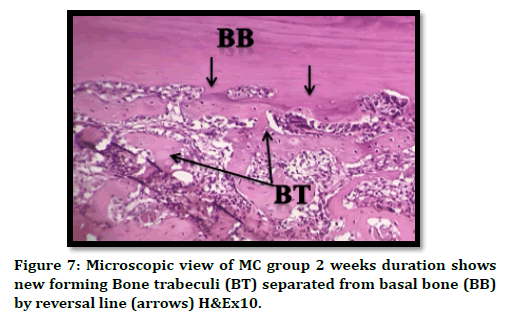
Figure 7. Microscopic view of MC group 2 weeks duration shows new forming Bone trabeculi (BT) separated from basal bone (BB) by reversal line (arrows) H&Ex10.
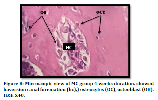
Figure 8. Microscopic view of MC group 4 weeks duration. showed haversion canal foremation (hc),) osteocytes (OC), osteoblast (OB). H&E X40.
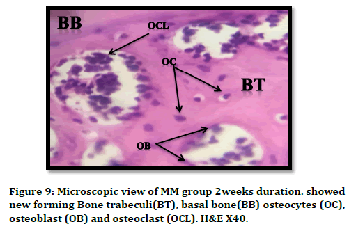
Figure 9. Microscopic view of MM group 2weeks duration. showed new forming Bone trabeculi(BT), basal bone(BB) osteocytes (OC), osteoblast (OB) and osteoclast (OCL). H&E X40.

Figure 10. View of 4weeks duration of bone defect treated with MC and MO show osteocytes (OC), osteoblast (OB) H & EX40.
Statistical finding
Table 1 revealed highly significant differences in osteocytes mean number between combination (MM) and control groups in both 2 and 4 weeks. Also, significant differences found between control & MO in 2 weeks, and between MC& MM in 4 weeks duration. Table 2 showed highly significant differences in osteoblasts mean number between MO and control groups in 2 weeks duration and between combination (MM) and control in 4 weeks duration. Table 3 illustrated highly significant differences in osteoclasts mean value between combination (MM) and control groups in 4 weeks. Also, significant differences seen between Mo and control groups in 4 weeks duration. Table 4 showed highly significant differences in trabecular area between combination (MM) and control groups in 4 weeks duration. Also, the result revealed significant differences between MO& control and MC&MM in 4 weeks duration.
| Study group | P - Value | ||||
|---|---|---|---|---|---|
| CONT | MO | MC | MM | ||
| Mean ± SD | Mean ± SD | Mean ± SD | Mean ± SD | ||
| Mean of osteocyte after two weeks | 24.35 ± 2.9 | 30.1 ± 4.4 | - | - | 0.033 |
| 24.35 ± 2.9 | - | 27.2 ± 3.9 | - | 0.264 | |
| 24.35 ± 2.9 | - | - | 33.25 ± 4.2 | 0.002 | |
| - | 30.1 ± 4.4 | 27.2 ± 3.9 | - | 0.256 | |
| - | 30.1 ± 4.4 | - | 33.25 ± 4.2 | 0.219 | |
| - | - | 27.2 ± 3.9 | 33.25 ± 4.2 | 0.026 | |
| Mean of osteocyte after four weeks | 35.45 ± 4.5 | 42.7 ± 7.4 | - | - | 0.065 |
| 35.45 ± 4.5 | - | 39.3 ± 3.3 | - | 0.308 | |
| 35.45 ± 4.5 | - | - | 47.2 ± 6.8 | 0.005 | |
| - | 42.7 ± 7.4 | 39.3 ± 3.3 | - | 0.366 | |
| - | 42.7 ± 7.4 | - | 47.2 ± 6.8 | 0.236 | |
| - | - | 39.3 ± 3.3 | 47.2 ± 6.8 | 0.046 | |
Table 1: Group comparisons differences by Post hoc tests for osteocytes mean value for all studied groups in each duration.
| Study group | P - Value | ||||
|---|---|---|---|---|---|
| CONT | MO | MC | MM | ||
| Mean ± SD | Mean ± SD | Mean ± SD | Mean ± SD | ||
| Mean of osteoblasts after two weeks | 34.75 ± 3.8 | 45.3 ± 5.4 | - | - | 0.005 |
| 34.75 ± 3.8 | - | 40.35 ± 6.1 | - | 0.099 | |
| 34.75 ± 3.8 | - | - | 38.8 ± 4.6 | 0.224 | |
| - | 45.3 ± 5.4 | 40.35 ± 6.1 | - | 0.142 | |
| - | 45.3 ± 5.4 | - | 38.8 ± 4.6 | 0.059 | |
| - | - | 40.35 ± 6.1 | 38.8 ± 4.6 | 0.635 | |
| Mean of osteoblasts after four weeks | 21.9 ± 2.2 | 25.7 ± 4.5 | - | - | 0.173 |
| 21.9 ± 2.2 | - | 26.35 ± 6.0 | - | 0.114 | |
| 21.9 ± 2.2 | - | - | 30.5 ± 3.1 | 0.005 | |
| - | 25.7 ± 4.5 | 26.35 ± 6.0 | - | 0.81 | |
| - | 25.7 ± 4.5 | - | 30.5 ± 3.1 | 0.091 | |
| - | - | 26.35 ± 6.0 | 30.5 ± 3.1 | 0.139 | |
Table 2: Group comparisons differences by Post hoc tests for osteoblasts mean value for all studied groups in each durations.
| Study group | P - Value | ||||
|---|---|---|---|---|---|
| CONT | MO | MC | MM | ||
| Mean ± SD | Mean ± SD | Mean ± SD | Mean ± SD | ||
| Mean of osteoclast after two weeks | 2.4 ± 0.7 | 1.5 ± 0.75 | - | - | 0.048 |
| 2.4 ± 0.7 | - | 1.65 ± 0.72 | - | 0.094 | |
| 2.4 ± 0.7 | - | - | 0.95 ± 0.5 | 0.003 | |
| - | 1.5 ± 0.75 | 1.65 ± 0.72 | - | 0.726 | |
| - | 1.5 ± 0.75 | - | 0.95 ± 0.5 | 0.209 | |
| - | - | 1.65 ± 0.72 | 0.95 ± 0.5 | 0.115 | |
Table 3: Group comparisons differences by Post hoc tests for osteoclasts mean value for all studied groups in 4 weeks duration.
| Study group | P - Value | ||||
|---|---|---|---|---|---|
| CONT | MO | MC | MM | ||
| Mean ± SD | Mean ± SD | Mean ± SD | Mean ± SD | ||
| Mean of trabecular area after four weeks | 0.41 ± 0.08 | 0.52 ± 0.09 | - | - | 0.039 |
| 0.41 ± 0.08 | - | 0.45 ± 0.06 | - | 0.437 | |
| 0.41 ± 0.08 | - | - | 0.56 ± 0.05 | 0.006 | |
| - | 0.52 ± 0.09 | 0.45 ± 0.06 | - | 0.167 | |
| - | 0.52 ± 0.09 | - | 0.56 ± 0.05 | 0.379 | |
| - | - | 0.45 ± 0.06 | 0.56 ± 0.05 | 0.032 | |
Table 4: Group comparisons differences by Post hoc tests for trabecular area for all studied groups in 4 weeks duration.
Discussion and Conclusion
Histological finding after 2 weeks postoperatively revealed deposition of new bone in the defect site in almost all experimental and control groups but in different rates among them. Microscopic examination of serial sections from the intervention site, performed 2 weeks after the start of the treatment with MO extract lead to increasing number of osteoblast significantly as compared to control group , this finding agree with Patel et al. and Marupanthorn et al. [6,13] who reveal that bone marrow derived cells cultured in MO extract enhanced osteogenic differentiation capacity and reduce osteoclastic activity as demonstrated by increased ALP staining and ALP activity, also agree with [14] who found that MO lead to increase osteoblast proliferation and regeneration capacity in the defect area after 16 days of consumption.
Increase in osteoblast activity and reduction in osteoclast activity in the group treated with MO which may cause increase in the trabecular area and reduction in bone marrow area when compared to control group after 2- and 4-weeks duration. This agrees with [15] who found that treatment of socket after tooth extraction with MO extract led to increase trabecular area after 5 weeks. Also conducted with [6] who find that MO extract treatment cause increase in osteocyte and osteoblast number and lead to reduction in glucose levels in Streptozotocin induced diabetic- ovariectomized (STZ-OVX) rats after 30 days of treatment.
Histological finding of Marine Collagen on bone healing revealed increase in osteoblast activity as compared to the control group. This result agrees with [16] who study the effect of Collagen from marine esponges on forehead bone healing in rats, after 14 days show that the treatment with MC lead to more osteoblast proliferation and connective tissue deposition as compared to control group. YAMADA study osteoblast cell in marine fish collagen peptide for 2,5,7,10, and 14 days, and approved that there was increase in collagen synthesis and deposition and elevate level of collagen hydroxylation [17]. In the same way [18,19] demonstrated that this MC/hydroxyapatite-based composite sponge mineralized with hydroxyapatite efficiently promoted the viability of osteoblast cells and significantly elevated osteoblastic alkaline phosphatase activity (ALP).
After 4 weeks there were noticed increase in trabecular area as compared with control group, this agree with [16] study the effect of marine sponge collagen on bone healing and found that after six weeks of treatment there was increase in level of connective tissue deposition. this finding also agrees with [20] approve that consumption of MC for 4 weeks lead to: Increase level of serum osteocalcin, increase of femoral bone diameter and length and increase level of femoral meniral content include Ca, P and Zinc.
In combination group there was increase number of osteoblast cells after 2 weeks duration as compared to control group, also bone trabecular areas were higher in combination than any of the other groups. At 4 weeks duration bone trabecular area increased obviously when compared to 2 weeks duration, bone trabeculi appear thicher with regular arranged osteocytes, no previous study found using MO and MC combination.
References
- Oryan A, Alidadi S, Moshiri A. Current concerns regarding healing of bone defects. Hard Tissue 2013; 2:13.
- Marsell R, Einhorn TA. The biology of fracture healing. Injury 2011; 42:551-555.
- Wang SC, Yang CK, Tu H, et al. Characterization and metabolic diversity of flavonoids in citrus species. Scientific Reports 2017; 7.
- Bais S, Singh GS, Sharma R. Antiobesity and hypolipidemic activity of Moringa oleifera leaves against high fat diet-induced obesity in rats. Adv Biol 2014; 2014:1–9.
- Anwar F, Latif S, Ashraf M, et al. Moringa oleifera: a food plant with multiple medicinal uses. Phytother Res 2007; 21:17-25.
- Patel C, Ayaz RM, Parikh P. Studies on the osteoprotective and antidiabetic activities of moringa oleifera plant extract. J Pharmacy 2015; 5:19-22
- Soekobagiono S, Alfiandy A, Dahlan A. RANKL expressions in preservation of surgical tooh extraction treated with Moringa (Moringa oleifera) leaf extract and demineralized freeze-dried bovine bone xenograft. Dent J 2017; 50:149–153.
- Langasco R, Cadeddu B, Formato M, et al. Natural collagenic skeleton of marine sponges in pharmaceutics: Innovative biomaterial for topical drug delivery. Mater Sci Eng C Mater Biol Appl 2017; 70:710–720.
- Cicciù M, Cervino G, Herford AS, et al. Facial bone reconstruction using both marine or non-marine bone substitutes: Evaluation of current outcomes in a systematic literature review. Mar Drugs 2018; 16:27.
- Cheung RC, Ng TB, Wong JH. Marine peptides: Bioactivities and applications. Mar Drugs 2015; 13:4006–4043.
- Velasco MA, Narváez-Tovar CA, Garzón-Alvarado DA. Design, materials, and mechanobiology of biodegradable scafolds for bone tissue engineering. BioMed Res Int 2015; 2015:729076.
- Liu C, Sun J. Hydrolyzed tilapia fish collagen induces osteogenic decentration of human periodontal ligament cells. Biomed Mater 2015; 10:065020.
- Marupanthorn K, Kedpanyapong W. The effects of moringa oleifera lam. Leaves extract on osteogenic differentiation of porcine bone marrow derived mesenchymal stem cells. 4th International conference on advances in agricultural, biological & ecological sciences (AABES-16) Dec 1-2, 2016 London (UK).
- Owolabi JO, Opoola E, Caxton-Martins EA. Healing and prophylactic effects of moringa oleifera leaf extract on lead induced damage to haematological and bone marrow elements in adult wistar rat models. Open Access Scientific Reports 2012; 1.
- EI behairy RA, Ramadan NA, El Rouby D, et al. Improvements of alveolar bone healing using Moringa oleifera leaf powder and extract biomimetic composite: an experimental study in dogs. Egyptian Dent J 2019; 65:2219-2232.
- Cruz MA, Fernandes KR, Parisi JR, et al. Marine collagen scaffolds and photobiomodulation on bone healing process in a model of calvaria defects. J Bone Mineral Metabolism 2020; 2020:18:1-9.
- Yamada S, Yamamoto K, Ikeda T, et al. Potency of fish collagen as a scaffold for regenerative medicine. BioMed Res Int 2014; 302932.
- Elango J, Zhang J, Bao B, et al. Rheological, biocompatibility and osteogenesis assessment of fish collagen scafold for bone tissue engineering. Int J Biol Macromol 2016; 91:51–59.
- AL-Ghaban NMH, Jasem GH. Histomorphometric evaluation of the effects of local application of red cloveroil (trifolium pratense) on bone. J Bagh College Dent 2020; 32:26-31.
- YaJun Xu, XiaoLong Han, Yong Li. Effect of marine collagen peptides on long bone development in growing rats. J Sci Food Agric 2010; 90:1485–1491.
Author Info
Areej Salim Al-Azzawi* and Nada MH Al-Ghaban
Department of Oral Histology, Dental Teaching Hospital, College of Dentistry, University of Baghdad, IraqCitation: Areej Salim Al-Azzawi, Nada MH Al-Ghaban, Histomorphometric Evaluation of the Effect of Local Application of Moringa Oliefera/Marine Collagen on Bone Healing in Rats, J Res Med Dent Sci, 2021, 9 (1): 225-230.
Received: 01-Dec-2020 Accepted: 26-Dec-2020
