Research - (2021) Volume 9, Issue 7
Histological Changes of Gall Bladder Mucosa and Its Correlation with Various Types of Cholelithiasis
Areeba Nasar1*, SadiqWadood Siddiqui2, Prerna Gupta3, Anas Ahmad Khan4 and Kamal Arora5
*Correspondence: Areeba Nasar, Department of Anatomy, United Institute of Medical Sciences, India, Email:
Abstract
Introduction: The gallbladder is a hollow, pear-shaped organ that stores and concentrates bile. Histologically, the gallbladder consists of three layers: mucosa, muscularis externa, and adventitia or serosa. Aim & Objective: To investigate the histological changes of gall bladder mucosa in relation to different gallstones whether it was cholesterol, pigment, or mixed stone. Material and Method: This study was done in the Department of Anatomy in association with the Department of Pathology and the Department of Surgery, SRMS-IMS, Bareilly. A total number of 104 specimens were selected from gallbladders after cholecystectomy with clinical and histopathological diagnosis of chronic calculus cholecystitis. Paraffin sections were stained with haematoxylin and eosin to demonstrate the general histology. The gallbladders were divided into groups depending on the type of gallstones found; cholesterol, pigment or mixed stones. Result: The histological changes like epithelial ulceration and antral metaplasia were found to be more obvious in gall bladders with cholesterol stones, whereas mucosal hyperplasia and muscular hypertrophy were more prominent in gallbladder with pigment stones. Conclusion: Gallstones are accompanied by major changes in the gallbladder epithelium. These changes were clearer in gallbladder mucosa with cholesterol stones may be due to the large size stones leading to more irritation to the mucosa.
Keywords
Gall Bladder (GB), Gall stones, Hematoxylin and Eosin (H & E)
Introduction
The gallbladder is a hollow, pear-shaped organ that stores and concentrates bile. It is in the fossa for the gallbladder which extends from the right end of the porta hepatis to the inferior border of the liver. The gallbladder is divided into three regions: the fundus, the body, and the neck. Neck continues as cystic duct [1]. Histologically, the gallbladder consists of three layers: mucosa, muscularis externa, and adventitia or serosa. It has no muscularis mucosa or submucosa. The mucosa consists of a single layer of columnar epithelial cells with brush border due to the presence of microvilli [2].The underlying lamina propria contains loose connective tissue, blood vessels and some diffuse lymphatic tissue. In the non-distended state, the gallbladder wall shows temporary mucosal folds which disappear during distention with bile. These mucosal folds resemble the villi in the small intestine, but they vary in size and shape and display an irregular arrangement. Between the mucosal folds are found diverticula or crypts that often form deep indentations in the mucosa. In cross section, these crypts in the lamina propria resemble tubular glands. However, there are no glands in the gallbladder, except in the neck region. The muscularis externa has numerous collagens and elastic fibres among the randomly oriented smooth muscle bundles. This layer is responsible for the expulsion of bile. A thick layer of dense connective tissue lying outside the muscularis externa is rich in adipose tissue and elastic fibres and contains large blood and lymph vessels and autonomic nerves. This connective tissue layer forms adventitia (where the gallbladder is attached to the liver) and serosa (where the gallbladder surface is exposed to the peritoneal cavity) [3,4]. Cholelithiasis is said to be more prevalent in fatty, fertile, fair, female of forty to fifty years [5]. Cholecystitis and cholelithiasis appear to be increasing in incidence over past couple of decades in India and western world due to increased intake of fatty and high calorie diet and increased consumption of alcohols [6]. The gallbladder mucus plays a regulatory role in cholelithiasis as it promotes the nucleation of stones. Mucus, calcium and lipids act in concert to form the gallstones [7]. Gallstones form when there is an imbalance or change in the composition of bile [8]. The composition of gallstones determines their classification as cholesterol gallstones (more than 70-80% of cholesterol), pigment gallstones (40-60% of calcium bilirubinate) or mixed gallstones (30-70% of cholesterol) [9]. The present study was designed to analyse the effects of chronic calculus cholecystitis on histology of gallbladder in cholecystectomy specimens and to correlate these changes with the different types of gallstones.
Materials and Methods
A cross sectional study was conducted in the Department of Anatomy in association with the Department of Pathology and the Department of Surgery, Shri Ram MurtiSmarak Institute of Medical Sciences, Bareilly. A total number of 104 specimens were selected from gallbladders after cholecystectomy with clinical and histopathological diagnosis of chronic calculus cholecystitis received in the Department of Pathology. The period of collection of specimens was january 2015 to june 2016. Specimen inclusion criteria was specimens of cholecystectomy with cholecystitis and cholelithiasis, specimens with histopathological confirmation of chronic calculous cholecystitis, specimens from male and female of any age group. Specimens excluded werethose without any clinical details, autolysed specimens, specimens of acalculous cholecystitis, specimens with prior diagnosis of malignancy.
Physical characteristics of stones namely cholesterol, pigment or mixed, based on morphology were noted as per the following parameters and clinical reports were reviewed. Yellow and whitish stones were identified as cholesterol stone. Black and dark brown stones were identified as pigment stones. Brownish yellow or green stones were identified as mixed stones.
The gall bladder specimens were collected in 10% formalin. The age range of the patients undergone cholecystectomy varied from 18 to 84 years. The gallbladders were divided into groups depending on the type of gallstones found, cholesterol, pigment or mixed stones. The full thickness tissue block about 1 cm x 0.5 cm was taken from the body part of each gallbladder fixed in 10% formalin. Tissues were subjected to formalin fixation, routine processing and paraffin embedding for histological examination. Paraffin sections was further processed for staining with hematoxylin and eosin (H&E) for studying general histology.
All microscopic findings were observed and recorded in a predesigned proforma. Statistical data was analysed by using appropriate software. Appropriate statistical tests of significance were applied wherever necessary (Table 1).
Table 1: On histological examination, following findings were recorded based on microscopic criteria.
| Normal epithelium | - |
| Mucosal erosion/ Epithelial ulceration | Focal or diffused |
| Mucosal hyperplasia | Pseudo stratification of epithelium and nuclear crowding with tall columnar cells |
| Haemorrhages and congestion | - |
| Antral metaplasia | Branched tortuous glands in lamina propia |
| Intestinal metaplasia | Presence of goblet cells |
| Presence of Rokitansky-Aschoff sinuses | Outpouching of gallbladder mucosa into muscle layer. |
| Smooth muscle hypertrophy and hyperplasia | Increased thickness of the muscularis externa |
| Cholesterolosis | Foamy macrophages |
Results
Majority of the gallbladders (61 cases-58.7%) had cholesterol stones, followed by 27 gallbladders (26.0%) with mixed stones and 16 gallbladders (15.4%) with pigment gallstones (Table 2). In the present work, the histological changes in the gallbladder of gallstone patients were evaluated and were correlated. Incidence of these changes differ according to the types of the stone found. As shown in table 3, most common histological findings were haemorrhage and congestion seen in 77 specimens (74%) followed by antral metaplasia found in 76 specimens (73.1%), epithelial hyperplasia observed in 65 specimens (62.5%) and intestinal metaplasia found in 54 specimens (51.9%). Normal epithelium was found in 11 specimens (10.6%) (Table 3).
Table 2: Distribution of types of gallstones in cholecystectomy specimens.
| Type of gallstones | No. of specimens (104) | % |
|---|---|---|
| Cholesterol | 61 | 58.7 |
| Pigment | 27 | 26 |
| Mixed | 16 | 15.4 |
Table 3: Histological findings in cholecystectomy specimens.
| Histological findings | No. (104) | % |
|---|---|---|
| Normal epithelium | 11 | 10.6 |
| Epithelial ulceration | 49 | 47.1 |
| Mucosal hyperplasia | 65 | 62.5 |
| Haemorrhage and congestion | 77 | 74 |
| Antral metaplasia | 76 | 73.1 |
| Intestinal metaplasia | 54 | 51.9 |
| Rokitansky – Aschoff Sinuses | 33 | 31.7 |
| Smooth muscle hypertrophy and hyperplasia | 51 | 49 |
| Cholesterolosis | 35 | 33.7 |
As shown in Table 4, antral metaplasia was seen in 55 cases (90.2%), epithelial ulceration in 43 cases (70.5%), Rokitansky–Aschoff sinuses in 25 cases (41%) and Cholesterolosis in 24 cases (39.3%) in gallbladders with cholesterol stones.
Table 4: Association between histological findings and types of gallstones.
| Histological findings | Type of gallstones | χ2 | p-value | |||||
|---|---|---|---|---|---|---|---|---|
| Cholesterol (No=61) | Pigment (No.=27) | Mixed (No.=16) | ||||||
| No. | % | No. | % | No. | % | |||
| Normal epithelium | 4 | 6.6 | 5 | 18.5 | 2 | 12.5 | 2.9 | 0.23 |
| Epithelial ulceration | 43 | 70.5 | 4 | 14.8 | 2 | 12.5 | 32.37 | 0.0001 |
| Mucosal hyperplasia | 35 | 57.4 | 25 | 92.6 | 5 | 31.2 | 17.78 | 0.0001 |
| Haemorrhages and congestions | 45 | 73.8 | 22 | 81.5 | 10 | 62.5 | 1.88 | 0.38 |
| Antral metaplasia | 55 | 90.2 | 12 | 44.4 | 9 | 56.2 | 22.6 | 0.0001 |
| Intestinal metaplasia | 30 | 49.2 | 18 | 66.7 | 6 | 37.5 | 3.86 | 0.14 |
| Rokitansky – Aschoff Sinuses | 25 | 41 | 4 | 14.8 | 4 | 25 | 6.31 | 0.04 |
| Smooth muscle hypertrophy and hyperplasia | 28 | 45.9 | 20 | 74.1 | 3 | 18.8 | 12.88 | 0.002 |
| Cholesterolosis | 24 | 39.3 | 6 | 22.2 | 5 | 31.2 | 2.5 | 0.28 |
Mucosal hyperplasia was seen in 25 cases (92.6%), haemorrhage and congestion in 22 cases (81.5%), smooth muscle hypertrophy and hyperplasia in 20 cases (74.1%) and intestinal metaplasia in 18 cases (66.7%) in gallbladders with pigment stones.
Epithelial ulceration was observed in 43 specimens (70.5%) with cholesterol gallstones, in 4 specimens (14.8%) with pigment and in 2 specimens (12.5%) with mixed gallstones. The difference was found to be statistically highly significant (p=0.0001). Mucosal hyperplasia was a common histological finding to be found in 25 out of 27 cases (92.6%) with pigment stones followed by 35 out of 61 cases (57.4%) with cholesterol stones and 5 out of 16 cases (31.2%) with mixed stones. The association of this mucosal response with type of stone was found to be highly significant (p= 0.0001). Haemorrhage and congestion was commonest finding in specimens with pigment stones (81.5%), though It was not found statistically significant (p<0.05). Rokitansky– Aschoff sinuses were found in 25 specimens (41.0%) with cholesterol gallstones followed by 4 specimens (14.8%) with pigment and 4 specimens (25.0%) with mixed gallstones. This association was found to be statistically significant (p=0.04). This indicates that cholesterol stones might be more potent stimulus than other types of stones for Rokitansky-Achoff sinuses formation.
Antral metaplasia was observed in 55 specimens (90.2%) with cholesterol gallstones, in 12 specimens (44.4%) with pigment and in 9 specimens (56.2%) with mixed gallstones. This association was found to be highly significant statistically (p=0.0001). Intestinal metaplasia was found in 30 specimens (49.2%) with cholesterol gallstones, in 18 specimens (66.7%) with pigment and in 6 specimens (37.5%) with mixed gallstones. This association was not statistically significant (p=0.14). Smooth muscle hypertrophy and hyperplasia was found predominantly in gallbladders with pigment stones (74.1%). This association was found to be statistically significant (Figures 1 to Figure 11).
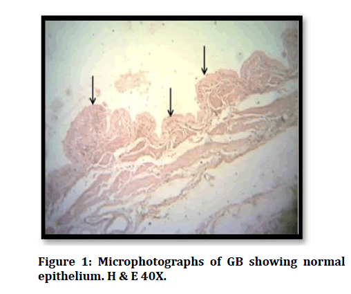
Figure 1: Microphotographs of GB showing normal epithelium. H & E 40X.
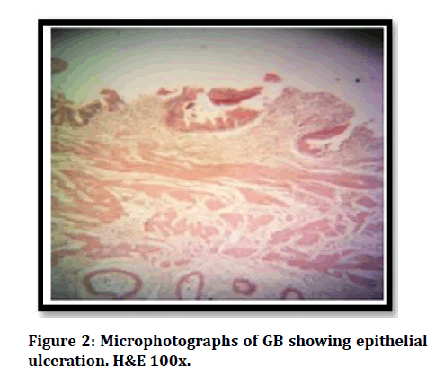
Figure 2: Microphotographs of GB showing epithelial ulceration. H&E 100x.
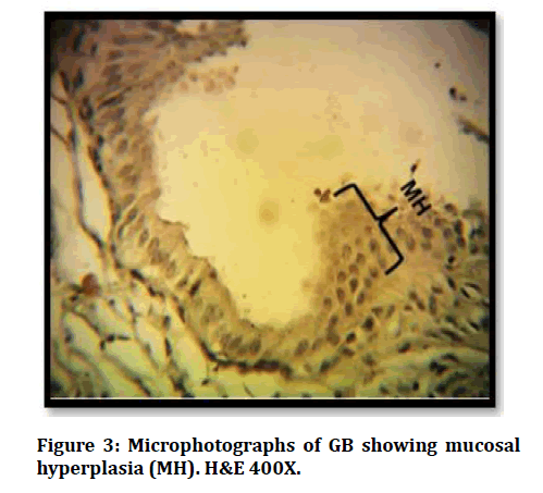
Figure 3: Microphotographs of GB showing mucosal hyperplasia (MH). H&E 400X.
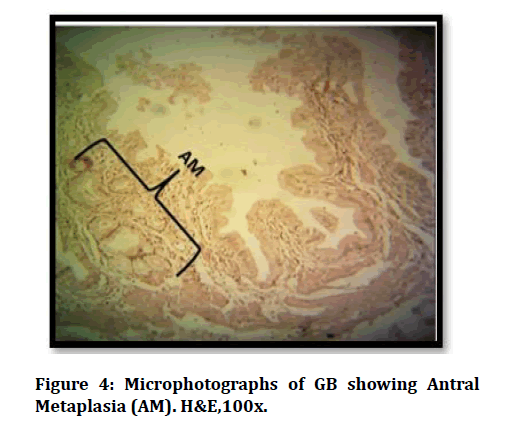
Figure 4: Microphotographs of GB showing Antral Metaplasia (AM). H&E,100x.
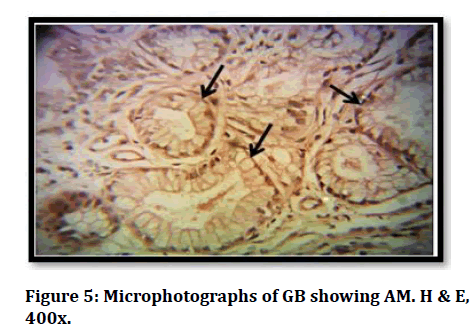
Figure 5: Microphotographs of GB showing AM. H & E, 400x.
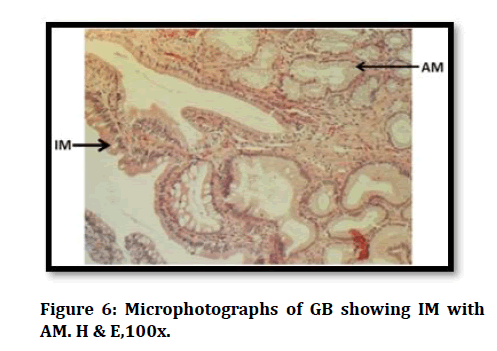
Figure 6: Microphotographs of GB showing IM with AM. H & E,100x.
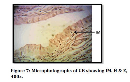
Figure 7: Microphotographs of GB showing IM. H & E, 400x.
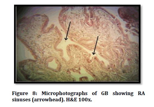
Figure 8: Microphotographs of GB showing RA sinuses (arrowhead). H&E 100x.
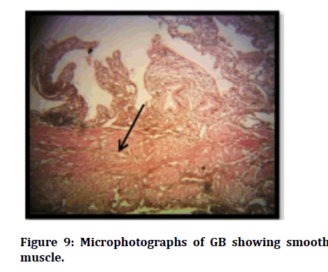
Figure 9: Microphotographs of GB showing smooth muscle.
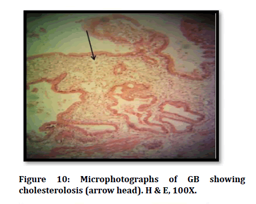
Figure 10: Microphotographs of GB showing cholesterolosis (arrow head). H & E, 100X.
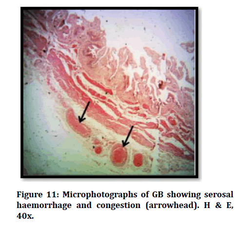
Figure 11: Microphotographs of GB showing serosal haemorrhage and congestion (arrowhead). H & E, 40x.
Discussion
In the present study, most common histological findings were haemorrhage and congestion seen in 77 specimens (74%) followed by antral metaplasia found in 76 specimens (73.1%), epithelial hyperplasia observed in 65 specimens (62.5%) and intestinal metaplasia found in 54 specimens (51.9%). Normal epithelium was found in 11 specimens (10.6%). Zaki et al. [10] studied histological changes in gallbladder mucosa of 40 patients who underwent cholecystectomy for gallstone disease with chronic cholecystitis. They found a positive correlation between gallstones and microscopic changes in the gallbladder epithelium. In the study done on 140 gallbladders electively resected for cholelithiasis by Khanna R et al [11], most common finding was epithelial hyperplasia, observed in 83 specimens (69%) followed by antral metaplasia found in 23 specimens (16.5%), epithelial ulceration found in 26 specimens (19%) and intestinal metaplasia observed in 22 specimens (15.5%) whereas normal epithelium was least frequent finding to be reported in 13 specimens (9%). Their findings were comparable to the findings of present study. Khanna et al. [11] observed that, epithelial hyperplasia was the most frequent change in 69% of gallbladder specimens with stone disease which is comparable to the present study in which it is 62.5%. Elfving et al. [12] proposed the hypothesis that primary cholelithiasis causes secondary hyperplasia because of mechanical irritation caused by the calculi. However, they found no association in the epithelial changes with respect to the character of the gallstones which contrasts with the present study. Baidya et al. [13] conducted a study on 396 cholecystectomy specimens. They observed epithelial hyperplasia to be the most frequent histological change in 183 gallbladder specimens (46.2%) followed by intestinal metaplasia in 112 specimens (28.2%). Normal epithelium was least common finding reported in 40 specimens (20%). They found positive correlation between histological changes of gallbladder mucosa and gallstones like the present study. Giri et al. [14] studied 526 cholecystectomy specimens of cholelithiasis. They reported that on histological examination, epithelial hyperplasia was observed in 195 specimens (37.07%), followed by intestinal metaplasia in 181 specimens (34.41%). Normal epithelium was the least frequent finding seen in 79 specimens (15.01%). Albores et al [15] suggested that a small number of hyperplasia of gallbladder evolves towards atypical hyperplasia and that may progress to in situ carcinoma which finally becomes invasive carcinoma. Tadashi et al. [16] studied 540 cholecystectomy specimens. They reported antral metaplasia to be the most common finding in 292 cases (54%). It was characterized by the presence of glands resembling gastric pyloric glands. It was comparable to present finding. No intestinal metaplasia was recognized, which contrasted with the present study. Khan et al. [17] conducted the study on 360 cholecystectomy specimens. They reported that pyloric gland metaplasia was the commonest finding observed in 73% of cholecystectomy specimens. Their findings were comparable to the findings of the present study. Yamamoto et al. [18] reported that metaplastic epithelium of the gallbladder is more susceptible to malignant transformation than normal. Kasprzak et al. [19] conducted a study on 37 gallbladder specimens from patients with cholelithiasis and analysed morphological alterations in the gallbladder mucosa. They reported that morphological lesions in the mucosa did not necessarily involve gallbladder epithelium. They observed absence of evident intestinal metaplasia traits in mucosa. Luma et al. [20] conducted the study on 22 cholecystectomy specimens. They stated that microscopic examination revealed cholesterolosis (66%) to be the most frequent lesion mainly in the corpus and fundus of the gallbladder. This contrasted with the findings of the present study in which cholesterolosis was present in 33.7% of specimens.
In the present study, antral metaplasia was seen in 55 cases (90.2%), epithelial ulceration in 43 cases (70.5%), Rokitansky–Aschoff sinuses in 25 cases (41%) and cholesterolosis in 24 cases (39.3%) in gallbladders with cholesterol stones. Mucosal hyperplasia was seen in 25 cases (92.6%), haemorrhage and congestion in 22 cases (81.5%), smooth muscle hypertrophy and hyperplasia in 20 cases (74.1%) and intestinal metaplasia in 18 cases (66.7%) in gallbladders with pigment stones.
Mathur et al. [21] conducted a study on 330 cholecystectomy specimens. They stated that hyperplasia (66%) and metaplasia (58%) were more common with mixed stones. Haemorrhage and congestion were the frequent change in the gallbladders with cholesterol (26.6%) and mixed (23.3%) stones reported by Zuhair et al. [22] in 30 gallbladders specimens. This contrasts with the present study in which haemorrhage and congestion was commonest finding in specimens with pigment stones (81.5%). They also reported prominent Rokitansky-Achoff sinuses mainly in the gallbladders with cholesterol stones (13.3%). They stated that formation of gallstones is usually associated with inward proliferation of the mucosa due to increase in the intraluminal pressure and weakening of the wall by distension leading to the formation of Rokitansky-Achoff sinuses. Their findings were like the results of present study, in which Rokitansky–Aschoff sinuses were found in 25 specimens (41.0%) with cholesterol gallstones followed by 4 specimens (14.8%) with pigment and 4 specimens (25.0%) with mixed gallstones. This association was found to be statistically significant (p=0.04). This indicates that cholesterol stones might be more potent stimulus than other types of stones for Rokitansky-Achoff sinuses formation. Khanna et al. [11] observed that gallbladders having pigment stones were devoid of Rokitansky-Aschoff sinuses. Similar observations were made by Baig et al. [23] in a study on 40 patients with cholelithiasis. They found a positive correlation of cholelithiasis with histological changes which is like the present study. In the present study, smooth muscle hypertrophy and hyperplasia was found predominantly in gallbladders with pigment stones (74.1%). This association was found to be statistically significant. Similarly, Zuhair M et al [22] studied 30 cholelithiasis specimens and reported that muscular hypertrophy was observed mainly in the gallbladders with pigment stone (26.6%). Similar observation was also made by Baig et al. [23]in their study of 40 cholecystectomy specimens.
Chang et al [24] studied 70 human gallbladders and observed pseudo pyloric mucous glands like the pyloric glands of the stomach within the lamina propria in majority (55.2%) of the gallbladders with cholesterol stones. They stated that it was due to the toxic effect of the lithogenic bile on the gallbladder mucosa. Intestinal metaplasia was regarded as a precancerous lesion in contrast to the pyloric gland metaplasia which is regarded as a benign condition. Fernandes et al. [25] observed that intestinal metaplasia is more common in 29 cholecystectomy specimens and regarded them as precancerous lesion, in contrast to the pyloric gland metaplasia which is regarded as a benign condition. Zuhair et al. [22] studied 30 cholelithiasis specimens, they observed that mucous gland metaplasia appeared as an oval shaped acinus within the lamina propria and is lined by pyramidal cells with flat basally located nuclei in 20 % of the gallbladder specimens having cholesterol stone. The metaplastic changes were less obvious in the gallbladders with pigment (6.6%) and mixed (13.3%) stones. These findings are like the findings of the present study.
In the present study, mucosal hyperplasia was a common histological finding to be found in 25 out of 27 cases (92.6%) with pigment stones followed by 35 out of 61 cases (57.4%) with cholesterol stones and 5 out of 16 cases (31.2%) with 94 mixed stones. The association of this mucosal response with types of stone was found to be highly significant (p=0.0001). There is an apparent increase of incidence of hyperplasia in the present study with regards to pigment stones as compared to the study conducted by Khanna R et al [11], in which incidence of hyperplasia was found to be 59% and by Zuhair et al. [22] in which incidence reported was 62.5% in gallbladders with pigment stones. They stated that cholelithiasis induces active proliferation of the epithelium in response to chronic irritation, so the surface epithelial cells look more crowded, taller than normal and have a pseudostratified appearance.
Conclusion
Gallstones are accompanied by major changes in the gallbladder epithelium. These changes were clearer in gallbladder mucosa with cholesterol stones. This may be due to the large size stones leading to more irritation to the mucosa. The predominant finding in gallbladder with cholesterol gallstones were antral metaplasia; with pigment stones was mucosa hyperplasia and with mixed stones were haemorrhage and congestion. This study also indicates that cholesterol stones might be more potent stimulus than other types of stones for Rokitansky-Achoff sinuses formation. Chronic inflammatory changes can occur in the gallbladder mucosa prior to appearance of macroscopic stones. Early detection of these microscopic features can contribute to clinical understanding and management of associated morbidities.
References
- Gray H. Gallbladder and biliary tree. In: Standring S. Gray's anatomy: The anatomical basis of clinical practice. 40th Edition. Philadelphia: Churchill Livingstone Elsevier; 2004; 1177-81.
- Kuver R, Klinkspoor JH, Osborne WR, et al. Mucous granule exocytosis and CFTR expression in gallbladder epithelium. Glycobiology 2000; 10:149- 57.
- Eroschenko VP. DiFiore's atlas of histology with functional correlations.11thed. Baltimore, Maryland, USA: Lippincot Williams & Wilkins 2008; 322.
- Henrikson RC, Mazurkiewicz JE. Methods in histology. Baltimore, Maryland, USA: Lippincott Williams & Wilkins; 1997; 512.
- Mohan H, Punia RP, Dhawan SB, et al. Morphological spectrum of gallstone disease in 1100 cholecystectomies in north India. Indian J Surg 2005; 67:140-42.
- Carey MC. Pathogenesis of gallstones. Am J Surg 1993; 165:410.
- Gad A. A histochemical study of human alimentary tract mucosubstance in health and disease: normal and tumours. B J Cancer 1969; 23:52-63.
- Thamil SR, Sinha P, Subramaniam PM, et al. A clinicopathological study of cholecystitis with special reference to analysis of cholelithiasis. Int J Basic Med Sci 2011; 2:68-72.
- Kim HJ, Kim JS, Kim KO, et al. Expression of MUC3, MUC5A, MUC6 and epidermal growth factor receptor in gallbladder epithelium according to gallstone composition. Korean J Gastroenterol 2003; 42:330-6.
- Zaki M, AI-Rafeidi A. Histological changes in the human gallbladder epithelium associated with gallstones. Oman Med J 2009; 24:8-13.
- Khanna R, Chansuria R, Kumar M, et al. Histological changes in gall bladder due to stone disease. Indian J Surg 2006; 68:201-4.
- Elfving G. Crypts and ducts in the gallbladder wall. Acta Pathol Microbiol Immunol Scand 1960; 49:1-45.
- Baidya R, Sigdel B, BaidyaNl. Histopathological changes in gallbladder mucosa associated with cholelithiasis. J Pathol Nepal 2012; 2:224-5.
- Giri S, Singh K. Spectrum of morphological alterations in cholecystectomy specimens due to cholelithiasis: A two years study. Int J Res Rev 2016; 3:42.
- Albores SJ, Molberg K, Henson DE. Unusual malignant epithelial tumours of the gallbladder. Semin Diagn Pathol 1996; 13:326-28.
- Tadashi T. Histopathologic features and frequency of gall bladder lesions in consecutive 540 cholecystectomies. Int J Clin Exp Pathol 2013; 6:91-6.
- Khan S, Jetley S, Husain M. Spectrum of histopathological lesions in cholecystectomy specimens: A study of 360 cases at a teaching hospital in South Delhi. Arch Int Surg 2013; 3:13-9.
- Yamamoto YM, Nakajo S, Tahara E. Carcinoma of the gallbladder: The correlation between histogenesis and prognosis. Virchows Arch Pathol Anat 1989; 414:83-90.
- Kasprzak A, Wojciech M, Wiesława B, et al. Histological alterations of gallbladder mucosa and selected clinical data in young patients with symptomatic gallstones. Pol J Pathol 2011; 1:41–9.
- Luma IA, Mohammed TT, Samir B, et al. Histochemical study of alkaline phosphatase enzyme in gallbladder containing cholesterol stones. Ann Coll Med Mosul 2013; 39:1-8.
- Mathur SK, Duhan A, Singh S, et al. Correlation of gallstone characteristics with mucosal changes in gallbladder. Tropical Gastroenterol 2012; 33:39-44.
- Zuhair M, Mumtaz R. Histological changes of gall bladder mucosa: correlation with various types of cholelithiasis. Iraqi J Comm Med 2011; 24:234-40.
- Baig SJ, Biswas S, Das S, et al. Histopathological changes in gallbladder mucosa in cholelithiasis: Correlation with chemical composition of gallstones. Trop Gastroenterol 2001; 23:25-7.
- Chang HJ, Suh JI, Kwon SY. Gallstone formation and gallbladder mucosal changes in mice fed a lithogenic diet. J Korean Med Sci 1999; 14:286-92.
- Fernandes JE, France MI, Suzuki RK, et al. Intestinal metaplasia in gall bladders: prevalance study. Sao Paulo Med J 2008; 126:131-51.
Author Info
Areeba Nasar1*, SadiqWadood Siddiqui2, Prerna Gupta3, Anas Ahmad Khan4 and Kamal Arora5
1Department of Anatomy, United Institute of Medical Sciences, Prayagraj, India2Department of Anatomy, Autonomous State Medical College, Pratapgarh, India
3Department of Anatomy, Integral Institute of Medical Sciences& Research, Lucknow, India
4Department of Community Medicine, United Institute of Medical Sciences, Prayagraj, India
5Department of Anatomy, SRMS IMS, Bareilly, India
Citation: AreebaNasar, SadiqWadood Siddiqui, Prerna Gupta, Anas Ahmad Khan, Neel Kamal Arora,Histological Changes of Gall Bladder Mucosa and Its Correlation with Various Types of Cholelithiasis, J Res Med Dent Sci, 2021, 9(7): 211-218
Received: 26-Jun-2021 Accepted: 12-Jul-2021
