Research - (2020) Volume 8, Issue 6
Factors Affecting Smile Attractiveness: An Eye Tracking Study
Murat Celikdelen1* and Ali Altug Bicakci2
*Correspondence: Murat Celikdelen, Private practice, Turkey, Email:
Abstract
Objective: The purpose of this study was to evaluate the effects of the buccal corridor width, gingival display, and upper midline deviation on smile aesthetics.
Methods: A frontal posed smile photograph (reference photograph) of a young female was rearranged using digital imaging software to produce artificially created photographs that exhibited different buccal corridor widths, midline deviations, and gingival displays. A total of 21 images were obtained with reference photograph. While an eye tracking device was recording, each image was evaluated and assigned aesthetic scores by 16 laypeople (8 males, 8 female). A total of 336 laypeople participated in the study. A 5-point Likert scale was used for scoring. One-way ANOVA, Welch's T-test, Tukey’s HSD test, and Tamhane's T2 test were used for statistical analysis. P values of less than 0.05 were considered statistically significant.
Results: The highest and lowest scores were assigned to 2- and 8-mm buccal corridor widths, respectively. Gingival displays of +2 and above, and +5 mm and above were scored significantly lower than the reference image (p<0.05) by female and male participants, respectively. There was a significant difference in 6-mm gingival display for males (p<0.05).
Conclusions: The different buccal corridor widths yielded no significant difference in attractiveness scores or focusing times for either gender. In the midline evaluation, even a deviation of 6 mm was not noticed by laypeople and was not found to be less attractive.
Keywords
Buccal corridor, Eye tracking, Gingival display, Midline deviation
Introduction
A smile is defined as a facial expression characterized by the upward curling of the mouth corners and is often used to express contentment, fun, or disdain. A smile is an important physical action for people because it affects the perceived attractiveness of individuals and thus plays a significant role in social interaction. In recent years, parallel to the increase in aesthetic expectations, orthodontic treatments have become more common in the society [1]. In addition to the malaligned teeth, dental conditions such as tooth impaction, supernumerary tooth, and tooth agenesis cause orthodontic problems by affecting the occlusion [2]. However, these are not the only reasons for applying to the orthodontists at the present time. Smile aesthetics affecting both personal and social attractiveness is a major concern of patients and one of the main reasons for applying to clinicians [3]. Therefore, patients undergoing orthodontic treatment have started to desire not only well-aligned teeth but also an attractive smile, and this topic has become a crucial criterion for whether the treatment is successful [4]. Parallel to this situation, the effect of smile on facial appearance has become an increasingly relevant topic in current orthodontic studies [5,6].
Beauty is a subjective phenomenon, and the statement "Beauty is in the eye of the beholder" succinctly expresses this situation. It is not possible to describe the ideal smile because individual variations, such as age, gender, or culture, can be decisive factors in beauty perception. Therefore, the researchers focused on a balanced smile rather than the ideal smile. A balanced smile results from the relationship among different smile components and requires understanding the elements that provide a balance between the teeth and soft tissues [7,8]. These components are the buccal corridor width, gingival display, and dental midline [9-13]. Furthermore, Sabri listed the following eight components as the major factors for a balanced smile: the lip line, smile arc, upper lip curvature, lateral negative space, smile symmetry, frontal occlusal plane, dental components, and gingival components [14].
Various studies have evaluated smile aesthetics [11,15,16] by examining photographs constructed using photo-editing software and making gradual changes to dental and facial structures in the photographs. In these score-based studies, participants rated the attractiveness level of photographs. However, no objective method has been developed for assessing whether participants notice the gradual changes, whether the changes affect their scores, or on which areas of the photographs participants focus.
Eye tracking is an objective method for detecting where a person's visual attention is concentrated. Eye trackers consisting of a camera and video-processing software obtain a quantitative measure of a person’s real-time visual attention [17]. The pupil is tracked using infrared or near-infrared light, and corneal reflection is utilized for visual attention recording [18].
In the present study, in addition to deriving and calculating attractiveness scores, we tried to determine whether there would be changes in focusing times depending on the variables through eye tracking device.
Materials and Methods
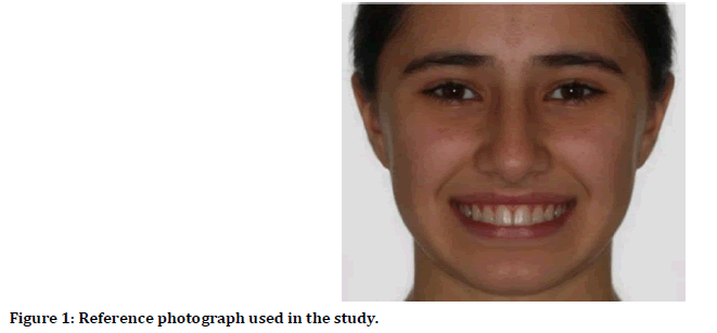
The frontal smile photographs of 15 female patients who had no striking elements on their face, such as asymmetry or scars, were obtained from department archive for inclusion in this study. The photographs were viewed by eight orthodontists who selected the photograph that they believed most represented the general facial features of our society. The photograph selected most frequently was used for this study as reference photo (Figure 1). Artificial photographs with a manipulated buccal corridor width, gingival display, and upper dental midline were created by using the reference photograph and Adobe Photoshop CS2 Software (Adobe Systems, San Jose, CA, USA).

Figure 1. Reference photograph used in the study.
Changes in buccal corridor width, gingival display, and dental midline
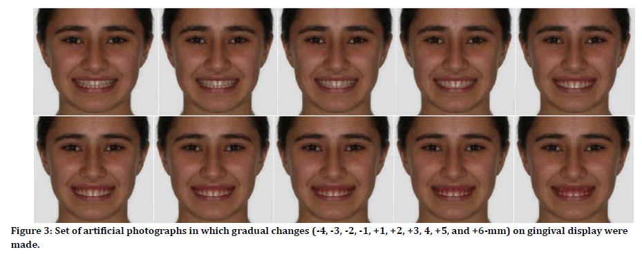
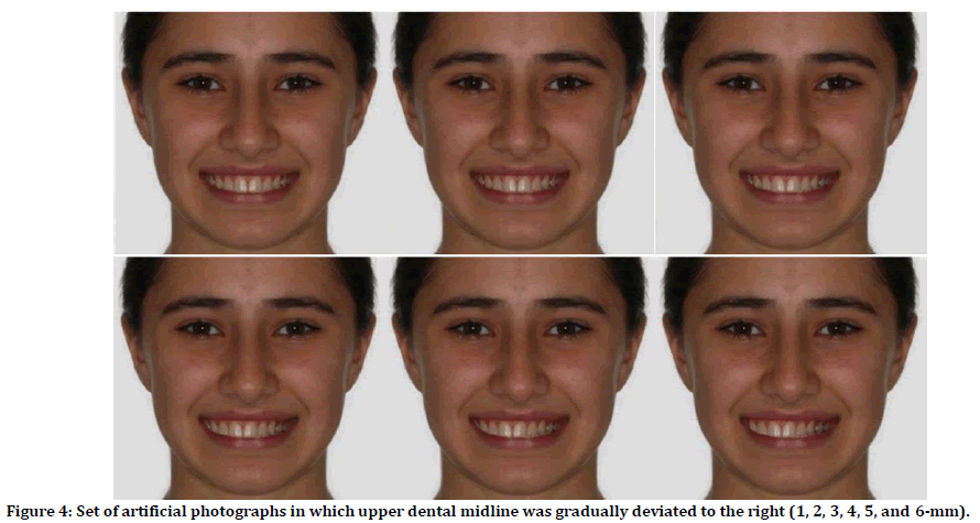
Four artificial photographs displaying 0-, 4-, 6-, and 8-mm buccal corridor widths were created by moving the buccolingual positions of the canine, premolar, and molar teeth on both sides (Figure 2). In addition, 10 artificial photographs exhibiting different gingival displays ranging from -4 mm to +6 mm were created according to the reference photograph (Figure 3). Finally, six artificial photographs displaying 1-, 2-, 3-, 4-, 5-, and 6-mm deviations were created by moving the upper dental midline to the right (Figure 4). During this procedure, the buccolingual positions of the left premolar and molar were adjusted and the natural dental arch was maintained. Namely, a total of 21 images were obtained with reference photograph.

Figure 2. Set of artificial photographs in which gradual changes (0, 4, 6, and 8-mm) on buccal corridor width were made.

Figure 3. Set of artificial photographs in which gradual changes (-4, -3, -2, -1, +1, +2, +3, 4, +5, and +6-mm) on gingival display were made.

Figure 4. Set of artificial photographs in which upper dental midline was gradually deviated to the right (1, 2, 3, 4, 5, and 6-mm).
An eye tracker (Smarttek Eye Navigator; Smarttek Software and Industrial Automation Company, Istanbul, Turkey) and 19.5'' computer monitor (Hewlett-Packard Company, Palo Alto, CA, USA) were used in this study. The system was placed on a table at the eye level of the participants. Each participant was seated 60 cm away from the computer screen and calibrated according to a 9-
point calibration with the eye tracker.
Each photograph was viewed and scored by 16 participants (eight males, eight female). Each layperson examined just 1 image. Since 21 photographs were used in the study, a total of 336 individuals (168 male and 168 female) participated in this study. They were included according to the following criteria: (A) being aged 18–25 years, (B) having no training in dentistry, (C) having no aesthetic medical or dental history, (D) having no mental or nervous disease, and (E) having no vision problems.
The participants were informed about how the program would be run and what they should do. They were told that a photograph would appear on the screen and disappear after 6 seconds, and that they were to examine the photograph during this interval. A 5-
point Likert scale was used for scoring as follows: 1=Strongly unattractive, 2=Unattractive, 3=Undecided, 4=Attractive, and 5=Strongly attractive.
After the presentation was completed, data recorded using the Smarttek Eye Navigator Studio software (Smarttek Software and Industrial Automation Company, Istanbul, Turkey) were analyzed. The lower limit of the focusing time was set at 50 milliseconds, and all areas where each participant focused for 50 milliseconds or more were identified. The photographs were divided into three main zones with the help of the used software: eye, nose, and mouth. For each zone, the total time of the participant's focus was calculated.
Statistical analysis
At the beginning of this study, it was determined that sample size to be used in the study yielded a power of 0.90 with a nondirectional alpha risk of 0.05 and an effect size of 0.35. One-way ANOVA was used to compare the means of quantitative variables between the groups. According to the homogeneity of variances, Welch's T-test, Tukey’s HSD test, and Tamhane's T2 test were used for statistical analysis. P values of less than 0.05 were considered statistically significant. Statistical analysis was performed using the Statistical Package for the Social Sciences (IBM SPSS Statistics for Windows, Version 19.0. Armonk, NY).
Results
Evaluation of changes in buccal corridor width
For the females, there was no statistically significant difference among the ages, scores, and focusing times for the eye, nose, or mouth zones. Similarly, there was no significant difference for these parameters for the males (Table 1).
| Number of subjects | Mean ± Standard deviation | F | P | |||
|---|---|---|---|---|---|---|
| Female | Age (year) | 0-mm | 8 | 19.75 ± 1.39 | 0.212 | 0.93 |
| 2-mmR | 8 | 20.25 ± 1.58 | ||||
| 4-mm | 8 | 19.75 ± 2.31 | ||||
| 6-mm | 8 | 19.75 ± 1.58 | ||||
| 8-mm | 8 | 20.38 ± 2.45 | ||||
| Score | 0-mm | 8 | 4.13 ± 0.83 | 1.236 | 0.314 | |
| 2-mmR | 8 | 4.38 ± 0.52 | ||||
| 4-mm | 8 | 3.50 ± 1.31 | ||||
| 6-mm | 8 | 3.88 ± 0.99 | ||||
| 8-mm | 8 | 3.50 ± 1.07 | ||||
| Focusing time on eyes (millisecond) | 0-mm | 8 | 1144.75 ± 994.29 | 2.071 | 0.106 | |
| 2-mmR | 8 | 1268.13 ± 1034..92 | ||||
| 4-mm | 8 | 1928.75 ± 693.21 | ||||
| 6-mm | 8 | 905.25 ± 569.63 | ||||
| 8-mm | 8 | 839.13 ± 881.87 | ||||
| Focusing time on nose (millisecond) | 0-mm | 8 | 575 ± 363.32 | 0.37 | 0.828 | |
| 2-mmR | 8 | 772.25 ± 815.42 | ||||
| 4-mm | 8 | 815.5 ± 505.62 | ||||
| 6-mm | 8 | 590.75 ± 355.77 | ||||
| 8-mm | 8 | 607.25 ± 440.64 | ||||
| Focusing time on mouth (millisecond) | 0-mm | 8 | 1124.25 ± 1093.88 | 1.156 | 0.347 | |
| 2-mmR | 8 | 733.13 ± 749.06 | ||||
| 4-mm | 8 | 670.13 ± 680.15 | ||||
| 6-mm | 8 | 1201.38 ± 927.11 | ||||
| 8-mm | 8 | 1482.38 ± 947.99 | ||||
| Male | Age (year) | 0-mm | 8 | 21.38 ± 2.39 | ||
| 2-mmR | 8 | 20.38 ± 2.2 | ||||
| 4-mm | 8 | 21.25 ± 1.67 | ||||
| 6-mm | 8 | 21.88 ± 1.36 | ||||
| 8-mm | 8 | 22.00 ± 2.14 | ||||
| Score | 0-mm | 8 | 3.38 ± 1.06 | 1.34 | 0.275 | |
| 2-mmR | 8 | 4 ± 0.76 | ||||
| 4-mm | 8 | 3.13 ± 0.83 | ||||
| 6-mm | 8 | 3.13 ± 1.25 | ||||
| 8-mm | 8 | 3 ± 0.93 | ||||
| Focusing time on eyes (millisecond) | 0-mm | 8 | 747.75 ± 746 | 1.462 | 0.235 | |
| 2-mmR | 8 | 1489.25 ± 480.4 | ||||
| 4-mm | 8 | 1819.25 ± 1342.62 | ||||
| 6-mm | 8 | 1222.38 ± 921.81 | ||||
| 8-mm | 8 | 1041.25 ± 1090.61 | ||||
| Focusing time on nose (millisecond) | 0-mm | 8 | 524 ± 524.29 | 0.952 | 0.446 | |
| 2-mmR | 8 | 542.63 ± 623.27 | ||||
| 4-mm | 8 | 484.25 ± 384.53 | ||||
| 6-mm | 8 | 246.38 ± 275.63 | ||||
| 8-mm | 8 | 241 ± 269.92 | ||||
| Focusing time on mouth (millisecond) | 0-mm | 8 | 1039.13 ± 545.98 | 2.354 | 0.073 | |
| 2-mmR | 8 | 770.75 ± 730.6 | ||||
| 4-mm | 8 | 494.13 ± 415.13 | ||||
| 6-mm | 8 | 1240.5 ± 1014.21 | ||||
| 8-mm | 8 | 1667.5 ± 1176.69 | ||||
R Reference photograph
Table 1: Evaluation of quantitative variables in different buccal corridor widths.
Evaluation of the changes in gingival display
For the females, there was no significant difference between the ages and focusing times regarding the different zones. However, a significant difference in scores was determined. Images of +6- and
+4-mm gingival displays were scored significantly lower than images of +1-, 0- (reference photograph), -2-, -3-, and -4-mm
gingival displays. Additionally, images of +5-, +3-, and +2-mm gingival displays were also scored significantly lower than the reference photograph (0-mm) (Table 2).
| Number of subjects | Mean ± Standard deviation | F | P | |||||||||||||
|---|---|---|---|---|---|---|---|---|---|---|---|---|---|---|---|---|
| Age (year) | -4 mm | 8 | 20 ± 1.31 | 0.245 | 0.99 | |||||||||||
| -3 mm | 8 | 21 ± 1.51 | ||||||||||||||
| -2 mm | 8 | 20.38 ± 1.6 | ||||||||||||||
| -1 mm | 8 | 20.38 ± 1.51 | ||||||||||||||
| 0 mmR | 8 | 20.25 ± 1.58 | ||||||||||||||
| +1 mm | 8 | 20.5 ± 1.41 | ||||||||||||||
| +2 mm | 8 | 20.13 ± 1.64 | ||||||||||||||
| +3 mm | 8 | 20.25 ± 1.49 | ||||||||||||||
| +4 mm | 8 | 20.5 ± 1.77 | ||||||||||||||
| +5 mm | 8 | 20 ± 2.2 | ||||||||||||||
| +6 mm | 8 | 20.38 ± 1.41 | ||||||||||||||
| Score | -4 mm | 8 | 4.13 ± 0.64 | 7.24 | <0.001* | -4 mm | -3 mm | -2 mm | -1 mm | 0 mmR | +1 mm | +2 mm | +3 mm | +4 mm | +5 mm | |
| -3 mm | 8 | 4.25 ± 0.71 | - | - | - | - | - | - | - | - | - | - | -4 mm | |||
| -2 mm | 8 | 4.25 ± 0.71 | 1 | - | - | - | - | - | - | - | - | - | -3 mm | |||
| -1 mm | 8 | 3.5 ± 0.76 | 1 | 1 | - | - | - | - | - | - | - | - | -2 mm | |||
| 0 mmR | 8 | 4.38 ± 0.52 | 0.921 | 0.787 | 0.787 | - | - | - | - | - | - | - | -1 mm | |||
| +1 mm | 8 | 4.25 ± 1.04 | 1 | 1 | 1 | 0.596 | - | - | - | - | - | - | 0 mm | |||
| +2 mm | 8 | 2.88 ± 0.83 | 1 | 1 | 1 | 0.787 | 1 | - | - | - | - | - | +1 mm | |||
| +3 mm | 8 | 2.88 ± 0.83 | 0.122 | 0.058 | 0.058 | 0.921 | 0.025** | 0.058 | - | - | - | - | +2 mm | |||
| +4 mm | 8 | 2.13 ± 0.99 | 0.122 | 0.058 | 0.058 | 0.921 | 0.025** | 0.058 | 1 | - | - | - | +3 mm | |||
| +5 mm | 8 | 2.88 ± 1.13 | <0.001** | <0.001** | <0.001** | 0.058 | <0.001** | <0.001** | 0.787 | 0.787 | - | - | +4 mm | |||
| +6 mm | 8 | 2.63 ± 0.92 | 0.122 | 0.058 | 0.058 | 0.921 | 0.025** | 0.058 | 1 | 1 | 0.787 | - | +5 mm | |||
| 0.025** | 0.010** | 0.010** | 0.596 | 0.004** | 0.010** | 1 | 1 | 0.982 | 1 | +6 mm | ||||||
| Focusing time on eyes (millisecond) | -4 mm | 8 | 1570.88 ± 899.9 | 0.313 | 0.976 | |||||||||||
| -3 mm | 8 | 1505.5 ± 721.32 | ||||||||||||||
| -2 mm | 8 | 1163.25 ± 923.41 | ||||||||||||||
| -1 mm | 8 | 1376.63 ± 722.41 | ||||||||||||||
| 0 mmR | 8 | 1268.13 ± 1034.92 | ||||||||||||||
| +1 mm | 8 | 1544.5 ± 919.56 | ||||||||||||||
| +2 mm | 8 | 1428.25 ± 1053.97 | ||||||||||||||
| +3 mm | 8 | 1376 ± 1319.87 | ||||||||||||||
| +4 mm | 8 | 1122.63 ± 855.17 | ||||||||||||||
| +5 mm | 8 | 1129.63 ± 1057.43 | ||||||||||||||
| +6 mm | 8 | 1040.5 ± 636.05 | ||||||||||||||
| Focusing time on nose (millisecond) | -4 mm | 8 | 345.13 ± 480.68 | 1.035 | 0.423 | |||||||||||
| -3 mm | 8 | 693.5 ± 344.53 | ||||||||||||||
| -2 mm | 8 | 591.5 ± 557.52 | ||||||||||||||
| -1 mm | 8 | 551.13 ± 824.24 | ||||||||||||||
| 0 mmR | 8 | 772.25 ± 815.42 | ||||||||||||||
| +1 mm | 8 | 226.25 ± 325.06 | ||||||||||||||
| +2 mm | 8 | 383 ± 467.59 | ||||||||||||||
| +3 mm | 8 | 161.75 ± 239.57 | ||||||||||||||
| +4 mm | 8 | 686 ± 708.97 | ||||||||||||||
| +5 mm | 8 | 506.63 ± 617.71 | ||||||||||||||
| +6 mm | 8 | 368.38 ± 328.79 | ||||||||||||||
| Focusing time on mouth (millisecond) | -4 mm | 8 | 1220.25 ± 1244.27 | 1.528 | 0.146 | |||||||||||
| -3 mm | 8 | 860 ± 601.79 | ||||||||||||||
| -2 mm | 8 | 839.88 ± 653.72 | ||||||||||||||
| -1 mm | 8 | 1361.13 ± 1024.57 | ||||||||||||||
| 0 mmR | 8 | 733.13 ± 749.06 | ||||||||||||||
| +1 mm | 8 | 918.63 ± 763.27 | ||||||||||||||
| +2 mm | 8 | 1199.13 ± 1287.36 | ||||||||||||||
| +3 mm | 8 | 1036.5 ± 1356.25 | ||||||||||||||
| +4 mm | 8 | 976.75 ± 924.77 | ||||||||||||||
| +5 mm | 8 | 1826.75 ± 1231.51 | ||||||||||||||
| +6 mm | 8 | 2197.13 ± 1142.9 | ||||||||||||||
**Significant at P<0.05 by Tukey HSD test
R Reference photograph
Table 2: Evaluation of different gingival visibilities in female participants.
For the males, there were significant differences in the scores and
focusing times concerning the oral zone. There was no significant difference between the ages. Images with +6- and +5-mm gingival displays were scored significantly lower than the reference photograph (0-mm). The focusing time for the +6-mm gingival
displays for the mouth was found to be significantly higher than that for images that had +1- and 0-mm (reference photograph) gingival displays (Table 3).
| Number of subjects | Mean ± Standard deviation | F | P | |||||||||||||
|---|---|---|---|---|---|---|---|---|---|---|---|---|---|---|---|---|
| Age (year) | -4 mm | 8 | 21.38 ± 1.77 | 1.062 | 0.401 | |||||||||||
| -3 mm | 8 | 20.88 ± 2.03 | ||||||||||||||
| -2 mm | 8 | 23.25 ± 1.69 | ||||||||||||||
| -1 mm | 8 | 22 ± 1.51 | ||||||||||||||
| 0 mmR | 8 | 20.38 ± 2.2 | ||||||||||||||
| +1 mm | 8 | 21.5 ± 2.39 | ||||||||||||||
| +2 mm | 8 | 20.75 ± 1.83 | ||||||||||||||
| +3 mm | 8 | 22 ± 2.39 | ||||||||||||||
| +4 mm | 8 | 20.88 ± 1.46 | ||||||||||||||
| +5 mm | 8 | 22 ± 2.67 | ||||||||||||||
| +6 mm | 8 | 21 ± 1.77 | ||||||||||||||
| Score | -4 mm | 8 | 3.25 ± 1.16 | 2.793 | 0.005* | -4 mm | -3 mm | -2 mm | -1 mm | 0 mmR | +1 mm | +2 mm | +3 mm | +4 mm | +5 mm | |
| -3 mm | 8 | 3 ± 1.07 | - | - | - | - | - | - | - | - | - | - | -4 mm | |||
| -2 mm | 8 | 3.75 ± 0.71 | 1 | - | - | - | - | - | - | - | - | - | -3 mm | |||
| -1 mm | 8 | 3.25 ± 0.89 | 0.995 | 0.916 | - | - | - | - | - | - | - | - | -2 mm | |||
| 0 mmR | 8 | 4 ± 0.76 | 1 | 1 | 0.995 | - | - | - | - | - | - | - | -1 mm | |||
| +1 mm | 8 | 3.75 ± 1.04 | 0.916 | 0.652 | 1 | 0.916 | - | - | - | - | - | - | 0 mm | |||
| +2 mm | 8 | 3.13 ± 1.25 | 0.995 | 0.916 | 1 | 0.995 | 1 | - | - | - | - | - | +1 mm | |||
| +3 mm | 8 | 2.63 ± 1.06 | 1 | 1 | 0.974 | 1 | 0.806 | 0.974 | - | - | - | - | +2 mm | |||
| +4 mm | 8 | 2.75 ± 1.16 | 0.974 | 1 | 0.48 | 0.974 | 0.2 | 0.48 | 0.995 | - | - | - | +3 mm | |||
| +5 mm | 8 | 2.25 ± 0.89 | 0.995 | 1 | 0.652 | 0.995 | 0.323 | 0.652 | 1 | 1 | - | - | +4 mm | |||
| +6 mm | 8 | 2.25 ± 0.89 | 0.652 | 0.916 | 0.114 | 0.652 | 0.030** | 0.114 | 0.806 | 1 | 0.995 | +5 mm | ||||
| 0.652 | 0.916 | 0.114 | 0.652 | 0.030** | 0.114 | 0.806 | 1 | 0.995 | 1 | +6 mm | ||||||
| Focusing time on eyes (millisecond) | -4 mm | 8 | 1339 ± 1114.4 | |||||||||||||
| -3 mm | 8 | 959.5 ± 888.05 | ||||||||||||||
| -2 mm | 8 | 939.75 ± 634.55 | ||||||||||||||
| -1 mm | 8 | 1390.5 ± 847.68 | ||||||||||||||
| 0 mmR | 8 | 1489.25480.4 | ||||||||||||||
| +1 mm | 8 | 1359.75 ± 1144.96 | ||||||||||||||
| +2 mm | 8 | 1051.38 ± 569.14 | ||||||||||||||
| +3 mm | 8 | 1030.13 ± 790.92 | ||||||||||||||
| +4 mm | 8 | 1303.88 ± 632.2 | ||||||||||||||
| +5 mm | 8 | 653.25 ± 455.39 | ||||||||||||||
| +6 mm | 8 | 605.13 ± 608.05 | ||||||||||||||
| Focusing time on nose (millisecond) | -4 mm | 8 | 413.25 ± 571.93 | 0.625 | 0.78 | |||||||||||
| -3 mm | 8 | 325.63 ± 431.22 | ||||||||||||||
| -2 mm | 8 | 342.25 ± 361.95 | ||||||||||||||
| -1 mm | 8 | 376.63 ± 164.76 | ||||||||||||||
| 0 mmR | 8 | 542.63 ± 623.27 | ||||||||||||||
| +1 mm | 8 | 395.13 ± 362.03 | ||||||||||||||
| +2 mm | 8 | 554.25 ± 393.36 | ||||||||||||||
| +3 mm | 8 | 364.13 ± 305.07 | ||||||||||||||
| +4 mm | 8 | 477 ± 460.05 | ||||||||||||||
| +5 mm | 8 | 1125.5 ± 1006.82 | ||||||||||||||
| +6 mm | 8 | 291.63 ± 350.66 | ||||||||||||||
| Focusing time on mouth (millisecond) | -4 mm | 8 | 1394.38 ± 1470.70 | 1.99 | 0.046* | -4 mm | -3 mm | -2 mm | -1 mm | 0 mmR | +1 mm | +2 mm | +3 mm | +4 mm | +5 mm | |
| -3 mm | 8 | 1455.88 ± 1086.9 | - | - | - | - | - | - | - | - | - | - | -4 mm | |||
| -2 mm | 8 | 1088.5 ± 973.56 | 1 | - | - | - | - | - | - | - | - | - | -3 mm | |||
| -1 mm | 8 | 1091.63 ± 935.68 | 1 | 1 | - | - | - | - | - | - | - | - | -2 mm | |||
| 0 mmR | 8 | 770.75 ± 730.6 | 1 | 1 | 1 | - | - | - | - | - | - | - | -1 mm | |||
| +1 mm | 8 | 904.75 ± 1052.82 | 0.975 | 0.952 | 1 | 1 | - | - | - | - | - | - | 0 mm | |||
| +2 mm | 8 | 1314.75 ± 375.53 | 0.996 | 0.99 | 1 | 1 | 1 | - | - | - | - | - | +1 mm | |||
| +3 mm | 8 | 1132.88 ± 828.32 | 1 | 1 | 1 | 1 | 0.991 | 0.999 | - | - | - | - | +2 mm | |||
| +4 mm | 8 | 1335 ± 831.12 | 1 | 1 | 1 | 1 | 1 | 1 | 1 | - | - | - | +3 mm | |||
| +5 mm | 8 | 1486.88 ± 1277.17 | 1 | 1 | 1 | 1 | 0.988 | 0.999 | 1 | 1 | - | - | +4 mm | |||
| +6 mm | 8 | 2672.13 ± 1033.67 | 1 | 1 | 0.999 | 0.999 | 0.937 | 0.985 | 1 | 1 | 1 | - | +5 mm | |||
| 0.292 | 0.362 | 0.075 | 0.076 | 0.012** | 0.027** | 0.214 | 0.094 | 0.233 | 0.401 | +6 mm | ||||||
**Significant at P<0.05 by Tukey HSD test
R Reference photograph
Table 3: Evaluation of different gingival visibilities in male participants.
Evaluation of changes in midline
For the females, there was a significant difference in the focusing times for the oral zone. There was no difference for the other parameters. The focusing times for the mouth zone were found to be significantly higher in the photograph which had a 5-mm
midline deviation (Table 4).
| Number of subjects | Mean ± Standard deviation | F | P | |||||||||
|---|---|---|---|---|---|---|---|---|---|---|---|---|
| Age (year) | 0-mmR | 8 | 20.25 ± 1.58 | 0.982 | 0.448 | |||||||
| 1-mm | 8 | 20.13 ± 1.25 | ||||||||||
| 2-mm | 8 | 21.88 ± 1.81 | ||||||||||
| 3-mm | 8 | 20.88 ± 2.47 | ||||||||||
| 4-mm | 8 | 21.13 ± 1.36 | ||||||||||
| 5-mm | 8 | 20.63 ± 1.6 | ||||||||||
| 6-mm | 8 | 20.63 ± 1.41 | ||||||||||
| Score | 0-mmR | 8 | 4.38 ± 0.52 | 2.154 | 0.064 | |||||||
| 1-mm | 8 | 3.88 ± 0.99 | ||||||||||
| 2-mm | 8 | 3.5 ± 1.31 | ||||||||||
| 3-mm | 8 | 3.75 ± 1.28 | ||||||||||
| 4-mm | 8 | 3.25 ± 1.16 | ||||||||||
| 5-mm | 8 | 2.88 ± 1.25 | ||||||||||
| 6-mm | 8 | 2.75 ± 1.04 | ||||||||||
| Focusing time on eyes (millisecond) | 0-mmR | 8 | 1268.13 ± 1034.92 | 0.716 | 0.639 | |||||||
| 1-mm | 8 | 1003.25 ± 623.35 | ||||||||||
| 2-mm | 8 | 1814.25 ± 1107.51 | ||||||||||
| 3-mm | 8 | 1525.38 ± 1018.32 | ||||||||||
| 4-mm | 8 | 1189.5 ± 1299.45 | ||||||||||
| 5-mm | 8 | 1229.13 ± 819.34 | ||||||||||
| 6-mm | 8 | 1788.63 ± 1251.74 | ||||||||||
| Focusing time on nose (millisecond) | 0-mmR | 8 | 772.25 ± 815.42 | 1.047 | 0.407 | |||||||
| 1-mm | 8 | 615.25 ± 520.96 | ||||||||||
| 2-mm | 8 | 411.75 ± 487.41 | ||||||||||
| 3-mm | 8 | 579.5 ± 672.78 | ||||||||||
| 4-mm | 8 | 439.75 ± 418.95 | ||||||||||
| 5-mm | 8 | 174.38 ± 209.81 | ||||||||||
| 6-mm | 8 | 302.88 ± 574.47 | ||||||||||
| Focusing time on mouth (millisecond) | 0-mmR | 8 | 733.13 ± 749.06 | 3.23 | 0.009* | 0-mmR | 1-mm | 2-mm | 3-mm | 4-mm | 5-mm | |
| 1-mm | 8 | 613.25 ± 808.79 | - | - | - | - | - | - | 0-mm | |||
| 2-mm | 8 | 1183.63 ± 1402.88 | 0.812 | - | - | - | - | - | 1-mm | |||
| 3-mm | 8 | 587.88 ± 457.07 | 0.372 | 0.259 | - | - | - | - | 2-mm | |||
| 4-mm | 8 | 1259 ± 923.2 | 0.773 | 0.96 | 0.239 | - | - | - | 3-mm | |||
| 5-mm | 8 | 2363.13 ± 1295.5 | 0.298 | 0.203 | 0.881 | 0.186 | - | - | 4-mm | |||
| 6-mm | 8 | 703.88 ± 1037.14 | 0.002** | 0.001** | 0.022** | 0.001** | 0.031** | - | 5-mm | |||
| 0.954 | 0.857 | 0.342 | 0.817 | 0.272 | 0.002** | 6-mm | ||||||
**Significant at P<0.05 by Tukey HSD test
R Reference photograph
Table 4: Evaluation of midline deviations in female participants.
For the males, there was a significant difference in the focusing times regarding the eye zone. There was no difference for the other parameters. In the photograph that had a 4-mm midline deviation, the focusing times for the eye zone were found to be significantly lower than those for the reference photograph (Table 5).
| Number of subjects | Mean ± Standard deviation | F | P | |||||||||
|---|---|---|---|---|---|---|---|---|---|---|---|---|
| Age (year) | 0-mmR | 8 | 20.38 ± 2.2 | 1.281 | 0.308 | |||||||
| 1-mm | 8 | 20.63 ± 0.92 | ||||||||||
| 2-mm | 8 | 21.38 ± 2.39 | ||||||||||
| 3-mm | 8 | 21.75 ± 2.05 | ||||||||||
| 4-mm | 8 | 21.25 ± 0.71 | ||||||||||
| 5-mm | 8 | 20.25 ± 1.04 | ||||||||||
| 6-mm | 8 | 20.25 ± 1.83 | ||||||||||
| Score | 0-mmR | 8 | 4 ± 0.76 | 1.828 | 0.113 | |||||||
| 1-mm | 8 | 3.13 ± 0.83 | ||||||||||
| 2-mm | 8 | 3.38 ± 0.74 | ||||||||||
| 3-mm | 8 | 3.63 ± 0.74 | ||||||||||
| 4-mm | 8 | 3.5 ± 1.31 | ||||||||||
| 5-mm | 8 | 2.75 ± 1.28 | ||||||||||
| 6-mm | 8 | 2.75 ± 0.89 | ||||||||||
| Focusing time on eyes (millisecond) | 0-mmR | 8 | 1489.25 ± 480.4 | 3.436 | 0.016* | 0-mmR | 1-mm | 2-mm | 3-mm | 4-mm | 5-mm | |
| 1-mm | 8 | 916 ± 806.76 | - | - | - | - | - | - | 0-mm | |||
| 2-mm | 8 | 1013.38 ± 689.71 | 0.916 | - | - | - | - | - | 1-mm | |||
| 3-mm | 8 | 1263.88 ± 1142.52 | 0.952 | 1 | - | - | - | - | 2-mm | |||
| 4-mm | 8 | 474.63 ± 430.91 | 1 | 1 | 1 | - | - | - | 3-mm | |||
| 5-mm | 8 | 1828.75 ± 1464.92 | 0.012** | 0.991 | 0.849 | 0.893 | - | - | 4-mm | |||
| 6-mm | 8 | 1164.88 ± 620.93 | 1 | 0.968 | 0.986 | 1 | 0.535 | - | 5-mm | |||
| 0.998 | 1 | 1 | 1 | 0.391 | 0.999 | 6-mm | ||||||
| Focusing time on nose (millisecond) | 0-mmR | 8 | 542.63 ± 623.27 | 1.282 | 0.307 | |||||||
| 1-mm | 8 | 700.13 ± 706.04 | ||||||||||
| 2-mm | 8 | 298.5 ± 151.84 | ||||||||||
| 3-mm | 8 | 670 ± 565.8 | ||||||||||
| 4-mm | 8 | 425.25 ± 236.7 | ||||||||||
| 5-mm | 8 | 259.75 ± 188.29 | ||||||||||
| 6-mm | 8 | 295.38 ± 274.14 | ||||||||||
| Focusing time on mouth (millisecond) | 0-mmR | 8 | 770.75 ± 730.6 | 2.433 | 0.06 | |||||||
| 1-mm | 8 | 614 ± 426.35 | ||||||||||
| 2-mm | 8 | 1440.38 ± 669.81 | ||||||||||
| 3-mm | 8 | 995.63 ± 738.55 | ||||||||||
| 4-mm | 8 | 2065.63 ± 1276.47 | ||||||||||
| 5-mm | 8 | 1405.13 ± 1693.53 | ||||||||||
| 6-mm | 8 | 1246.63 ± 1291.51 | ||||||||||
**Significant at P<0.05 by Tamhane's T2 test
R Reference photograph
Table 5: Evaluation of midline deviations in male participants.
Discussion
In most studies evaluating smile aesthetics, new images have been created by making gradual changes to the dental and environmental structures of smile photographs by using computer software. Each participant scored such photographs according to their attractiveness levels [11,15,16]. Thus, it was possible to determine the attractiveness level of each photograph, but it could not be determined whether the participants noticed the changes to be evaluated in the photographs. In the other part of the studies, the participants were asked to score smile photographs that belonged to different patients and the norms of the ideal smile were tried to determine by evaluating the top-rated photographs [19,20]. In studies using different patients' photographs, variables that may affect smile aesthetics may differ for each photograph. This makes it impossible to evaluate the effect of the examined factor on smile aesthetics by isolating it from other variables. Therefore, our study was carried out using an eye tracking device combined with a scoring method to investigate the effects of the gingival display, buccal corridor width, and dental midline on smile aesthetics. Because we presumed that we could make a more accurate comparison by making changes only to the examined item and by keeping other variables constant, just one patient’s photograph was used.
Studies using images created by making changes in the same reference photograph have derived contradictory outcomes in eye tracking results. Because of the types of methods used in these studies, their participants might have attempted to determine changes in the photographs and, thus, might have focused on the changes. Barton et al. stated that if participants are asked to reevaluate the images which have been evaluated previously, there will be changes at the eye movement pattern [21]. To eliminate this drawback in our study, each participant evaluated only one photograph. Thus, conflicting results for focusing times caused by participants’ attempting to recognize changes were avoided. Wang et al. used the same method [22].
Having been used in different fields such as physiology, neurology, psychology, and plastic surgery, eye tracking is a relatively new method in orthodontics [22-25]. In the cited studies, the duration of displaying photographs to participants has varied. Richards et al., Dindaroglu et al. [23] and Wang et al. [22] have respectively used 3, 4, and 10 seconds for the photograph display time. In consistent with other studies, the display time for the photographs used in our study was 6 seconds. In addition, previous researchers have divided the examined face into various regions for making calculations. Whereas Johnson et al.24 used nine regions, Dindaroglu et al. used four zones: eye, nose, upper lip, lower lip–
chin tip. Wang et al. [22] divided the face into three parts: eye, nose, and mouth. Similarly, we designated three zones of the face for our study.
Based on the present results, the photograph exhibiting a 2-mm buccal corridor width (reference photograph) reached the highest score, followed sequentially by the photographs having 0-, 6-, 4-,
and 8-mm buccal corridor widths (Table 1). When the focusing times for the mouth were evaluated, the images receiving the longest and shortest focus were in the photographs having 8- and 4-mm buccal corridor widths, respectively. Even if the 4-mm
photograph were an exception, the focusing time for the mouth increased as the score decreased.
It was observed that buccal corridor widths of up to 8 mm did not significantly affect the perception of either the male or female participants (Table 1). In the literature, studies report that buccal corridor width affects attractiveness scores [26-28]. In these studies, all study photographs were shown to each participant for evaluating their attractiveness. However, participants who felt that they must make a comparison might have tended to like some photographs and dislike others. However, liking and comparing are distinct. In our study, just one photograph was shown to each participant. We believe that this is why the findings of our study differ from those of previous studies, and our findings are more consistent and accurate in revealing the impact of buccal corridor width on smile attractiveness.
For the female participants who evaluated the different gingival displays, there was a significant difference between the scores. There was no significant difference for the other parameters (Table 2). The reference photograph (0-mm) received the highest score. Compared with the reference photograph, +2-mm and above gingival displays were scored significantly lower. The females did not like excessive gingival displays, and relatively high scores were assigned to photographs in which upper teeth were covered by the upper lip. Similar data have been presented previously in the literature [29]. The excessive gingival display can be caused by reasons such as deep bite and short upper lip length [30]. Even though it cannot be treated upper lip shortness with orthodontic treatment, orthodontic treatment can treat the deep bite problem by intruding the anterior teeth and extruding the posterior teeth. In this way, both the problem of the deep bite is eliminated, and the low angle skeletal pattern is corrected by increasing the posterior dentoalveolar height [31].
For the male participants who evaluated the different gingival displays, there were significant differences between the scores and focusing times for the mouth. The reference photograph (0 mm) received the highest score, whereas the +5 and +6-mm gingival displays received the lowest scores, with the differences being significant (Table 3). Compared with the females, a gummy smile was less disturbing for the male participants. When the focusing times for the mouth were evaluated, the images receiving the longest and shortest focus were the photographs having +6 and 0-
mm (reference photograph) gingival displays, respectively. The focusing time for the +6-mm gingival display was statistically longer than those for the 0- and +1-mm gingival displays. The highest scores were assigned to the photographs with the shortest focusing times for the mouth. The lowest scores were assigned to the photographs receiving the longest focusing times. As the attractiveness decreased, the focusing time for the mouth extended.
For the females who evaluated the midline deviations, a significant difference was determined between the focusing times for the mouth. The focusing time for the photograph with a 5-mm midline deviation was significantly longer than those for the others. Although a 5-mm midline deviation had a longer focusing time, the photograph with a 6-mm midline deviation was scored the lowest (Table 4). For the males, no significant difference was determined in either the scores or the focusing times for the mouth. In addition, there was no regular trend in the correlation between the scores and times (Table 5). In consistent to this finding, Pinho et al. stated that laypeople did not notice midline shifts.16 Kokich et al. reported that both general dentists and laypeople were unable to detect even a 4-mm midline deviation.11 Springer et al. reported that a midline deviation of up to 3.2 mm was found to be acceptable by laypeople [32].
Conclusion
Different buccal corridor widths yielded no significant difference in the attractiveness scores or focusing times for either gender. However, the focusing times for the mouth tended to increase as the scores decreased. Gingival displays of +2 and above, and +5 mm and above were considered disturbing by the females and males, respectively. In the midline evaluation, even a deviation of 6 mm was not noticed by laypeople and was not found to be less attractive.
References
- Oz AA, Oz AZ, Canli E, et al. The comparison of color stability of different esthetic brackets. J Ondokuz Mayis University Faculty Dent 2012; 13:7-12.
- Celebi F, Taşkan MM, Turkal M, et al. Dental anomaly prevalence in middle black sea population. Cumhuriyet Dent J 2015; 18:343-350.
- Shaw WC, Rees G, Dawe M, et al. The influence of dentofacial appearance on the social attractiveness of young adults. Am J Orthod 1985; 87:21-26.
- Isik F, Nalbantgil D, Tabakoglu Ç, et al. The evaluation of smile aesthetics following extraction and non-extraction orthodontic therapies. Turk J Orthod 2005; 18:243-251.
- Parekh SM, Fields HW, Rosenstiel S. The acceptability of variations in smile arc and buccal corridor space. Orthod Craniofacial Res 2007; 10:15–21.
- Pithon MM, Santos AM, Couto FS, et al. Perception of the aesthetic impact of mandibular incisor extraction treatment on laypersons, dental professionals, and dental students. Angle Orthod 2012; 82:732-738.
- Gracco A, Cazzani M, D’Elia L, et al. The smilebuccal corridors: Aaesthetic value for dentists and laypersons. Prog Orthod 2006; 7:56-65.
- Gul-e-Erum, Fida M. Changes in smile parameters as perceived by orthodontists, dentists, artists, and laypeople. World J Orthod 2008; 9:132-140.
- Arnett GW, Bergman RT. Facial keys to orthodontic diagnosis and treatment planning–Part II. Am J Orthod Dentofacial Orthop 1993; 103:395-411.
- Hulsey CM. An aesthetic evaluation of lip-teeth relationships present in the smile. Am J Orthod 1970; 57:132-144.
- Kokich VO Jr, Kiyak HA, Shapiro PA. Comparing the perception of dentists and lay people to altered dental aesthetics. J Esthet Dent 1999; 11:311-324.
- Mackley RJ. An evaluation of smiles before and after orthodontic treatment. Angle Orthod 1993; 63:183-189.
- Wong NK, Kassim AA, Foong KW. Analysis of aesthetic smiles by using computer vision techniques. Am J Orthod Dentofacial Orthop 2005; 128:404–411.
- Sabri R. The eight components of balanced smile. J Clin Orthod 2005; 39:155-167.
- McLeod C, Fields HW, Hechter F, et al. Aesthetics and smile characteristics evaluated by laypersons. Angle Orthod 2011; 81:198–205.
- Pinho S, Ciriaco C, Faber J, et al. Impact of dental asymmetries on the perception of smile aesthetics. Am J Orthod Dentofacial Orthop 2007; 132:748-753.
- Duchowski A. A breadth-first survey of eye tracking applications. Behav Res Methods Instrum Comput 2002; 34:455-470.
- Baker RS, Fields HW, Beck FM, et al. Objective assessment of the contribution of dental aesthetics and facial attractiveness in men via eye tracking. Am J Orthod Dentofacial Orthop 2018; 153:523-533.
- Krishnan V, Daniel ST, Lazar D, et al. Characterization of posed smile by using visual analog scale, smile arc, buccal corridor measures, and modified smile index. Am J Orthod Dentofacial Orthop 2008; 133:515–523.
- Hata K, Arai K. Dimensional analyses of frontal posed smile attractiveness in Japanese female patients. Angle Orthod 2016; 86:127-134.
- Barton JJ, Radcliffe N, Cherkasova MV, et al. Information processing during face recognition: the effects of familiarity, inversion, and morphing on scanning fixations. Perception 2006; 35:1089–1105.
- Wang X, Cai B, Cao Y, et al. Objective method for evaluating orthodontic treatment from the lay perspective: An eye-tracking study. Am J Orthod Dentofacial Orthop 2016; 150:601-610.
- Dindaroğlu F, Doğan S, Amado S, et al. Visual perception of faces with unilateral and bilateral cleft lip and palate: An eye-tracking study. Orthod Craniofac Res 2017; 20:44-54.
- Johnson EK, Fields HW, Beck FM, et al. Role of facial attractiveness in patients with slight-to-borderline treatment need according to the aaesthetic component of the index of orthodontic treatment need as judged by eye tracking. Am J Orthod Dentofacial Orthop 2017; 151:297-310.
- Richards MR, Fields HW, Beck FM, et al. Contribution of malocclusion and female facial attractiveness to smile aesthetics evaluated by eye tracking. Am J Orthod Dentofacial Orthop 2015; 147:472-482.
- Moore T, Southard KA, Casko JS, et al. Buccal corridors and smile aesthetics. Am J Orthod Dentofacial Orthop 2005; 127:208–213.
- Martin AJ, Buschang PH, Boley JC, et al. The impact of buccal corridors on smile attractiveness. Eur J Orthod 2007; 29:530–537.
- Ioi H, Nakata S, Counts AL. Effects of buccal corridors on smile aesthetics in Japanese. Angle Orthod 2009; 79:628-633.
- Silberberg N, Goldstein M, Smidt A. Excessive gingival display-Etiology, diagnosis, and, treatment modalities. Quintessence Int 2009; 40:809-818.
- Celebi F. The double-sided intrusion springs. APOS Trends Orthod 2018; 8:230-233.
- Celebi F, Arici N, Canli E. The effects of non-extraction fixed orthodontic treatment on the vertical mandibular bone level. Indian J Orthod Dentofacial Res 2017; 3:48-52.
- Springer NC, Chang C, Fields HW, et al. Smile aesthetics from the laysperson’s perspective. Am J Orthod Dentofacial Orthop 2011; 139:e91-e101.
Author Info
Murat Celikdelen1* and Ali Altug Bicakci2
1Private practice, Giresun, Turkey2Department of Orthodontics, Faculty of Dentistry, Cumhuriyet University, Sivas, Turkey
Citation: Murat Celikdelen, Ali Altug Bicakci, Factors Affecting Smile Attractiveness: An Eye Tracking Study, J Res Med Dent Sci, 2020, 8 (6): 56-70.
Received: 03-Aug-2020 Accepted: 11-Sep-2020
