Review Article - (2023) Volume 11, Issue 1
Evaluation of Two Rotary Systems in Removing Root Canal Filling Material Using Different Ultrasonic Irrigation Techniques (SEM Study)
Ali A Al-Mutlak* and Hussain F Al-Huwaizi
*Correspondence: Ali A Al-Mutlak, Department of Conservative Dentistry, College of Dentistry, University of Baghdad, Iraq, Email:
Abstract
Aim: The first step in the retreatment of failed endodontically treated teeth is the removal of obturation materials from the root canals system. This study aimed to assess the effectiveness of two ultrasonic irrigation techniques using two different file systems in non-surgical endodontics retreatment.
Materials and methods: Forty eight extracted maxillary first molars palatal roots were prepared using size X3 (protaper next, dentsply) and obturated using a single cone technique using a bioceramic sealer. After two weeks of storage at 37°C and 100% humidity, the teeth were randomly divided into two groups (n=24) based on the type of instrument used for retreatment: Reciproc (R25) and Wave One Gold (WOG), then each group was subdivided according to the irrigation technique used: Conventional Needle Irrigation (CNI), Passive Ultrasonic Irrigation (PUI) and Continuous Ultrasonic Irrigation (CUI). Subsequently, the roots were then split and the sections parts were examined under SEM and scored according to somma classification. The results were analyzed statistically by the Kruskal-Wallis test and Mann-Whitney U test.
Results: Both groups show an amount of residual obturation materials covering the dentinal tubeless. CUI significantly reduced the amount of residual obturation materials (P<0.05).
Conclusion: None of the removal approaches successfully removed all of the root canal filling materials. CUI was found to improve the removal of root filling material in both groups.
Keywords
Continues ultrasonic irrigation, Passive ultrasonic irrigation, Non-surgical treatment, Reciproc, Wave one gold
Introduction
Root canal treatment plays a major role in restoring and preserving damaged teeth, cleaning and shaping the root canal system and filling the entire root canal space with a three dimensional obturating material is the objective of root canal treatment [1]. Although root canal treatment occasionally is a companied with a high success rate, failure may occur [2]. The first retreatment option is nonsurgical retreatment. Retreatment of failed endodontically treated teeth requires the complete elimination and clearance of old root canals filling materials and exposing the hidden bacteria and its by-products [3]. The removal of gutta-percha and sealer from the root canals system is accomplished by many techniques including various instruments, such as stainless steel hand files and nickel titanium, solvents and heat. The removal of root canal filling materials using stainless steel and nickel titanium files had been reported to not completely remove the entire root canal filling materials [4]. Solvents also come with limitations as more gutta-percha and sealer fine particles remain inside dentinal tubules [5]. To overcome the limitation of previously mentioned methods researchers have suggested using ultrasonic irrigation after instrumentation, to improve the removal of old filling material and sealer from the root canal system [6]. Continues ultrasonic irrigation tends to be more effective in penetration of curved and lateral canals than passive ultrasonic irrigation [7,8]. Continuous irrigation depends on the dynamics and flow of the fluid within the canal that will improve root canal disinfection. Activation of the irrigation solution plays a critical role in facilitating penetration of the fluid to canal irregularities and thus improving the overall cleaning and disinfection processes [9]. The recruiting of continuous ultrasonic irrigation seems to be effective in improving the retreatment procedure [10]. This study aims to test and research the hypothesis that using continuous ultrasonic irrigation will improve the retreatment procedure and compare it with passive ultrasonic irrigation and conventional needle irrigation using two types of file systems.
Materials and Methods
Ethical approval was obtained from the ethical committee at the Baghdad University (288521/2021). A total of 48 human maxillary first molar teeth were selected for this study. The teeth were without root decay, visible cracks, internal resorption, previous endodontic treatment and the teeth were with mature and closed apex and root length was at least 15 mm and maximum apical diameter of ISO size #20. The teeth were then cleaned with cumine and washed under tap water and kept in distilled water solution. The crown of each tooth was removed at the level of the cementum enamel junction (palatal roots with a minimum length of 15 mm were selected). Each root canal was initially negotiated with a #10 stainless steel K-file (M ACCESS™, Dentsply Maillefer, Switzerland) until the file was barely visible through the apex, then the working length was then determined by subtracting 0.5 mm. The root canals were prepared with protaper next rotary system in crown down using an endodontic micro motor, with the speed set to 300 rpm and a torque of 2.0 Ncm. The instrumentation started with X1 followed by X2 and X3 to the full working length. After each file and before switching to the next file in the instrumentation sequence, apical patency was checked with a #10 stainless steel K-file and the canals were irrigated with 1.0 ml of 5.25% NaOCl delivered by a 5.0 ml disposable syringe with a 27 gauge side vented needle. At the end of the preparation, the samples were irrigated with 2 ml distilled water to prevent the prolonged effect of sodium hypochlorite and dried with paper point size X3. PTN X3 GP points (30/07) were used to obturate the dried canals, using the single cone technique and bio ceramic sealer. The obturated teeth were removed from the heavy body and wrapped with moist cotton and placed individually in test tubes. The tubes were arranged in a tray and placed in an incubator at 37°C for two weeks in 100% for the complete set of the sealer and aging of the filling material.
The 48 root samples were randomly divided into two groups of eight samples each. Group A samples have retreated with reciproc (size 25, 0.08 taper) and group B samples retreated with wave one gold (size 25, 0.07 taper). The files were mounted on the endo motor at reciprocating motion (30°CCW, 150°CW). The files were used with slight apical pressure with an in and out action in a crown down manner to clean the cervical, middle and apical thirds of the canal. The obturation materials were removed by 3 strokes until reaching the working length and after each stroke, the canal was irrigated with 1 ml distilled water.
After the removal of gutta-percha, the two main groups were then subdivided into three subgroups according to the method of irrigation used. The first subgroup was irrigated with a 5 ml disposable syringe with a 27 gauge side vented needle. The needle moved in the root canal up and down 2-3 mm and a flow of 5 ml distilled water was in a total time of 60 seconds. The flow rate was approximately 0.08 ml/sec. The second subgroup was irrigated with passive ultrasonic irrigation using a piezoelectric ultrasonic unit (Woodpecker, Guilin, Guangxi, China) set at a power setting of 3 with an E2 ultrasonic endo tip inside the canal, with intermittent flush consisting of three 20 second cycles of ultrasonic activation, such that each canal was irrigated with passive ultrasonic irrigation for 1 minute. Irrigation of a total of 5 ml of distilled water was carried out between cycles at a flow rate of about 0.08 ml/sec [11]. The third subgroup was irrigated with continuous ultrasonic irrigation using stainless steel E2 ultrasonic endo tip with the same procedure of passive expect it with continues flush for 60 seconds so that each canal will be subjected to 1 minute of continuous ultrasonic irrigation. Irrigation of a total of 5 ml of irrigants’ distilled water was carried out at a flow rate of about 0.08 ml/sec.
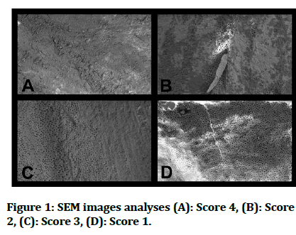
The residual obturation materials were evaluated using scanning electron microscopy after sectioning each sample longitudinally. Each sample was examined at the center of coronal, middle and apical thirds. The samples were imaged first under 1X to 3X magnifications. The images of SEM were obtained and analyzed according to the scale defined by somma and his coworkers [12]. Score 0: There is no or very little residual debris on the dentinal surface (0%–25%), presence of 25% to 50% residual debris on the surface, 2: Moderate residual debris presence (50%–75%) and 3: The entire or nearly entire surface (75%–100%) is covered with residual debris (Figure 1). Statistical analysis for the data obtained from the SEM images was done using the statistical package for the social sciences (SPSS Inc, Chicago, IL version 26).

Figure 1: SEM images analyses (A): Score 4, (B): Score 2, (C): Score 3, (D): Score 1.
Results
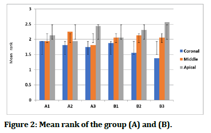
Two examiners scored the data, both examiners had a high agreement (weighted kappa=0.86) in the inter examiner analysis. Mean rank was used during the statistical analysis because of the nonparametric nature of the data. The residual of obturation materials was found to be greater in the apical third of all groups and decreased when moving towered the coronal third (Figure 2). Mann Whitney Wilcoxon test was used to compare the two main groups; no statistically significant differences were found (Table 1). Kruskal Wallis test was used to compare the three subgroups within each group, statistically, significant differences were recorded (Table 2). Additional compression was made using Mann- Whitney U tests to identify the difference between subgroups. In group A, statistically significant differences were recorded between subgroups (A1, A3) and between (A2, A3). While in group B significant differences were recorded between subgroups (B1, B3) only (Table 3).
| Subgroups | Test^ | Coronal | Middle | Apical |
|---|---|---|---|---|
| 1 | P value | 0.854 | 1 | 0.602 |
| 2 | p value | 0.777 | 0.48 | 0.814 |
| 3 | p value | 0.206 | 0.902 | 0.777 |
Table 1: Compression between the two main groups using Mann Whitney Wilcoxon test.
| Groups | Coronal | Middle | Apical | |
|---|---|---|---|---|
| A | P value | 0.098 | 0.032 | 0.288 |
| B | P value | 0.027 | 0.025 | 0.392 |
Table 2: Compression between subgroups within each main group using Kruskal Wallis test
Figure 2: Mean rank of the group (A) and (B).
| Groups | Subgroups | Coronal | Middle |
|---|---|---|---|
| A | 1 vs 2 | - | 0.626 |
| 1 vs 3 | - | 0.02 | |
| 2 vs 3 | - | 0.039 | |
| B | 1 vs 2 | 0.254 | 0.263 |
| 1 vs 3 | 0.008 | 0.007 | |
| 2 vs 3 | 0.118 | 0.114 |
Table 3: Compression between subgroups within each main group using Mann Whitney U tests.
Discussion
In non-surgical root canal retreatment, the complete removal of filling materials is essential to ensure the clearance of dentinal tubules because their blockage will reduce or prevent the activity of irrigation solutions and intra canal medicaments from reaching all the root canal spaces [13]. Many non-surgical retreatment methods are used for removing the root canal obturation materials: However, the complete removal of these materials had been not achieved according to previous studies [14,15]. The main disadvantage of bioceramic is that it is difficult to remove during endodontic retreatment due to chemical and micromechanical bonding between the sealer and the dentine surface. Additionally, the hardness of the sealer after setting may increase its adherence and resistance to dislocation from dentine, making it difficult to remove during secondary endodontic treatment. Regardless of the file system used during the retreatment process, the majority of residual materials were found in the apical third. This founding match the result of previous research [16,17].
Anatomical variations tend to be higher at the apical which could explain the previous statement [18]. Another reason for this may be the compaction and penetration of the obturating materials occurs at the middle and apical thirds of the canal resulting in more debris being deposited in the dentinal tubules. In addition, there are differences in tip sizes and tapers between instruments used for initial preparation and those used for canal retreatment. However, other research come out with an inconsistent result like faus matoses and his colleagues, which found that remnants at the middle and coronal thirds were substantially higher than that found at the apical third because the taper of the files used in retreatment differs from that used in primary shaping, the efficiency in the coronal and middle thirds may be lower than in the apical third [19].
In this study, there was no statistical difference between the two reciprocating systems used for retreatment (R25, WOG) in removing GP and sealer.
Reciproc file (R25) is distinguished by an S-shaped cross section with sharp cutting edges and a large chip space, which enhances cutting efficiency and hence the retreatment ability of the instrument. The efficacy of the RB file for removing obturating materials from the apical third had shown to be less than that at the coronal third according to Romeiro, who explained that it might be related to the size of the file used (R 25/08) and suggested that higher percentage of obturation materials removal and more touches on canal walls can be achieved if two sizes apical enlargement after initial preparation is done [20]. This suggestion comes in with agreement with the founding of the research, who found that increasing size from 25 to 40 helps improve the elimination of GP and sealer from the root canal spaces [21].
While for the WOG system, which possesses a parallelogram cross section and is made of more flexible NiTi gold wire. The retreating potential of the system at the coronal and middle thirds is similar to other examined systems. while for the apical third, where more residual materials were found, this could be associated with the system used which is with apical size 25 and 07 taper, additional apical enlargement may require, according to a recent study, which stated that the efficacy of WOG improved after instrumentation with a larger tip size file (35) in curved root canals [22]. Additionally, according to previous research, WOG's design does not enable enough area for debris to be removed, reducing its cutting performance [23].
When comparing the subgroups within each group, all irrigation protocols showed an increasing percentage of remanent of root canal filling materials when advancing from the coronal towards the apical direction. This finding is consonant with the findings of a previous study [24]. A significant difference was found within both groups when comparing conventional needle irrigation and continuous ultrasonic irrigation, the last showed a superior result in the sealer and guttapercha removal. This finding is consonant with the findings of previous studies. The ineffectiveness of traditional irrigation is due to the existence of an air bubble inside the canal (known as vapor lock), which prevents the irrigant from properly reaching the apical third. The use of CNI does not generate enough pressure to overcome the vaper lock and to deliver the irrigant solution to all canal areas. Another study mentioned the lack of effectiveness of the CNI is due to that the deliver potential of this method is limited to only 1 mm further than the needle's tip [25]. The use of CUI in both groups had shown to have a better result in removing remanent of obturation materials which can be explained that the use of CUI provides enough force to overcome the vapor lock and that there will be a continuous exchange of irrigant solution due to the use of CUI. The concept of CUI is based on a continuous flow of irrigants instead of intermittently replenished through needle irrigation to provide an advantage over intermittent irrigation [26]. The CUI is based primarily on the activation of the irrigation that is delivered through an ultrasonically energized tip connected directly to the ultrasonic unit which ultrasonic activation and delivered the irrigant solution at the same time [27]. Ultrasonic irrigation mechanism of action is based on the transmission of acoustic energy from an oscillating file in which the file motion is likely to be impeded as the root canal narrows toward the apical portion. The acoustic flow and cavitation, these two characteristics of the ultrasonic activated instruments promote the cleaning action of the irrigant solution and enhance the irrigation result. The energy is transmitted using ultrasonic waves, which might cause acoustic streaming of the irrigant, resulting in greater irrigant volume and improved penetration [28].
When comparing the subgroups within group A, a significant difference was found when comparing between PUI and CUI, the last show superior removal potential of remanent of obturation materials. These results are consistent with the results of a previous study [29]. This finding explains that CUI had shown sufficient force to overcome the vapor lock. This finding might be related to the CUI technique's constant solution exchange and optimal activation of the solution as it flows through the ultrasonically energized file.
When comparing between PUI and CUI in group B, no significant difference was reported. This can be explained by the fact that the taper of the file in group B (25/07) can influence the flow of irrigants and thus on the cleaning efficiency of the irrigation technique [30].
Conclusion
Within the limitations of this in vitro SEM study, the following conclusions can be made; none of the retreatment methods is capable of complete removal of the obturation materials from the entire root canals system walls. There was no significant difference among the two tested systems (RB and WOG) in the efficacy of removal of obturation materials from walls of the root canals. CNI alone is an unreliable method in the removal of root canal obturation materials regardless of the retreatment system used. The use of CUI significantly improves the process of removal of obturation filling materials from the root canal walls when compared to CNI. There was a significant difference between the subgroups of reciproc group between PUI and CUI, where the last showed superior results.
References
- Lee S, Tan S, Ab Aziz ZACJAoDUoM. Is profile alone sufficient to remove gutta-percha during endodontic re-treatment? Ann Dent UM 2005; 12:1-8. [Crossref][Googlescholar][Indexed]
- Kang M, Jung HI, Song M, et al. Outcome of nonsurgical retreatment and endodontic microsurgery: A meta-analysis. Clin Oral Investig 2015; 19:569-582. [Crossref][Googlescholar][Indexed]
- Olcay K, Ataoglu H, Belli SJJoe. Evaluation of related factors in the failure of endodontically treated teeth: A cross sectional study. J Endod 2018; 44:38-45. [Crossref][Googlescholar][Indexed]
- de Azevedo Rios M, Villela AM, Cunha RS, et al. Efficacy of 2 reciprocating systems compared with a rotary retreatment system for gutta-percha removal. J Endod 2014; 40:543-546. [Crossref][Googlescholar][Indexed]
- Horvath S, Altenburger M, Naumann M, et al. Cleanliness of dentinal tubules following gutta-percha removal with and without solvents: A scanning electron microscopic study. Int Endod J 2009; 42:1032-1038. [Crossref][Googlescholar][Indexed]
- Martins MP, Duarte MAH, Cavenago BC, et al. Effectiveness of the protaper next and reciproc systems in removing root canal filling material with sonic or ultrasonic irrigation: A micro computed tomographic study. J Endod 2017; 43:467-471. [Crossref][Googlescholar][Indexed]
- Castelo Baz P, Martin Biedma B, Cantatore G, et al. In vitro comparison of passive and continuous ultrasonic irrigation in simulated lateral canals of extracted teeth. J Endod 2012; 38:688-691. [Crossref][Googlescholar][Indexed]
- Castelo Baz P, Varela Patino P, Cantatore G, et al. In vitro comparison of passive and continuous ultrasonic irrigation in curved root canals. J Clin Exp Dent 2016; 8:e437. [Crossref][Googlescholar][Indexed]
- Gulabivala K, Ng Y, Gilbertson M, et al. The fluid mechanics of root canal irrigation. Physiol Meas 2010; 31:R49. [Crossref][Googlescholar][Indexed]
- Jamleh A, Suda H, Adorno CGJDmj. Irrigation effectiveness of continuous ultrasonic irrigation system: An ex vivo study. Dent Mater J 2018; 37:1-5. [Crossref][Googlescholar][Indexed]
- Van der Sluis L, Versluis M, Wu M, et al. Passive ultrasonic irrigation of the root canal: A review of the literature. Int Endod J 2007; 40:415-426. [Crossref][Googlescholar][Indexed]
- Somma F, Cammarota G, Plotino G, et al. The effectiveness of manual and mechanical instrumentation for the retreatment of three different root canal filling materials. J Endod 2008; 34:466-469. [Crossref][Googlescholar][Indexed]
- Cavenago B, Ordinola Zapata R, Duarte M, et al. Efficacy of xylene and passive ultrasonic irrigation on remaining root filling material during retreatment of anatomically complex teeth. Int Endod J 2014; 47:1078-1083. [Crossref][Googlescholar][Indexed]
- Nevares G, Diana S, Freire LG, et al. Efficacy of protaper NEXT compared with reciproc in removing obturation material from severely curved root canals: A micro computed tomography study. J Endod 2016; 42:803-808. [Crossref][Googlescholar][Indexed]
- Rodig T, Reicherts P, Konietschke F, et al. Efficacy of reciprocating and rotary NiTi instruments for retreatment of curved root canals assessed by micro CT. Int Endod J 2014; 47:942-948. [Crossref][Googlescholar][Indexed]
- Hegde V, Murkey LJE. Evaluation of residual root canal filling material after retreatment of canals filled with hydrophilic and hydrophobic obturating system: An in vitro scanning electron microscopy study. Endodontol 2017; 29:47. [Googlescholar][Indexed]
- Ma J, Al-Ashaw AJ, Shen Y, et al. Efficacy of protaper universal rotary retreatment system for gutta-percha removal from oval root canals: A micro computed tomography study. J Endod 2012; 38:1516-1520. [Crossref][Googlescholar][Indexed]
- Ferreira J, Rhodes J, Pitt Ford TJIEJ. The efficacy of gutta-percha removal using profiles. Int Endod J 2001; 34:267-274. [Crossref][Googlescholar][Indexed]
- Faus Matoses V, Pasarin Linares C, Faus Matoses I, et al. Comparison of obturation removal efficiency from straight root canals with protaper gold or reciproc blue: A micro computed tomography study. J Clin Med 2020; 9:1164. [Crossref][Googlescholar][Indexed]
- Romeiro K, de Almeida A, Cassimiro M, et al. Reciproc and reciproc Blue in the removal of bioceramic and resin based sealers in retreatment procedures. Clin Oral Investig 2020; 24:405-416. [Crossref][Googlescholar][Indexed]
- de Deus G, Belladonna F, Zuolo A, et al. Effectiveness of reciproc Blue in removing canal filling material and regaining apical patency. Int Endod J 2019; 52:250-257. [Crossref][Googlescholar][Indexed]
- Bago I, Plotino G, Katic M, et al. Evaluation of filling material remnants after basic preparation, apical enlargement and final irrigation in retreatment of severely curved root canals in extracted teeth. Int Endod J 2020; 53:962-973. [Crossref][Googlescholar][Indexed]
- Tocci L, Plotino G, Al-Sudani D, et al. Cutting efficiency of instruments with different movements: A comparative study. J Oral Maxillofac Res 2015; 6. [Googlescholar][Indexed]
- Grischke J, Muller Heine A, Hulsmann MJCoi. The effect of four different irrigation systems in the removal of a root canal sealer. Clin Oral Investig 2014; 18:1845-1851. [Crossref][Googlescholar][Indexed]
- Boutsioukis C, Lambrianidis T, Verhaagen B, et al. The effect of needle insertion depth on the irrigant flow in the root canal: Evaluation using an unsteady computational fluid dynamics model. J Endod 2010; 36:1664-1668. [Crossref][Googlescholar][Indexed]
- Mozo S, Llena C, Forner L. Review of ultrasonic irrigation in endodontics: Increasing action of irrigating solutions. Med Oral Patol Oral Cir Bucal 2012; 17:e512. [Crossref][Googlescholar][Indexed]
- Curtis TO, Sedgley CMJJoe. Comparison of a continuous ultrasonic irrigation device and conventional needle irrigation in the removal of root canal debris. J Endod 2012; 38:1261-1264. [Crossref][Googlescholar][Indexed]
- Ahmad M, Ford TP, Crum L, et al. Ultrasonic debridement of root canals: Acoustic cavitation and its relevance. J Endod 1988; 14:486-493. [Crossref][Googlescholar][Indexed]
- Miguens Vila R, Castelo Baz P, Aboy Pazos S, et al. Does the use of use of continuous or passive ultrasonic irrigation protocols improve the removal of smear layer? A scanning electron microscopic study. 2021. [Crossref][Googlescholar]
- Van Der Sluis L, Wu MK, Wesselink PJIEJ. A comparison between a smooth wire and a K file in removing artificially placed dentine debris from root canals in resin blocks during ultrasonic irrigation. Int Endod J 2005; 38:593-596. [Crossref][Googlescholar][Indexed]
Author Info
Ali A Al-Mutlak* and Hussain F Al-Huwaizi
Department of Conservative Dentistry, College of Dentistry, University of Baghdad, IraqCitation: Ali A Al-Mutlak, Hussain F Al-Huwaizi, Evaluation of Two Rotary Systems in Removing Root Canal Filling Material Using Different Ultrasonic Irrigation Techniques (SEM Study), J Res Med Dent Sci, 2023, 11 (01): 121-126.
Received: 02-Nov-2022, Manuscript No. JRMDS-23-66432; , Pre QC No. JRMDS-23-66432 (PQ); Editor assigned: 07-Nov-2022, Pre QC No. JRMDS-23-66432 (PQ); Reviewed: 21-Nov-2022, QC No. JRMDS-23-66432; Revised: 29-Dec-2022, Manuscript No. JRMDS-23-66432 (R); Published: 06-Jan-2023

