Research - (2021) Volume 9, Issue 4
Evaluation of the Cause and Pattern of Teeth Extraction in the College of Dentistry, Mustansiriyah University
*Correspondence: Noor Mohammed Al-Noori, Department of Oral Surgery and Periodontology, College of Dentistry, Mustansiriyah University, Baghdad, Iraq, Email:
Abstract
The aim of this study was to find the reasons for teeth extraction in patients attending department of Oral Surgery in college of dentistry and cross relation of factors such as age, gender, etc. Material and Methods: This study was carried out in oral surgery department of college of dentistry, Mustansiriyah University, Baghdad- Iraq. The data collected from the case sheets of under graduate student of college of dentistry, fourth and fifth stages. Results: There were 1964 patients had 2789 extracted teeth occupied in this study with mean age (39.96) years, 1151 patients were males and 813 of them were females. 1995 from teeth as retained roots (71.5%). (58.6%) were maxillary teeth and (41.4%) were mandibular teeth. There were 1099 (39.4%) out of 2789 teeth diagnosed with chronic periapical lesion and 896 teeth with necrotic pulps (32.13%). Conclusion: Caries and its progression until reach necrosis and periapical lesions and formation of retained roots that unable to treat by other ways except extraction was the prevalent cause of extraction in this study.
Keywords
Caries, Extraction, Retained root
Introduction
Tooth extraction has adverse social and psychological impression on the patient and also cause masticatory dysfunction [1]. So permanent teeth requires retention as long as possible for functional and esthetics effect [2]. Permanent teeth extraction takes place because of different reasons: dental caries, periodontal disease, trauma, orthodontic treatments, prosthetic indications and eruption problems [1]. Tooth extraction can be related to nonclinical factors include socioeconomic, low education, poor oral hygiene and patients demand [2].
The aim of this study was to find the reasons for teeth extraction in patients attending department of Oral Surgery in college of dentistry and cross relation of factors such as age, gender, systemic disease, position of tooth in dental jaw, diagnosis and chief complaint.
Materials and Methods
The cross sectional study was carried out during one academic year in oral surgery department of college of dentistry, Mustansiriyah University, Baghdad- Iraq.The data collected from the case sheets of under graduate student of college of dentistry, fourth and fifth stage.
Inclusion criteria
Patients requiring extraction. Patients age 12 years and more, Excluding surgical extraction. 1964 patients (1151 male and 813 female) were included in the study with age (12-80) years with mean age (39.96) included in this study. Information as age, gender, chief complaint (pain, prosthetic reasons, orthodontic reasons, swelling and checkup), systemic disease (blood pressure problems, diabetes mellitus, thyroid problems, heart disease, respiratory disease, etc.), social habits (smoking and drinking), previous extraction, tooth number, maxillary or mandibular jaw (right and left), anterior or posterior position and diagnosis (acute pulpitis, chronic pulpitis, chronic periapical lesion, necrotic pulp, supernumerary tooth, periodontitis, retained deciduous and orthodontic purpose). Obtained data were statistically analyzed by using descriptive statistics and chi-square test. Data analysis were performed by excel version 10, SPSS version 24.0. Statistical significance was considered for p < 0.05 in all cases.
Results
Age and gender: there were 1964 patients occupied in this study with mean age (39.96) years, 1151 patients (58.6%) were male with mean age (40.72) years and 813 of them (41.4%) were female with mean age (38.89) years. Distributions of patients according to age appear as: (31-40) years in the first (488) patients, (21-30) years were second (472) patients and (41-50) were third (420) patients. So the young adults and middle aged groups show more extraction than older patients (Table 1).
Table 1: Distributions of patients according to age and gender.
| Gender | Total | |||
|---|---|---|---|---|
| Female | Male | |||
| Age groups | ≤20 | 69 | 81 | 150 |
| 21-30 | 204 | 268 | 472 | |
| 31-40 | 201 | 287 | 488 | |
| 41-50 | 181 | 239 | 420 | |
| 51-60 | 100 | 170 | 270 | |
| 61-70 | 55 | 83 | 138 | |
| 71-80 | 3 | 23 | 26 | |
| Total | 813 | 1151 | 1964 | |
Social habits
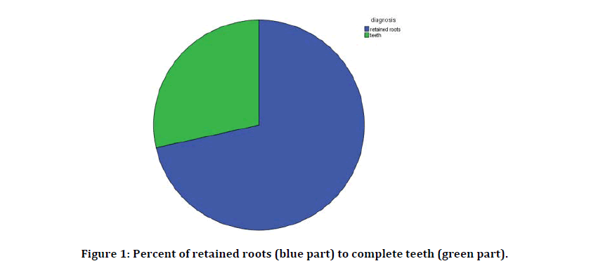
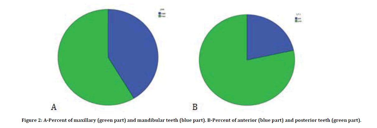
There were 500 patients out of 1964 patients (25.5%) with social habits (were smoker). Systemic disease: there were 196 patients (9.98%) out of 1964 with control systemic diseases, 131 patients of them with blood pressure problems (hypertension), 43 had diabetes mellitus, 9 had heart disease, 7 asthmatic patients and 6 with thyroid problems. But all these patients with control to their diseases and were monitored during extractions.Chief complaint: there were 780 patients came with pain, 603 patients for prosthetic reasons, 562 patients came for checkup, 18 patients for orthodontic reasons and 1 because of swelling (Table 2). Teeth and Jaws: 1964 patients had 2789 extracted teeth, with mean (1.42) tooth for each patient. 1995 teeth as retained roots (71.5%) (Figure 1). 1636 of them were maxillary teeth (58.6%) and 1153 of them were mandibular teeth (41.4%) (Figure 2A). There were 2187 posterior teeth (78.4%) and 602 were anterior teeth (21.6%) (Figure 2B). Upper first premolar (322) were the most tooth to be extracted, Upper second premolar (262) is second in the list followed by upper first molar (257) and lower first molar (239) (Table 3).
Figure 1: Percent of retained roots (blue part) to complete teeth (green part).
Figure 2: A-Percent of maxillary (green part) and mandibular teeth (blue part). B-Percent of anterior (blue part) and posterior teeth (green part).
Table 2: Number of patient according to chief complaint.
| Chief complaint | No. of patients |
|---|---|
| Check up | 562 |
| Ortho. | 18 |
| Pain | 780 |
| Pros. | 603 |
| Swelling | 1 |
| Total | 1964 |
Table 3: The counting numbers of teeth extraction.
| Maxilla | Mandible | Total | Percent | |
|---|---|---|---|---|
| Central Incisor | 100 | 70 | 170 | 6.1 |
| Lateral Incisor | 153 | 63 | 216 | 7.7 |
| Canine | 134 | 60 | 194 | 7 |
| First Premolar | 322 | 159 | 481 | 17.3 |
| Second Premolar | 262 | 180 | 442 | 15.8 |
| First Molar | 257 | 239 | 496 | 17.8 |
| Second Molar | 171 | 157 | 328 | 11.8 |
| Third Molar | 204 | 186 | 390 | 14 |
| Deciduous & Supernumerary | 38 | 33 | 71 | 2.5 |
Diagnosis
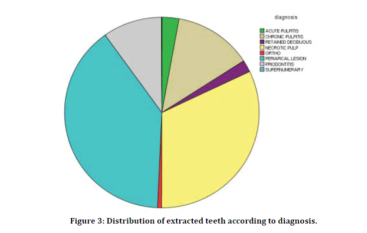
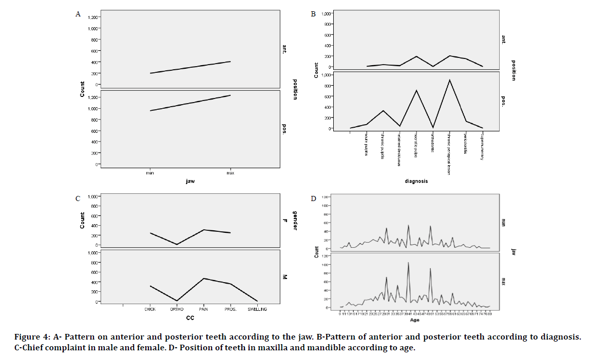
There were 1099 (39.4%) out of 2789 teeth diagnosed with chronic periapical lesion, 896 teeth with necrotic pulps (32.13%), 364 teeth with chronic pulpitis, 274 teeth with periodontitis, 78 teeth with acute pulpitis, 57 teeth were retained deciduous, 18 teeth extracted for orthodontic purpose and 2 were supernumerary teeth. So, 2438 (87.4%) teeth were extracted because of caries sequelae and 351 (12.6%) due to other reasons. (Figure 3) and (Tables 4 and 5). Other teeth: 49.7% of the patients had problems in their other teeth, 301 with missing teeth (M), 100 patient with caries (C) and 50 patients with retained roots (RR). 408 patients had (M, C and RR), 294 patients had both (M and C), 187 had both (M and RR) and 103 had both (C and RR). Pattern of distribution. The distribution pattern of teeth that extracted showed no difference between male and female, but extraction more in male. There was no difference in pattern of anterior or posterior teeth according to maxilla and mandible but posterior teeth more extracted than anterior (Figure 4A). There was change in pattern of anterior and posterior teeth according to diagnosis; periodontitis more in anterior teeth and chronic pulpitis more in posterior teeth, but retained roots with chronic periapical lesions and necrotic pulp were the winner in anterior and posterior (Figure 4B). Chief complaint in male and female toke the same pattern and the pain was more in the both genders (Figure 4C). The pattern of teeth extraction according to age is the same in both maxilla and mandible, but the count more in maxilla (Figure 4D).
Figure 3: Distribution of extracted teeth according to diagnosis.
Figure 4: A- Pattern on anterior and posterior teeth according to the jaw. B-Pattern of anterior and posterior teeth according to diagnosis. C-Chief complaint in male and female. D- Position of teeth in maxilla and mandible according to age.
Table 4: distribution of extracted teeth according to diagnosis.
| Frequency | Percent | |
|---|---|---|
| Diagnosis | ||
| Acute Pulpitis | 78 | 2.8 |
| Chronic Pulpitis | 364 | 13.1 |
| Retained Deciduous | 57 | 2 |
| Necrotic Pulp | 896 | 32.1 |
| Ortho | 18 | 0.6 |
| Periodical Lesion | 1099 | 39.4 |
| Periodontitis | 274 | 9.8 |
| Supernumerary | 2 | 0.1 |
| Total | 2789 | 100 |
Table 5: distribution of diagnosis according to age.
| Age | Acute pulpitis | Chronic pulpitis | Retained deciduous | Necrotic pulp | Ortho. | Periodical lesion | Period |
|---|---|---|---|---|---|---|---|
| ≥20 | 6 | 20 | 41 | 30 | 12 | 41 | 1 |
| 21-30 | 24 | 116 | 3 | 36 | 6 | 182 | 15 |
| 31-40 | 25 | 81 | 0 | 137 | 0 | 225 | 20 |
| 41-50 | 11 | 43 | 3 | 152 | 0 | 157 | 54 |
| 51-60 | 2 | 31 | 0 | 89 | 0 | 84 | 64 |
| 61-70 | 5 | 13 | 0 | 47 | 0 | 38 | 35 |
| 71-80 | 0 | 3 | 0 | 5 | 0 | 11 | 7 |
| Total | 73 | 307 | 47 | 586 | 16 | 737 | 196 |
Discussion
The present study demonstrated the reasons for extractions in the college of dentistry. The caries and its consequence were the main cause of teeth loses. This result in the same line of other that found caries was the cause of extraction [3]. The peak extraction result was chronic periapical lesion due to neglecting of caries and in adequate preventive dentistry, dietary bad habit was the most cause of caries and low socioeconomic of patients cause progression of caries until reaching periapical lesion. In this study, nearly most of the teeth that extracted (87.4%) were due to dental caries and its consequences. Periodontal disease was the second cause (9.8%). This agree with other studies regarding caries was the main cause but with widely different in percent, Shah et al found that (50.51%) of extraction was due to caries and its sequelae [4], and Manekar et al. also found the same results [5]. In my study first molars (17.8%) were more extracted followed by first premolars (17.3%) and then the second premolars (15.8%). Shah et al found that the first molar extracted more than others (19.4%) followed by third molars (16%) [4]. This due to a numerous details: 1. First molars were erupted firstly in oral cavity and 2. First molars with a larger surface area than others with surface anatomy that creating good area for accumulation of plaque and subsequent caries [6]. There was a male prevalence (58.6%), which is in disagreement with other studies that founded female more prominent [7-10]. But this result in agreement with the Alhadi et al that found Males comprised 53.6% of patients [11]. Teeth extraction were greater in male patients may be due to their deficiency of time and attentiveness to do restoration, and this accentual by Da'ameh et al. [12]. Lesolang et al. proposed that this condition may be due to the variances in diet and looking for treatment difference between two genders [13]. Saliva and tooth morphology affected by genetic factors, dietary habits, education and socioeconomically situation, as well as dental care and lifestyle issues like smoking, should be considered [2]. In this study, the mean number of teeth extracted per a patient was 1.42 less than 1.9 founded by Jafarian et al. [14], and higher than the 1.3 found by Corbet et al. [15]. These variations are due to organizational differences and different age of population sample. Accordingly, evaluation of tooth extraction records between different nations is troubled with difficulty [14]. In this study maxillary posterior teeth more extracted than others, mandibular posterior teeth were the second, followed by maxillary anterior and then mandibular anterior teeth. And this disagreement with other study that found mandibular teeth more extracted than maxillary [16,17]. Periodontal disease were the second cause of extraction, mandibular anterior teeth were the first followed by maxillary posterior. These disagree with other study that found periodontal disease the main cause of extraction and anterior teeth in both jaws were in the first line [18]. The mean age of this study was (39.96) with range (12-80), (31-40) years with more extractions followed by (21-30) years and (41- 50) years were third. This in line of other study that showed mean age (36.85), the age-group 20-29 years with most extraction followed by the age group 30-39 years. The result was due to the high prevalence of caries during this portion of life [19].
Conclusion
Caries and its progression until reach necrosis and periapical lesions and formation of retained roots that unable to treat by other ways except extraction was the prevalent cause of extraction in this study. Young adults and middle aged groups (21-50) years, show more extraction than others. In this study males were prominence than females. First molars and first premolars were the most extracted teeth. Chronic disease, smoking, dietary habits and low socioeconomic; factors affect oral and dental health and lead to more teeth extractions. Consequently, application of well-organized preventive programs to reduce and overcome caries is mandatory. This program should focus on young age to decrease numbers of extractions.
References
- Rahaf Al-Safadi, Riham Al-Safadi, Abdulrahman Al-Lahim, et al. Patterns of and reasons for permanent tooth extractions in a Saudi Population. IJETST 2019; 6:6811-6821.
- TaÅ?söker M, MenziletoÄ?lu D, BaÅ?türk F, et al. Investigation of tooth extraction reasons in patients who applied to a dental faculty. Meandros Med Dent J 2018; 19:219-225.3
- Al Ameer HM, Awad S. Reasons for permanent teeth extraction in Al-Madinah Al-Munawarah. J Adv Med Med Res 2017; 1-6.
- Shah A, Faldu M, Chowdhury S. Reasons for extractions of permanent teeth in western India: A prospective study. Int J Applied Dent Sci 2019; 5:180-184.
- Manekar VS, Kende P, Kulkarni S. Tooth mortality: An analysis of reasons underlying the extraction of permanent teeth. World J Dent 2015; 6:93-96.
- Katoma M, Siziya S, Sichilima A. The most frequently extracted teeth type: a retrospective cross-section study at Ndola teaching hospital, low cost dental clinic, Zambia. Asian Pac J Health Sci 2017; 4:98-101.
- Bikash D, Bishnu SP, Rajani S. To find out the causes of extraction of teeth in the patients coming to the department of oral and maxillofacial surgery at Kantipur dental college teaching hospital and research center from January 2014 to December 2018. Annals Clin Med Case Reports 2019; 2:1-5.
- Saheeb BD, Sede MA. Reasons and pattern of tooth mortality in a Nigerian Urban teaching hospital. Annals African Med 2013; 12:110.
- Okoje VN. Indications for permanent teeth exodontia: A comparative analysis of two tertiary institutions in South-Western Nigeria. J Dent Oral Health 2018; 5:1-8.
- Salman SMA, Ahmed S, Bari Ya, et al. Common factors leading to tooth extraction-A cross sectional study in a tertiary care hospital. Acta Scientific Dent Sci 2019; 3:23-28.
- Alhadi Y, Rassem AH, Al-Shamahy HA, et al. Causes for extraction of permanent teeth in general dental practices in Yemen. Universal J Pharm Res 2019; 4:1-6.
- Da'ameh DA. Reasons for permanent tooth extraction in the North of Afghanistan. J Dent 2006; 34:48-51.
- Lesolang RR, Motloba DP, Lalloo R. Patterns and reasons for tooth extraction at the Winterveldt Clinic: 1998-2002: Scientific. South Af Dent J 2009; 64:214-218.
- Jafarian M, Etebarian A. Reasons for extraction of permanent teeth in general dental practices in Tehran, Iran. Med Principles Practice 2013; 22:239-244.
- Corbet EF, Davies WI. Reasons given for tooth extraction in Hong Kong. Community Dent Health 1991; 8:121–130.
- Yadav A, Karikal A. Reasons Underlying the extraction of permanent teeth in patients attending ABSMIDS. Nitte University J Health Sci 2016; 6.
- Taiwo AO, Ibikunle AA, Braimah RO, et al. Tooth extraction: Pattern and etiology from extreme Northwestern Nigeria. Eur J Dent 2017; 11:335.
- Chrysanthakopoulos NA. Periodontal reasons for tooth extraction in a group of Greek army personnel. J Dent Res Dent Clin Dent Prospects 2011; 5:55-60.
- Nasreen T, Haq ME. Factors of tooth extraction among adult patients attending in exodontia department of Dhaka dental college and hospital. Bangladesh J Orthodont Dentofac Orthop 2011; 2:7-10.


