Research - (2022) Volume 10, Issue 8
Evaluation of Root Canal Morphology of Maxillary First and Second Premolars by Cone-Beam Computed Tomography in Saudi Population
Arwa S Alnoury1*, Lina O Bahanan2, Mohanad A Alhedbany3, Mustafa E Abuzinadah3, Hanadi M Khalifah4 and Abrar A Tounsi5
*Correspondence: Arwa S Alnoury, Department of Restorative Dentistry, Faculty of Dentistry, King Abdulaziz University, Jeddah 21589, Saudi Arabia, Email:
Abstract
Objectives: To estimate the prevalence of different root canal morphological variants in maxillary first and second premolars, and to analyze differences across gender and nationality in western Saudi Arabia individuals using cone-beam computed tomography (CBCT) who presented for dental care during February 2021- April 2021. Methods: Maxillary first and second premolars were analyzed independently to determine the number of roots and canal configuration using Vertucci's classification. The distribution by gender and nationality was analyzed using Fisher’s exact test with Monte Carlo simulation. Inter-examiner and intra-examiner reliability were evaluated using Cohen's kappa coefficient (1.0; P<0.001 and 0.9; P<0.001, respectively). Results: Maxillary first premolars were double-rooted in 57.8% and type IV was the most frequent (63.6%), followed by type II (18.3%). Second premolars were single-rooted in 77.6%, with type I being the most frequent (37.8%) in females (48.2%); while type IV being most frequent in males (33.8%). In both first and second premolars, females had a significant predilection to single root pattern. Differences across nationality were statistically significant with respect to canal configuration of maxillary first premolars. Conclusion: There is a high heterogeneity in the maxillary premolar root canal configuration among the studied Saudi population, with significant gender disparities supporting fewer roots and canals in females.
Keywords
Cone-beam computed tomography, Maxillary, Premolars, Root canal morphology, Saudi Arabia
Introduction
Correct identification of the root canal morphology of the tooth is a crucial clinical step for a successful endodontic treatment [1]. Several configurations for tooth hard tissue repository have been identified in normal human dentition. These variations concern the number, shape, and symmetry of the roots and root canals, and are determined using a standardized classification system with several clinical implications [2–4]. However, several factors have been identified to contribute in the development of such variations, such as ethnicity, developmental anomalies, age, and dental conditions such as trauma, caries, and restorative procedures [2].
Various invasive and conservative techniques have been used to analyze the root canal morphology, such as staining and clearing, conventional or digital dental radiographs, micro tomography, and more recently cone-beam computed tomography (CBCT) using threedimensional images with enhanced quality and accuracy[5]. Additionally, CBCT is convenient for large sample studies. One of the most frequently used classifications is that elaborated by Vertucci et al., which determined eight morphological types of root canal ranging from singlecanal throughout the tooth (type I) to three distinct canals (type VIII). The intermediate types include various anastomoses and bifurcations of the canals at different levels [6,7]. The maxillary first premolars are reported to be principally two-rooted; however, cases of three-rooted configurations are commonly reported among the anatomic variations [8,9]. For maxillary second premolars single-rooted canal was reported to be the most common configuration in different populations including Saudi [10], Spanish [9], and Chinese [11]; however, canal types IV and V were equally common in the Saudi population reported in 23% each, while type I was predominant in the Spanish (47.2%) and Chinese (55.1%) populations.
In this study, the authors used CBCT to estimate the prevalence of different root canal morphological variants in maxillary first and second premolars among consecutive sample of individuals in western Saudi Arabia, and to analyze the differences across gender and nationality. The authors also evaluated symmetry and asymmetry in relation to number of roots and root morphology in both premolars.
Materials and Methods
A cross-sectional study was conducted at the oral and maxillofacial radiology department of the faculty of dentistry, King Abdulaziz University, Jeddah, Saudi Arabia, between February 2021 and April 2021. This study is ethically approved by the research ethic committee of King Abdulaziz University faculty of dentistry (Proposal #: 182-12-20).
The study included consecutive CBCT images that were carried out for various dental indications in adult patients. The first CBCT images that fulfilled the inclusion and exclusion criteria were selected in the study.
The CBCT images included in the study were acquired using the i-CAT (Imaging Sciences International, Hatfield, PA, USA). For standardization, all CBCT images were obtained with voxel size of 0.125 μm and a field of view of 8X8 cm. On Demand 3D Imaging Software (Cyber med, Seoul, South Korea) was used to analyze the CBCT images in three planes (axial, sagittal, and coronal).
The inclusion criterion was the presence of at least one or more fully-developed-root maxillary first or second premolar. Previously endodontically treated teeth, teeth with posts, teeth with immature apexes, and unclear or distorted CBCT images were excluded. The patient ID, gender and nationality (Saudi or non-Saudi) were recorded for each CBCT image.
The total sample was analyzed independently by two dentists to determine the following parameters for each included premolar:
Number of roots (one, two, or three) for the maxillary first and second premolars.
Canal configuration for the maxillary first and second premolars, using Vertucci's classification [7], and comprising eight types defined in Box 1.
To ensure the reliability of the results, the inter- and intra-examiner reliability were assessed. Ten randomly selected CBCT images were evaluated by the examiners according to the parameters specified for the study. The same CBCT images were evaluated after 1 week for the intra-examiner reliability.
Vertucci's classification of root canal morphology [7]
Type I: Single canal throughout.
Type II: Two canals merging into one at the canal terminus.
Type III: One canal dividing into two at the mid canal, then reuniting at the canal terminus.
Type IV: Two distinct canals throughout.
Type V: One canal dividing into two canals at the canal terminus.
Type VI: Two distinct canals merging in the body of the root then separating again at the canal terminus.
Type VII: One canal dividing into two, then reuniting then redividing again at the canal terminus.
Type VIII: Three distinct canals throughout.
Statistical methods
Inter-examiner and intra-examiner reliability were evaluated using Cohen's kappa coefficient (1.0; P<0.001 and 0.9; P<0.001, respectively). Descriptive statistics including frequency, percentages and mean and standard deviation (SD) were conducted to summarize the sample variables. Fisher’s exact test with Monte Carlo simulation were used to compare the number of roots and root morphology of maxillary first and second premolars across gender and nationality. All statistical analyses were conducted using statistical software SAS, Version 9.4 (Cary, NC: SAS Institute Inc. 2013). The significance level was set at a P-value <0.05.
Results
Participant characteristics
The authors analyzed CBCT images from 269 patients, which included 723 teeth (383 maxillary first and 340 maxillary second premolars). Female patients included 54.6%, and the mean (SD) age was 33.9 (12.7) years. Majority of the participants were Saudi citizens (65.8%). The overall distribution of root canal morphology showed that type IV was the most prevalent type (46.3%), followed by type I (23.0%), and type II (22.4%) (Table 1).
| Characteristics | Frequency (%) |
|---|---|
| Gender* | |
| Male | 122 (45.4) |
| Female | 147 (54.6) |
| Age (mean ± SD) | 33.9 ± 12.7 |
| Nationality* | |
| Saudi | 177 (65.8) |
| Non-Saudi | 92 (34.2) |
| Maxillary premolars | |
| First right premolar | 198 (27.4) |
| Second right premolar | 176 (24.3) |
| First left premolar | 185 (25.6) |
| Second left premolar | 164 (22.7) |
| Morphology | |
| Type I | 166 (23.0) |
| Type II | 162 (22.4) |
| Type III | 20 (2.7) |
| Type IV | 335 (46.3) |
| Type V | 17 (2.4) |
| Type VI | 6 (0.8) |
| Type VII | 3 (0.4) |
| Type VIII | 12 (1.7) |
| Additional types | 2 (0.3) |
Table 1: Sample characteristics (N=269).
Root numbers in maxillary first and second premolars
Regarding maxillary first premolars, two-rooted and single-rooted variants were generally predominant, representing 57.8% and 40.1% of the total teeth, respectively; while three-rooted represented only 2.1%. In females, single-rooted and two-rooted variants were equally common (50% each), while two-rooted premolars were evidently more frequent in males (65.6%) than single-rooted ones (30.2%). The gender difference was statistically significant (P<0.0001). However, no significant difference was observed across nationality.
Maxillary second premolars were single-rooted in majority of cases (77.6%), followed by two-rooted form (22.1%), while three-rooted were marginal (0.3%). Single-rooted canals were more frequent in females compared to males (86.0% vs. 69.7%, respectively; P=0.0009). There was no significant difference across nationality (Table 2).
| Number of roots | ||||
|---|---|---|---|---|
| One | Two | Three | P-value† | |
| Maxillary first premolar | ||||
| Gender | ||||
| Male | 58 (30.2) | 126 (65.6) | 8 (4.2) | <0.0001* |
| Female | 95 (50.0) | 95 (50.0) | 0 | |
| Total | 153 (40.1) | 221 (57.8) | 8 (2.1) | |
| Nationality | ||||
| Saudi | 102 (41.5) | 138 (56.1) | 6 (2.4) | 0.7 |
| Non-Saudi | 52 (38.2) | 82 (60.3) | 2 (1.5) | |
| Total | 154 (40.3) | 220 (57.6) | 8 (2.1) | |
| Maxillary second premolar | ||||
| Gender | ||||
| Male | 122 (69.7) | 52 (29.7) | 1 (0.6) | 0.0009* |
| Female | 141 (86.0) | 23 (14.0) | 0 (0.0) | |
| Total | 263 (77.6) | 75 (22.1) | 1 (0.3) | |
| Nationality | ||||
| Saudi | 169 (79.0) | 44 (20.5) | 1 (0.5) | 0.6 |
| Non-Saudi | 95 (75.4) | 31 (24.6) | 0 (0.0) | |
| Total | 264 (77.6) | 75 (22.1) | 1 (0.3) | |
| †Fisher’s exact test with Monte Carlo simulation | ||||
| *Statistically significant | ||||
Table 2: Frequency distribution of root numbers in maxillary first and second premolars by gender and nationality [n (%)].
Root morphology in maxillary first premolar by gender and nationality
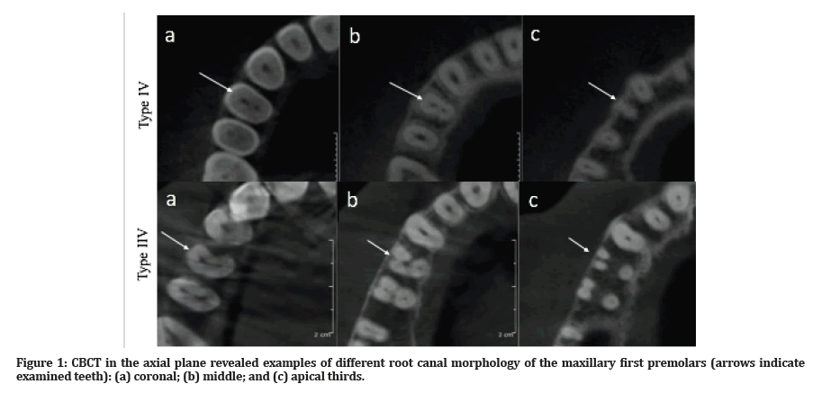
Type IV was the most prevalent in both males and females (68.3% and 58.9%, respectively), followed by type II (16.2% and 20.5%, respectively), and type I (5.7% and 13.2%, respectively). However, type VIII was only observed in males (4.7%). The gender difference was statistically significant (P=0.001). The most common types in Saudi participants were type IV (63.0%), type II (17.5%) and type I (12.6%), while in non-Saudis, the most common types included type IV (63.9%), type II (19.9%), and type III (8.1%). The difference was statistically significant (P=0.0004) (Table 3). Figures 1 and 2 show examples of maxillary first premolar root canal types detected in axial and coronal section, respectively.
| Types of root morphology | ||||||||||
|---|---|---|---|---|---|---|---|---|---|---|
| Type I | Type II | Type III | Type IV | Type V | Type VI | Type VII | Type VIII | Additional types | P-value† | |
| Gender | ||||||||||
| Male | 11 (5.7) | 31 (16.2) | 6 (3.1) | 131 (68.3) | 1 (0.5) | 2 (1.0) | 1 (0.5) | 9 (4.7) | 0 | 0.001* |
| Female | 25 (13.2) | 39 (20.5) | 6 (3.2) | 112 (58.9) | 3 (1.6) | 4 (2.1) | 1 (0.5) | 0 | 0 | |
| Total | 36 (9.4) | 70 (18.3) | 12 (3.1) | 243 (63.6) | 4 (1.1) | 6 (1.6) | 2 (0.5) | 9 (2.4) | 0 | |
| Nationality | ||||||||||
| Saudi | 31 (12.6) | 43 (17.5) | 1 (0.4) | 155 (63.0) | 3 (1.2) | 4 (1.6) | 2 (0.8) | 7 (2.9) | 0 | 0.0004* |
| Non- Saudi | 6 (4.4) | 27 (19.9) | 11 (8.1) | 87 (63.9) | 1 (0.7) | 2 (1.5) | 0 | 2 (1.5) | 0 | |
| Total | 37 (9.7) | 70 (18.3) | 12 (3.1) | 242 (63.3) | 4 (1.1) | 6 (1.6) | 2 (0.5) | 9 (2.4) | 0 | |
| †Fisher’s exact test with Monte Carlo simulation | ||||||||||
| *Statistically significant | ||||||||||
Table 3: Frequency distribution of root morphology in maxillary first premolar by gender nationality [n (%)].

Figure 1: CBCT in the axial plane revealed examples of different root canal morphology of the maxillary first premolars (arrows indicate examined teeth): (a) coronal; (b) middle; and (c) apical thirds.

Figure 2:CBCT in the coronal plane revealed examples of different root canal morphology of the maxillary first premolars: (a) Type II (2-1); (b) Type III (1-2-1); and (c) Type IV (2).
Root morphology in maxillary second premolar by gender and nationality
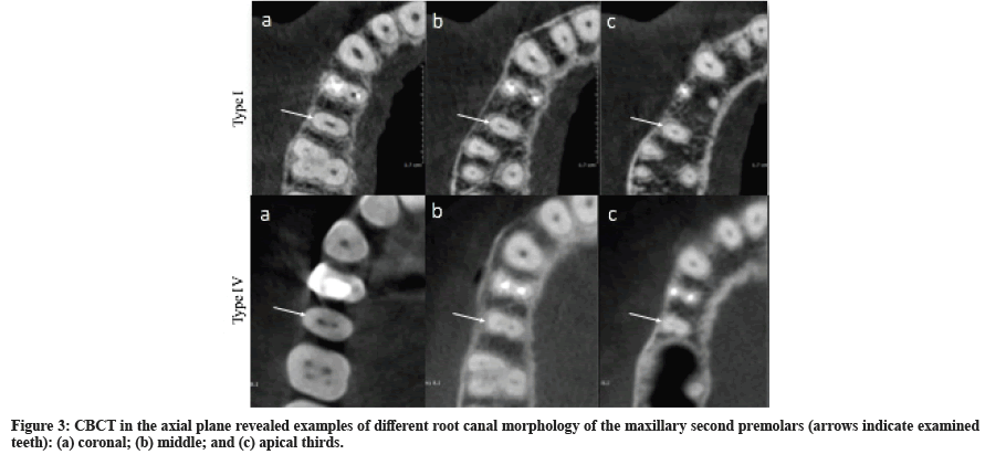
The most common types in males were type IV (33.8%), type II (28.6%), and type I (28.0%); while in females the most common types were type I (48.2%), type II (25.6%), and type IV (20.1%). The gender difference was statistically significant (P=0.002). No statistically significant difference across nationality was observed (P=0.9) (Table 4). Figures 3 shows examples of maxillary second premolar root canal types detected in axial.
| Types of root morphology | ||||||||||
|---|---|---|---|---|---|---|---|---|---|---|
| Type I | Type II | Type III | Type IV | Type V | Type VI | Type VII | Type VIII | Additional types | P-value† | |
| Gender | ||||||||||
| Male | 49 (28.0) | 50 (28.6) | 3 (1.7) | 59 (33.8) | 9 (5.1) | 0 (0.0) | 1 (0.6) | 2 (1.1) | 2 (1.1) | 0.002* |
| Female | 79 (48.2) | 42 (25.6) | 5 (3.1) | 33 (20.1) | 4 (2.4) | 0 (0.0) | 0 (0.0) | 1 (0.6) | 0 (0.0) | |
| Total | 128 (37.8) | 92 (27.1) | 8 (2.4) | 92 (27.1) | 13 (3.8) | 0 (0.0) | 1 (0.3) | 3 (0.9) | 2 (0.6) | |
| Nationality | ||||||||||
| Saudi | 80 (37.4) | 58 (27.1) | 5 (2.3) | 56 (26.2) | 12 (5.6) | 0 (0.0) | 0 (0.0) | 3 (1.4) | 0 (0.0) | 0.9 |
| Non- Saudi | 49 (38.9) | 34 (26.9) | 3 (2.4) | 36 (28.6) | 1 (0.8) | 0 (0.0) | 1 (0.8) | 0 (0.0) | 2 (1.6) | |
| Total | 129 (37.9) | 92 (27.1) | 8 (2.4) | 82 (27.1) | 13 (3.8) | 0 (0.0) | 1 (0.3) | 3 (0.8) | 2 (0.6) | |
| †Fisher’s exact test with Monte Carlo simulation | ||||||||||
| *Statistically significant | ||||||||||
Table 4: Frequency distribution of root morphology in maxillary second premolar by gender and nationality [n (%)].

Figure 3:CBCT in the axial plane revealed examples of different root canal morphology of the maxillary second premolars (arrows indicate examined teeth): (a) coronal; (b) middle; and (c) apical thirds.
Root morphology in maxillary first premolar by gender and nationality
The number of roots was symmetrical in 75.0% and 74.7% of maxillary first and second premolars pairs, respectively. Root canal morphology was symmetrical in 31.0% and 53.8% of maxillary first and second premolars pairs, respectively. The highest symmetry rate was observed in type IV in both first (65.6%) and second (87.9%) maxillary premolar pairs (Table 5).
| Parameter | Maxillary first premolars | Maxillary second premolars | ||||
|---|---|---|---|---|---|---|
| Number of pairs | Symmetry | Asymmetry | Number of pairs | Symmetry | Asymmetry | |
| Number of roots | 100 (100.0) | 75 (75.0) | 25 (25.0) | 91 (100.0) | 68 (74.7) | 23 (25.3) |
| One | 78 (78.0) | 53 (67.9) | 25 (32.1) | 65 (71.4) | 43 (66.2) | 22 (33.8) |
| Two | 21 (21.0) | 21 (100.0) | 0 | 26 (28.6) | 25 (96.2) | 1 (3.8) |
| Three | 1 (1.0) | 1 (100.0) | 0 | 0 (0.0) | 0 | 0 |
| Morphology | 100 (100.0) | 31 (31.0) | 69 (69.0) | 91 (100.0) | 49 (53.8) | 42 (46.2) |
| Type I | 43 (43.0) | 7 (16.3) | 36 (83.7) | 37 (40.7) | 11 (29.7) | 26 (70.3) |
| Type II | 24 (24.0) | 2 (8.3) | 22 (91.7) | 19 (20.8) | 8 (42.1) | 11 (57.9) |
| Type III | 0 | 0 | 0 (0.0) | 1 (1.1) | 0 (0.0) | 1 (100.0) |
| Type IV | 32 (32.0) | 21 (65.6) | 11 (34.4) | 33 (36.3) | 29 (87.9) | 4 (12.1) |
| Type V | 0 | 0 | 0 | 1 (1.1) | 1 (100.0) | 0 |
| Type VI | 0 | 0 | 0 | 0 | 0 | 0 |
| Type VII | 0 (0.0) | 0 | 0 | 0 | 0 | 0 |
| Type VIII | 1 (1.0) | 1 (100.0) | 0 | 0 | 0 | 0 |
| Type VIII | 0 | 0 | 0 | 0 | 0 | 0 |
Table 5: Symmetry of maxillary first and second premolars [n (%)].
Discussion
Summary of findings
This CBCT-based study demonstrated that Vertucci's type IV and type I were the most common root canal morphology of maxillary first and second premolars, respectively. In maxillary first premolars, two-rooted and single-rooted variants were the most frequent, while single-rooted variant was predominant in maxillary second premolars. Root numbers and root canal morphology of maxillary first and second premolars showed significant diffrence across gender. However, root canal morphology of the maxillary first premolar showed significant difference by nation.
Morphology and number of roots in maxillary first premolar
In this study population, two-rooted anatomy was the most frequent, observed in approximately 58% of the participants, and was more frequent in males (65.6%) compared to females (50%). The morphological classification showed type IV to be the most frequent, found in approximately two-thirds of the participants, regardless of gender or nationality. On the other hand, significant differences across gender and nationality were observed in less common types, notably type VIII which was only observed in males. In addition, type III which was the third most common type in non-Saudi participants, instead of type I in Saudis.
Other local studies have reported some variations in the maxillary first premolars. For example, a study by Alqedairi et al. used CBCT to analyze the number of roots and canal configuration of 334 maxillary first premolars. The findings were consistent with those from the present study, showing double-rooted (75.1%), and type IV (70.6%) to be the predominant pattern. Similar to the findings in this study, type I was the second most common (11%), followed by type II (8.4%) [12]. Furthermore, Alqedairi et al. reported 2.3% versus 0% three-rooted teeth in males versus females, respectively, which is consistent with 4.2% versus 0% the findings in this study. However, the authors did not find a significant difference across gender regarding the canal configuration, while the difference was statistically significant in the present study. In another study by Atieh [13], 246 extracted maxillary first premolars were analyzed visually and radiographically. Results showed the predominance of double-rooted teeth accounting for 81% of the sample, while 18% were single-rooted. Although, the author did not use Vertucci's classification, it was found that the most common type (63%) had two separate root canals from the pulp chamber to the apex, corresponding to Vertucci's type IV. The second most prevalent type (27%) had two fused roots, which corresponds to Vertucci's type II. However, type I was observed in only 9%, which is not consistent with the findings in the present study (23%).
International data generally agree for the commonness of double- and single-rooted teeth, with type IV being often the most frequent canal configuration. However, some exceptions have been noted. A study among the Yemeni population, which analyzed 250 maxillary first premolars using staining and digital photography, showed that 55% of the teeth were single-rooted and 44.4% were double-rooted. However, the canal system configuration was consistent with findings in this study, showing type IV to be the most common (56%), followed by type I and III [14]. In a Turkish population, an in vitro study of 653 extracted maxillary first premolars showed 54% double-rooted and 45% single-rooted teeth [15], which is consistent with the pattern observed in this study. Furthermore, although authors did not classify the morphology using Vertucci's classification, they reported that 68.5% had “two separate root canals that remained separate with two apical foramina”, which corresponds to Vertucci's type IV. In North India, Gupta et al. analyzed 250 extracted maxillary first premolar teeth and found that 54% were single-rooted, which is inconsistent with the data in this study and with other Saudi studies. However, similar to this study findings, type IV configuration was the most frequent, accounting for 33.2% of the cases, followed by type I (23.2%) [16]. In Europe, similar observations were reported in Spanish and German populations, where double-rooted pattern was the most common in maxillary first premolars, followed by single-rooted, and type IV being the most frequent in both single- and double-rooted teeth [9,17]. Type IV was also the most common (42%) in a North American population, followed by type I (28%) and type II (26%) [18].
Morphology and number of roots in maxillary second premolar
Regarding the maxillary second premolar, approximately 78% were single-rooted, more frequently in females than in males (86.0% vs. 69.7%, respectively), and the most common canal configuration was type I (aproximately, 38%), notably among females (48.2%), while type IV was the most common among males (33.8%). For both genders, type II was the second most common. While all gender differences were statistically significant. However, no significant differences were observed by nation.
In the study by Alqedairi et al., of the 318 maxillary second premolars analyzed, 85.2% were single-rooted with no notable difference across gender (87% in males versus 83% in females). Additionally, the most frequent canal configuration was type I (49%), irrespective of gender, followed by type II (26%) and type IV (12%) [12]. Concordantly, maxillary second premolars were found to be single-rooted in 83% of the Spanish and German populations; however, while Vertucci’s type I was predominant in the Spanish population [9], types V (29%) and IV (25%) were most frequent in the German population [17].
Variations across gender and nationality
The authors observed significant differences across gender in the number of roots and canal configuration. In both maxillary premolars, the single-rooted pattern was more frequent among females. Regarding the canal morphology, type VIII was only observed in males in maxillary first premolars. However, in maxillary second premolars, type IV was the most frequent in males, while type I was the most frequent in females. The gender predilection has been previously reported in several studies, such as the previously cited German study [17], showing generally fewer number of roots and root canals in females than in males. In a study by Martins et al., which analyzed 12,325 teeth from 670 Portuguese individuals, the percentage of single-rooted teeth was significantly higher in females including maxillary first and second premolars [19]. Another study from Turkey demonstrated that females exhibited more frequently single-rooted and single canal maxillary first and second premolars than males, and the differences were statistically significant [20]. However, a Saudi study that examined 5,254 maxillary and mandibular permanent teeth, in 100 males and 108 females, showed no significant difference in the number of roots or canals between males and females. By focusing on premolars, results showed a significantly higher percentage of one canal in females (42.4%), compared to males (34.1%), in the Saudi population. The authors concluded that there were significant gender disparities, with respect to the number of canals, which were only observed in certain groups of teeth [21].
With respect to the nationality, there was no difference in the root numbers in either tooth, whereas a statistically significant difference was observed in the canal configuration of maxillary first premolars. Ethnically wise, the Saudi population comprises a great diversity, notably in the Western region of the country, where this study has been conducted as demonstrated by geneticsbased ethnic analysis. The Eastern region also comprises great ethnic diversity, while more limited diversity was observed in the Central and Northern regions of the country [22]. The ethnic group mixture among the Saudi population may explain the absence of significant difference between the Saudi citizens and non-Saudi participants with regards to the morphological types and number of roots in the maxillary premolars.
Number of roots and canal morphology symmetry
Symmetry in root number was equally observed in threequarters of the maxillary first and second premolars pairs, and a double-rooted premolar was symmetrical in almost all cases (96.2-100%). On the other hand, symmetry in root canal morphology was relatively low, and was more frequent in type IV, probably due to its high prevalence. By comparison, Guneser and Unver observed symmetry in the root and canal morphology among 93% and 98% of mandibular first and second premolar pairs, among a population of Turkish patients [23]. Likewise, Li et al. observed 80.2% and 81.8% symmetry in the number of roots and 72.3% and 73.2% in root morphology, in maxillary first and second premolar pairs respectively. However, Li et al. observed a higher symmetry in type I in maxillary second premolars, as this was the most prevalent root morphology in the studied population [24].
Conclusions
There is a high heterogeneity in the maxillary premolar root canal configuration among the studied Saudi population. This is more noticeable in the first premolar, where single- and double-rooted patterns compete with each other, while Vertucci's type IV remains the most common canal configuration. In second premolars, single-rooted pattern is predominant. In both first and second premolars, a significant gender disparity is observed in the number of roots as well as in the canal configuration supporting fewer roots and canals in females. On the other hand, differences across nationality were less significant.
References
- Vertucci FJ. Root canal morphology and its relationship to endodontic procedures. Endod Top 2005;10:3–29.
- Ahmed HMA, Hashem AA. Accessory roots and root canals in human anterior teeth: A review and clinical considerations. Int Endod J 2016; 49:724–36.
- Ahmed HMA, Abbott P V. Accessory roots in maxillary molar teeth: A review and endodontic considerations. Aust Dent J 2012; 57:123–31.
- Versiani MA, Ordinola-Zapata R, Keleş A, et al. Middle mesial canals in mandibular first molars: A micro-CT study in different populations. Arch Oral Biol 2016; 61:130–7.
- Grover C, Shetty N. Methods to study root canal morphology: A review. Endo 2012; 6:171–82.
- Ahmed HMA, Versiani MA, De-Deus G, et al. A new system for classifying root and root canal morphology. Int Endod J 2017; 50:761–770.
- Vertucci F, Seelig A, Gillis R. Root canal morphology of the human maxillary second premolar. Oral Surg Oral Med Oral Pathol 1974; 38:456–464.
- Ahmad IA, Alenezi MA. Root and root canal morphology of maxillary first premolars: a literature review and clinical considerations. J Endod 2016; 42:861–872.
- Abella F, Teixidó LM, Patel S, et al. Cone-beam computed tomography analysis of the root canal morphology of maxillary first and second premolars in a spanish population. J Endod 2015; 41:1241–1247.
- Elnour M, Khabeer A, AlShwaimi E. Evaluation of root canal morphology of maxillary second premolars in a Saudi Arabian sub-population: An in vitro microcomputed tomography study. Saudi Dent J 2016; 28:162–168.
- Yan Y, Li J, Zhu H, et al. CBCT evaluation of root canal morphology and anatomical relationship of root of maxillary second premolar to maxillary sinus in a western Chinese population. BMC Oral Health 2021; 21:358.
- Alqedairi A, Alfawaz H, Al-Dahman Y, et al. Cone-beam computed tomographic evaluation of root canal morphology of maxillary premolars in a Saudi population. Biomed Res Int 2018; 2018:1–8.
- Atieh MA. Root and canal morphology of maxillary first premolars in a Saudi population. J Contemp Dent 2008; 9:46–53.
- Senan EM, Alhadainy HA, Genaid TM, et al. Root form and canal morphology of maxillary first premolars of a Yemeni population. BMC Oral Health 2018; 18:94.
- Özcan E, Çolak H, Hamidi MM. Root and canal morphology of maxillary first premolars in a Turkish population. J Dent Sci 2012; 7:390–4.
- Gupta S, Sinha D, Gowhar O, et al. Root and canal morphology of maxillary first premolar teeth in north Indian population using clearing technique: An in vitro study. J Conserv Dent 2015; 18:232.
- Bürklein S, Heck R, Schäfer E. Evaluation of the root canal anatomy of maxillary and mandibular premolars in a selected German population using cone-beam computed tomographic data. J Endod 2017; 43:1448–1452.
- Guo J, Vahidnia A, Sedghizadeh P, et al. Evaluation of root and canal morphology of maxillary permanent first molars in a north american population by cone-beam computed tomography. J Endod 2014; 40:635–639.
- Martins JNR, Marques D, Francisco H, et al. Gender influence on the number of roots and root canal system configuration in human permanent teeth of a Portuguese subpopulation. Quintessence Int 2018; 49:103–111.
- Evlice B, Duyan H. Canal configuration of maxillary premolars in Cukurova population: A CBCT analysis. Balk J Dent Med 2021; 25:147–152.
- Mashyakhy M, Gambarini G. Root and root canal morphology differences between genders: A comprehensive in-vivo CBCT study in a saudi population. Acta Stomatol Croat 2019; 53:231–246.
- Khubrani YM, Wetton JH, Jobling MA. Extensive geographical and social structure in the paternal lineages of Saudi Arabia revealed by analysis of 27 Y-STRs. Forensic Sci Int Genet 2018; 33:98–105.
- Güneşer MB, ÜNVER T. Prevalence of bilateral symmetry in root and root canal anatomy of mandibular premolars: A cone-beam computed tomography study. Clin Dent Res 2016; 40:66–72.
- Li Y, Bao S, Yang X, et al. Symmetry of root anatomy and root canal morphology in maxillary premolars analyzed using cone-beam computed tomography. Arch Oral Biol 2018; 94:84–92.
Indexed at, Google Scholar, Cross Ref
Indexed at, Google Scholar, Cross Ref
Indexed at, Google Scholar, Cross Ref
Indexed at, Google Scholar, Cross Ref
Indexed at, Google Scholar, Cross Ref
Indexed at, Google Scholar, Cross Ref
Indexed at, Google Scholar, Cross Ref
Indexed at, Google Scholar, Cross Ref
Indexed at, Google Scholar, Cross Ref
Indexed at, Google Scholar, Cross Ref
Indexed at, Google Scholar, Cross Ref
Indexed at, Google Scholar, Cross Ref
Indexed at, Google Scholar, Cross Ref
Indexed at, Google Scholar, Cross Ref
Indexed at, Google Scholar, Cross Ref
Indexed at, Google Scholar, Cross Ref
Indexed at, Google Scholar, Cross Ref
Indexed at, Google Scholar, Cross Ref
Indexed at, Google Scholar, Cross Ref
Indexed at, Google Scholar, Cross Ref
Indexed at, Google Scholar, Cross Ref
Author Info
Arwa S Alnoury1*, Lina O Bahanan2, Mohanad A Alhedbany3, Mustafa E Abuzinadah3, Hanadi M Khalifah4 and Abrar A Tounsi5
1Department of Restorative Dentistry, Faculty of Dentistry, King Abdulaziz University, Jeddah 21589, Saudi Arabia2Department of Dental Public Health, Faculty of Dentistry, King Abdulaziz University, Jeddah 21589, Saudi Arabia
3Faculty of Dentistry, King Abdulaziz University, Jeddah 21589, Saudi Arabia
4Oral and Maxillofacial Radiology Division, Faculty of Dentistry, King Abdulaziz University, Jeddah 21589, Saudi Arabia
5Department of Periodontics and Community Dentistry, College of Dentistry, King saud university, Saudi Arabia
Citation: Arwa S Alnoury, Lina O Bahanan, Mohanad A Alhedbany, Mustafa E Abuzinadah, Hanadi M Khalifah, Abrar A Tounsi, Evaluation of Root Canal Morphology of Maxillary First and Second Premolars by Cone-Beam Computed Tomography in Saudi Populations, J Res Med Dent Sci, 2022, 10 (8):83-90.
Received: 06-Aug-2022, Manuscript No. jrmds-22-71361; , Pre QC No. jrmds-22-71361(PQ); Editor assigned: 08-Aug-2022, Pre QC No. jrmds-22-71361(PQ); Reviewed: 22-Aug-2022, QC No. jrmds-22-71361; Revised: 24-Aug-2022, Manuscript No. jrmds-22-71085(R); Published: 31-Aug-2022
