Research - (2020) Advances in Dental Surgery
Evaluation of Knowledge and Perspective of Endodontic Residents and General Dentist Towards the Endodontic Application of CBCT in Saudi Arabia
Areen Aljuhani1*, Smita Dutta1 and Ayman Mandorah2
*Correspondence: Areen Aljuhani, College of Dentistry, Qassim University, Saudi Arabia, Email:
Abstract
Objective: This study is aiming to evaluate the knowledge, skills, and the awareness of CBCT importance in endodontic treatment and diagnosis among the Endodontic residents and General Dentists in Saudi Arabia. Methodology: This is a cross-sectional survey carried out among Saudi General Dentists and Endodontic residents. The questionnaire was administered to 99 participants. This survey consists fourteen closed-ended questions formulated and validated by the Endodontics Committee in Qassim University. Results: On analyzing the response to the questionnaire it was found that 39 General Dentist chose limited FOV and 11 for full FOV, while all Endodontic residents chose limited FOV. About 10 participants rate the accuracy and specificity of CBCT verses digital radiography as equally accurate and specific, and 80 rate as thrice accurate and specific. Around 81 participants think that the true size, location be appreciated with CBCT, 5 participants thinks no, while 13 participants says I don’t know. Conclusion: This study reveals that information and applicability of CBCT in varied clinical dental specifications is furnished by few dental colleges. However, to churn the maximum benefit of the CBCT, its uses, advantages, contraindications, and interpretation. More efforts and ideas need to be incorporated in teaching curriculum that fall well within the limits of the institute.
Keywords
Cone beam computed tomography, Interpretation, Endodontic residents, Questionnaire survey, Root canal treatment, Endodontic diagnosis
Introduction
Cone Beam Computed Tomography (CBCT) is not a long past invented diagnostic imaging modality which produces accurate threedimensional (3D) image construction [1,2]. CBCT 3-D anatomic representation has overcome the limitation of two-dimensional (2D) radiographs such as overlapping of osseous structures [1,2,3] Low-definition imaging of anatomic structure being assessed which may impair the accuracy of diagnosis [3].
CBCT applications in dentistry have eased the image interpretation thus improving the diagnosis and treatment planning in most dentistry fields such as the dental implant, location and the number of root canals, teeth impaction, orthognathic surgeries tumors.
The American Association of Endodontics, along with the American Academy of Oral and Maxillofacial Radiology has provided evidencebased guidelines regarding the applications of CBCT in endodontics, As CBCT can provide a small field of view image with low dose and high resolution to be applicable in endodontic diagnosis, treatment planning, and after treatment assessment.
It provides information about the pulp chamber size, morphology of the tooth, location and number of canals, degree of calcification, direction and curvature, fractures, and iatrogenic defects. [4-8]
CBCT imaging should be carried out by an appropriately qualified and well-trained dentist. Therefore, dentists should make a wise decision in the prescription of CBCT examinations by consulting recommendations from CBCT evidence-based guidelines [4,5,9,10].
Literature search shows no published paper of knowledge and skills on CBCT interpretation in endodontic treatment procedures in Saudi Arabia; therefore, this study is aiming to compare the knowledge, skills, and the awareness of CBCT importance in endodontic treatment and diagnosis among the Endodontic residents and General Dentists in Saudi Arabia [11,12].
Materials and Methods
This is a cross-sectional study, questionnairebased survey conducted among Saudi General Dentists and Endodontic residents. The questionnaire was administered to 99 participants of two groups:50 General dentists and 49 Endodontic residents. Participants were offered to fill the questionnaires online using a version designed to be accessible on mobile phones and computers. participation in this survey was voluntary. Therefore , consent was assumed by the voluntary choice of participating, and this study was approved by the ethical approval committee (Ethics committee of Qassim University). This survey consists fourteen closedended questions were formulated and validated by the Endodontics Committee In Qassim University. The questionnaire collected data regarding the participant's gender, specialty, educational level, and Data that evaluate knowledge and skills of CBCT interpretation in endodontic treatment procedures.
Statistical analysis
The collected data was analyzed with IBM, SPSS statistics software 23.0 Version. To describe the data, descriptive statistics like frequency analysis, and percentage analysis were used.
Results
About 150 applicants were invited to participate in this study but we received responses from only 99 applicants, out of which 50 (50.5%) were general dentists and 49 (49.5%) were endodontic residents. Among the total, 50 (51%) were males and 48 (49%) were females. 1 (1%) choose OPG as the choice of method for endodontic diagnosis, 16 (16.2%) chooses conventional methods for diagnosis, 63 (63.6%) chooses digital methods of diagnosis and 19 (19.2%) chooses CBCT as the choice of endodontic diagnosis.
Around 48 (48.5%) participants of the survey had undergone training and 51 (51.5%) had not undergone any training nor did they attend any workshops. 8 (8.1%) would advise CBCT imaging for endodontic procedures whereas 63 (63.6%) would do it frequently and 28 (28.3%) would never advice.
65 (65.7%) of the participants have access to CBCT at workplace on-site, whereas 34 (34.3%) do not have it. About 26 (26.3%) have access to CBCT at workplace off-site and 73 (73.7%) do not have access. A maximum of 82 (82.8%) did not choose CBCT for its cost, 13 (13.1%) don’t choose because of the radiation exposure and 4 (4%) don’t choose because of lack of installation space.
The field of view (FOV) for CBCT in case of General Dentist is 39 for limited FOV and 11 for full FOV and in case of Endodontic resident is 49 for limited FOV and 0 for full FOV as shown below along with the graphical representation (Table 1 and Figure 1).
| Count | CBCT fields of view (FOV) | Total | ||
|---|---|---|---|---|
| Limited FOV CBCT | Full FOV CBCT | |||
| Skill | General dentist | 39 | 11 | 50 |
| Endodontic resident | 49 | 0 | 49 | |
| Total | 88 | 11 | 99 | |
Table 1: Skills *CBCT fields of view (FOV).
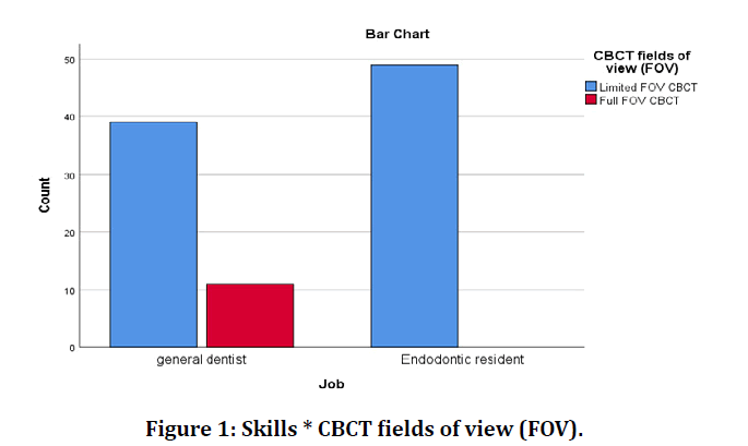
Figure 1: Skills * CBCT fields of view (FOV).
About 74 number of participants of this survey chooses CBCT for surgical re-treatment, followed by 53 in case of missing canals, 50 for internal and external resorption, 48 for dental trauma, 34 for calcified cases and differential diagnosis and 16 in case of non-surgical treatments (Figure 2).
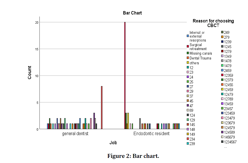
Figure 2: Accuracy and specificity of CBCT.
About 10 participants rate the accuracy and specificity of cone beam computed tomography verses digital radiography as equally accurate and specific, 80 participants rate the accuracy and specificity of cone beam computed tomography verses digital radiography as thrice accurate and specific and 2 participants rate the accuracy and specificity of cone beam computed tomography verses digital radiography (Table 2 and Figure 3).
| Count | Accuracy and specificity of CBCT | Total | ||||
|---|---|---|---|---|---|---|
| Equally accurate and specific | Thrice accurate and specific | Less accurate and specific | I don't Know | |||
| Job | General dentist | 5 | 36 | 2 | 7 | 50 |
| Endodontic resident | 5 | 44 | 0 | 0 | 49 | |
| Total | 10 | 80 | 2 | 7 | 99 | |
Table 2: Accuracy and specificity of CBCT.
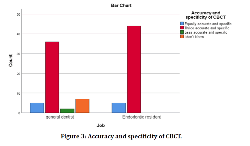
Figure 3: Accuracy and specificity of CBCT.
Around 81 participants think that the true size, location, and extent of a periapical lesion be appreciated with cone beam computed tomography, 5 participants think no, while 13 participants say I don’t know (Table 3 and Figure 4).
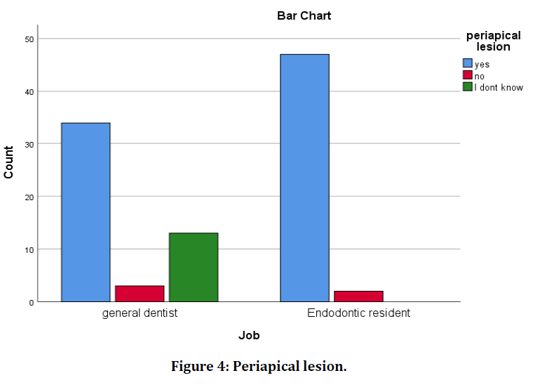
Figure 4: Periapical lesion.
| Count | periapical lesion | Total | |||
| yes | no | I dont know | |||
| Job | General dentist | 34 | 3 | 13 | 50 |
| Endodontic resident | 47 | 2 | 0 | 49 | |
| Total | 81 | 5 | 13 | 99 | |
Table 3: Periapical lesion.
77 participants think that Cone beam computed tomography detect radio lucent lesions before lingual and buccal plates are de-mineralized, followed by 21 who opted for I do not know and 1 opts for no (Table 4).
| Count | Radioluscent lesion | Total | |||
|---|---|---|---|---|---|
| Yes | No | I don’t know | |||
| Job | General dentist | 30 | 1 | 19 | 50 |
| Endodontic resident | 47 | 0 | 2 | 49 | |
| Total | 77 | 1 | 21 | 99 | |
Table 4: Radioluscent lesion.
86 participants agree CBCT is an indispensable diagnostic modality in for modern endodontic practice, 4 disagrees and 9 opts I don’t know (Figure 5).
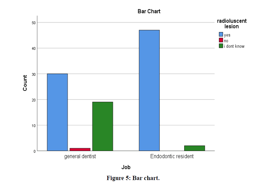
Figure 5: Bar chart.
Discussion
For Diagnosis, planning, execution, and evaluation of success of endodontic treatment, radiology is essential [13]. The radical change for dental and maxillofacial radiology is represented by Cone Beam Computed Tomography (CBCT). The use of CBCT has many clinical applications in the diagnosis of denture [11]. A detailed threedimensional teeth evaluation, maxillofacial skeletal structure and relation between the anatomical structures can be obtained via the CBCT. It is an extremely helpful tool in case of imaging the third molar extraction, sinus pathology, to detect vertical root fracture, maxillofacial surgery, evaluation of tumors, orthodontic cases, forensic dentistry and temporomandibular joint disorders [14].
SEDENTEXCT, a multinational project supported by EURATOM published the guidelines on the use of CBCT [15]. Recent research showed that there was less awareness about the clinical applications about the CBCT among the practitioners. Aditya et al. found in their study that CBCT is still not very frequently used by dental specialists due to less availability of the technique, high cost, or inability of case selection for CBCT imaging by the dentists [16]. The CBCT is less commonly used by the dental specialists to diagnose due to its cost, lack of installation space, resolution limitations and radiation exposure. In the past years very, limited literature is available about the awareness of the radiographic imaging in dentistry [17].
In the year 2008, the first CBCT was installed in the Kingdom of Saudi Arabia. The source of knowledge about the CBCT was available via., the postgraduate studies in Saudi Arabia [18]. In recent times the use of CBCT in radiographic imaging in dentistry field is gaining importance in Saudi Arabia as well. However, a clear literature stating about the knowledge and awareness about CBCT among the dental practitioners in Saudi Arabia is not available until now [19]. Thus, this subject gained our interest to study and survey the dental practitioners about the knowledge and awareness about CBCT in Saudi Arabia.
About 74 participants of this survey answered that they chose CBCT for surgical re-treatments. Only 48 participants had got trained for the use of CBCT. Yalcinkaya SE et al reported in his study 100% awareness among the participants about CBCT [13]. Ghoncheh et al. reported 72.2% using CBCT to evaluate the implants [20]. Lavanya et al reported that about 68.2% were partially aware of the technologies used in CBCT [21]. A study in Turkey [22] reported that there was less knowledge about the CBCT among their dental students and a study in South India [23] reported the same.
According to this study, only 19 (19.2%) of the dental practitioners chooses CBCT as the choice of endodontic diagnosis. About 9,000 members of the American Association of Oral and Maxillofacial Surgery (AAOMS) uses CBCT since 2007 [24]. In this study about 80 participants rate the accuracy and specificity of cone beam computed tomography verses digital radiography as thrice accurate and specific. The field of view (FOV) for CBCT in case of General Dentist is 39 for limited FOV whereas in case of Endodontic resident is 49 for limited FOV. Ghanbarnezhad et al. reported to measure doses of NewTom VGi , by changing field of view (FOV) size from 8×8 cm2 (height × diameter) to 6×6 cm2 [25]. 34 General Dentists and 47 endodontic residents thinks that the true size, location and extent of a periapical lesion be appreciated with cone beam computed tomography. 30 General dentists and 47 endodontic residents thinks that Cone beam computed tomography detect radio lucent lesions before lingual and buccal plates are de-mineralized.
However, with this survey it gave a hint that the endodontic residents had more knowledge about the CBCT when compared to the general dentists.
Rai S et al reported that Dentists including specialists from other specialities must gain more knowledge about indications and contraindications of digital imaging and CBCT for accurate diagnosis and better management of patients [26-28].
Conclusion
This research showed that specialized training influenced the result outcome as endodontic residents had more awareness about the CBCT when compared to the general dentists.
Although dental colleges in Saudi Arabia gives knowledge about the use of the CBCT in varied clinical applications in dentistry. The emerging need of acquiring skill, knowledge and useage of CBCt through various programs, workshops and firsthand experience need to be considered promptly. Also, proper teachings need to be given on the interpretation of data retrieved from CBCT.
Financial Support and Sponsorship
Nil.
Conflicts of Interest
None.
References
- Scarfe W, Farman A. What is cone-beam CT and how does it work? Dent Clin North Am 2008; 52:707-730.
- Venkatesh E, Venkatesh Elluru S. Cone beam computed tomography: basics and applications in dentistry. J Istanbul University Faculty Dent 2017; 51.
- Sethi P, Tiwari R, Das M, et al. Two dimensional versus three-dimensional imaging in endodontics-An updated review. J Evolution Med Dent Sci 2016; 5:6287-6293.
- Scarfe WC, Levin MD, Gane D, et al. Use of cone beam computed tomography in endodontics. Int J Dent 2009; 2009.
- Patel S, Durack C, Abella F, et al. European society of endodontology position statement: The use of CBCT in endodontics. Int Endodont J 2014; 47:502-504.
- Pinsky H, Dyda S, Pinsky R, et al. Accuracy of three-dimensional measurements using cone-beam CT. Dentomaxillofac Radiol 2006; 35:410-416.
- Scarfe W, Levin M, Gane D, et al. Use of cone beam computed tomography in endodontics. Int J Dent 2009; 2009:1-20.
- Patel S. New dimensions in endodontic imaging: Part 2. Cone beam computed tomography. Int Endodont J 2009; 42:463-475.
- Horner K, Islam M, Flygare L, et al. Basic principles for use of dental cone beam computed tomography: Consensus guidelines of the European academy of dental and maxillofacial radiology. Dentomaxillofac Radiol 2009; 38:187-195.
- Meena N. Applications of cone beam computed tomography in endodontics: A review. Dentistry. 2014; 4.
- Balabaskaran K, Srinivasan A. Awareness and attitude among dental professional towards CBCT. J Dent Med Sci 2013; 10:55-59.
- Janani K, Sandhya R. A survey on skills for cone beam computed tomography interpretation among endodontists for endodontic treatment procedure. Indian J Dent Res 2019; 30:834-838.
- Yalcinkaya SE, Berker YG, Peker S, et al. Knowledge and attitudes of Turkish endodontists towards digital radiology and cone beam computed tomography. Niger J Clin Pract 2014; 17:471-478.
- Lopes IA, Tucunduva RM, Handem RH, et al. Study of the frequency and location of incidental findings of the maxillofacial region in different fields of view in CBCT scans. Dentomaxillofac Radiol 2017; 46:20160215
- Sedentexct, European commission, radiation protection N 172: Cone beam ct for dental and maxillofacial radiology. Evidence based guidelines, SEDENTECT Project, Luxembourg, Europe, 2012.
- Aditya A, Lele S, Aditya P. Current status of knowledge, attitude, and perspective of dental practitioners toward cone beam computed tomography: A survey. J Oral Maxillofac Radiol 2015; 13:54-57.
- Tyndall DA, Rathore S. Cone-beam CT diagnostic applications: Caries, periodontal bone assessment, and endodontic applications. Dent Clin North Am 2008; 52:825-841.
- Almohiy H, Alshahrani I. Cone beam computed tomography (CBCT) in dentistry-students’ and interns’ perspective in Abha, Saudi Arabia. Ann Trop Med Public Health 2019; 22.
- Miles DA. The future of dental and maxillofacial imaging. Dent Clin North Am 2008; 52:917-928.
- Ghoncheh Z, Panjnoush M, Kaviani H, et al. Knowledge and attitude of iranian dentists towards cone-beam computed tomography. Front Dent 2019; 16:379-385.
- Lavanya R, Babu DB, Waghray S, et al. A questionnaire cross-sectional study on application of CBCT in dental postgraduate students. Pol J Radiol 2016; 81:181-189.
- Kamburoglu K, Kursun S, Akarslan ZZ. Dental students' knowledge and attitudes towards cone beam computed tomography in Turkey. Dentomaxillofac Radiol 2011; 40:439-443.
- Ramakrishnan P, Shafi FM, Subhash A, et al. Asurvey on radiographic prescription practices in dental implant assessment among dentists in Kerala, India. Oral Health Dent Manag 2014; 13:826-830.
- American Dental Association. US department of health and human services. The selection of patients for dental radiographic examinations. American Dental Association, Chicago 2004.
- Ghanbarnezhad Farshi R, Mesbahi A, Johari M, et al. Dosimetry of critical organs in maxillofacial imaging with cone-beam computed tomography. J Biomed Phys Eng 2019; 9:51-60.
- Rai S, Misra D, Dhawan A, et al. Knowledge, awareness, and aptitude of general dentists toward dental radiology and CBCT: A questionnaire study. J Indian Acad Oral Med Radiol 2018; 30:110-115.
- Ganguly R, Ramesh A. Systematic interpretation of CBCT scans: Why do it? J Mass Dent Soc 2014; 62:68-70.
- Garlapati K, Babu DBG, Chaitanya NCSK, et al. Evaluation of preference and purpose of utilisation of cone beam computed tomography (CBCT) compared to orthopantomogram (OPG) by dental practitioners–A cross-sectional study. Pol J Radiol 2017; 82:248-251.
Author Info
Areen Aljuhani1*, Smita Dutta1 and Ayman Mandorah2
1College of Dentistry, Qassim University, Saudi Arabia2Faculty of Dentistry, Taif University, Saudi Arabia
Citation: Areen Aljuhani, Smita Dutta, Ayman Mandorah, Evaluation of Knowledge and Perspective of Endodontic Residents and General Dentist Towards the Endodontic Application of CBCT in Saudi Arabia, J Res Med Dent Sci, 2020, 8 (7): 459-464.
Received: 07-Oct-2020 Accepted: 10-Nov-2020 Published: 17-Nov-2020
