Research - (2022) Volume 10, Issue 11
Effect of Three Filling Techniques Using Gutta-Percha and Bioceramic-Based Sealer on Apical Leakage Levels in Roots with Different Apical Foramen Sizes
Esma Dinger2* and Emre Bodrumlu1
*Correspondence: Esma Dinger, Faculty of Dentistry, Bolu Abant Izzet Baysal University, Bolu, Turkey, Email:
Abstract
Aim: This in-vitro study aimed to evaluate the effect of three different filling techniques on apical leakage in roots with various apical foramen diameters, using gutta-percha and bioceramic-based root canal sealers. Method: A total of 160 extracted maxillary anterior teeth were divided into four main groups of teeth based on the apical foramen diameters. The apical foramen diameters of 0.30 mm, 0.40 mm, 0.50 mm, and 0.80 mm were prepared using a 30 #K, 40 #K, 50 #K, and 80 #K type hand file, respectively. Following root canal preparation was completed with adjusted rotary system and root canals were filled using gutta-percha and Well Root ST bio ceramic sealer. The samples were then divided into 3 subgroups as filling techniques the single cone, the lateral condensation, and the thermoplastic injection. The amount of apical leakage of the samples was evaluated using the dye penetration technique. Result: The amount of apical leakage varied according to apical diameter and filling technique used. The greatest leakage occurred in the teeth filled using the single-cone technique with an apical diameter of 0.80 mm (80.74 ± 0.45) and the least leakage occurred in the teeth filled by using the single cone technique with an apical diameter of 0.30 mm (2.821 ± 0.28). There was a significant difference between the groups (p<0.05) Conclusion: In teeth with smaller apical diameters, less apical leakage occurred. The filling technique affects the amount of apical leakage differently depending on the apical diameter of the tooth.
Keywords
Apical diameter size, Apical leakage, Bioceramic based root canal sealer, Root canal filling techniques, Single cone technique, Lateral condensation technique, Thermoplastic injection technique
Introduction
The main purpose of endodontic treatment is to eliminate the dental lesions and clean the bacterial smear for effective canal filling, therefore reducing the possibility of reinfection to keep the teeth functional. Root canal treatment is one of the endodontic treatments, based on the three-dimensional hermetic filling of the canals by forming an apical plug at the root apex, after biomechanical shaping and disinfection of the canals [1].
Microorganisms and their products are major etiological factors of pulpal or pulp-induced persistent periapical diseases [2]. Especially the cleaning of bacteria and their products from the root canal system is one of the important stages of endodontic treatment. The removal of microorganisms from the root canal system is achieved by disinfecting and shaping the root canals. Therefore, the root canal filling is achieved by a hermetic sealing from the coronal to the apical of the canal to prevent leakage [3].
Canal shaping and apical plug formation may cause difficulties in the following cases: i. When the root apex is not closed, in young and permanent teeth where root development has stopped due to inflammation or necrosis caused by caries or trauma, ii. When resorption is present, in teeth with periapical lesions, iii. When apical construction points are distorted due to apical resection or over-instrumentation, iv. When dentin walls are thin and apical foramens are wide [2,4].
In cases where the apical foramen is large in diameter for various reasons, the filling materials potentially come into contact with a greater surface area along the canal walls, increasing the possibility of leakage. However, the tendency of a large apical width to leak compared to a smaller apical width has not been precisely specified [2]. Due to apical micro leakage, bacteria can spread from areas that are not completely filled in the apical, causing both persistent infections and apical pathologies.
Many techniques and canal filling materials have been used to fill the root canals. Gutta-percha and root canal sealers are the most common filling materials used to plug the root canal void. With the development of technology, many new root canal sealers and guttapercha filling techniques have been developed. Bioceramic root canal sealers are among the materials whose usage and popularity have increased recently.
In our study was chose sealer (Well Root-ST, Werom, Gangwon-Do, South Korea) which is used as a calcium silicate-based, bioactive, bioceramic root canal sealer. Well Root-ST (Werom, Gangwon-Do, South Korea) shows more durability, less apical leakage, less toxicity and more regenerative effect in root canals compared to other root canal sealers. It has been stated by the manufacturer and studies that it is bioactive and can support root development, and it is reported that it is a canal sealer that can be preferred in teeth with open apex and presence of perforation.
There is no root canal filling technique that provides full hermetic occlusion in root canal treatment. However, the applied superiority of each technique in studies may vary depending on used the root canal sealer, the anatomical canal width of the tooth, preparation techniques and washing procedures. In filling techniques, different methods have also been developed to evaluate the sealing of root canal filling materials and leakage at the apical [5,6]. Currently, there are no data on apical leakage of teeth prepared with different apical diameters, and filled with gutta-percha and bioceramic root canal sealer [2].
This study aimed to evaluate the effect of three different filling techniques with two different sealer materials (gutta-percha and bioceramic-based sealar) on apical leakage in roots with four different apical foramen diameters.
Materials and Methods
In our study, 160 single-root and single-canal maxillary incisors containing central, lateral and canine with similar root morphology, without any restorative procedures, without fractures, cracks in the crown and roots, without caries, with closed apexes, were used. (The teeth extracted as a result of the consent/approval obtained from the patients were used in the study.) In the study, the teeth with straight root morphology were used and the completion of development of each root was analyzed by the radiographic examination. Dental calculus, organic debris and soft tissues on the teeth were cleaned with a cavitron (Cavitron, Siemens, Germany) and periodontal curette (#3-4 Gracey, Nordent, USA) and the teeth were placed in saline at room temperature until they were used in the study.
To standardize the root length of the teeth, they were cut with diamond fissure bur (ISO 806314, 014, Meisinger, Germany) using water cooling at an average distance of 15 mm from the apex. An endodontic access cavity was opened on the roots under water cooling using a diamond fissure bur (ISO 806314, 012, Meisinger, Germany) and a diamond rond bur (ISO 806314, 021, Meisinger, Germany).
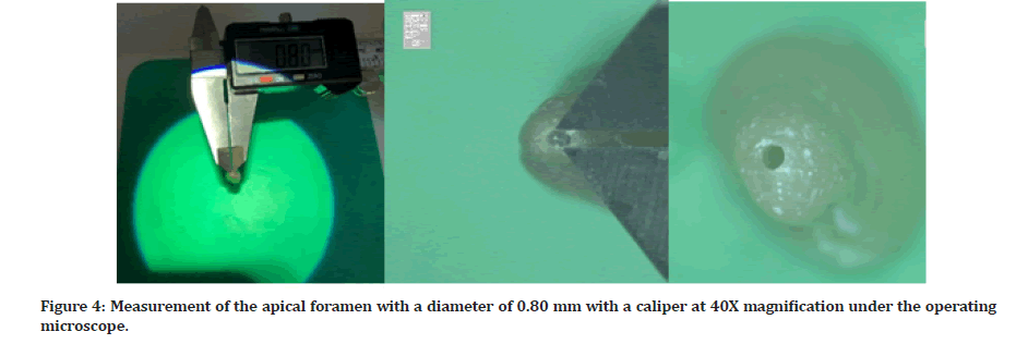
To determine the working length, a size 10 K-type file (Dentsply Maillefer, Ballaigues, Switzerland) was introduced in the root canal until the file was visible at the apical foramen and working lengths were measured. Four main groups, each consisting of 36 teeth, and a positive and negative control group consisting of 8 teeth, were formed in proportion to the feeling of clenching teeth and the width of the natural apical foramen. In this direction, the diameters of the apical foramen of the teeth in the 4 main groups were expanded to 0.30 mm, 0.40 mm, 0.50 mm and 0.80 mm with hand files of the type 30 #K, 40 #K, 50 #K, 80 #K, respectively, until they became visible at the apical foramen. In this way, apical foramens with different diameters were obtained. Digital images were obtained under 40x magnification with the help of an operating microscope so that the expanded diameters of the apical foramens were examined (Figures 1 to Figure 4).

Figure 1: Measurement of the apical foramen with a diameter of 0.30 mm with a caliper at 40X magnification under the operating microscope.

Figure 2: Measurement of the apical foramen with a diameter of 0.40 mm with a caliper at 40X magnification under the operating microscope.

Figure 3: Measurement of the apical foramen with a diameter of 0.50 mm with a caliper at 40X magnification under the operating microscope.

Figure 4: Measurement of the apical foramen with a diameter of 0.80 mm with a caliper at 40X magnification under the operating microscope.
The new working length was determined to be 1 mm shorter than the working length of the files placed at the level of the apical foramen until they became visible at the apical foramen. The X-Smart Endomotor (Dentsply Maillefer) and Protaper Universal rotary file systems (F3, F4, F5) (Dentsply Maillefer) were used for the application. Root canals were shaped with crown down technique diameter of 0.30 mm group 1, up to F4; diameter of 0.40 mm group 2, up to F5; diameter of 0.50 mm group 3, up to F5 (shortened by 1 mm) and diameter of 0.80 mm group 4, up to F5 (shortened by 4 mm) respectively. Each root canal file was irrigated with 2 ml of 5,25% sodium hypochlorite (NaOCl) solution (microwave, Melikgazi, Antalya, Turkey) and dried, before using. Completing the preparation, the smear layer was removed, with 5 ml of 17% EDTA (EDTA Melikgazi microwave, Antalya, Turkey) by immersing for 1 minute in canals. Then canals were irrigated with 2 ml of 5,25% NaOCl (sodium hypochlorite microwave, Melikgazi, Antalya, Turkey) and were dried. Finally, the root canals were washed with 5 ml of distilled water and dried with paper cones.
After irrigation was completed, 4 main groups consisting of teeth with different apical diameters, were divided into three subgroups, according to the filling techniques.
The samples were filled with only bioceramic based root canal sealer (Well-Root ST, Vericom, Gangwon-Do, South Korea) using the single cone technique, the lateral condensation technique and the thermoplastic injection technique. In the single cone technique and the lateral condensation technique, the main cone was used for the root canal filling. The filling was adjusted to the apical, and the feeling of tug back was controlled.
A total of 16 teeth with an apical diameter of 0.30 mm, 0.40 mm, 0.50 mm, and 0.80 mm were separated as the control group. The samples of the negative control group were filled with the root canal filling sealer and gutta-percha. The positive control group samples were left blank. After the root canal fillings were completed, the mouths of the canals were closed with temporary filling material (Caviton G-C Dental Industrial Corp., Tokyo, Japan).
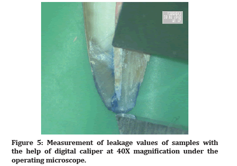
All the filled samples were covered with a double layer of nail polish, except for the apical 2 mm parts. The specimens in the positive control group were covered with nail polish, except for the apical 2 mm, while the specimens in the negative control group were completely covered with nail polish. Each group was placed in a test tube with 2% methylene blue solution, centrifuged at 3000 rpm at 3G for 3 minutes, and then the samples were washed and separated into two with a diamond separator in the longitudinal direction. Dye penetration level from the apical foramen to the coronal end of the canal. Dye penetration level formed from apical to coronal was measured with a micrometer (caliper) by taking digital images at 40x magnification under the operating microscope (Figure 5). The analysis of the data was performed with the statistical analysis program (SPSS 19.0, SPSS Inc. Chicago, IL, USA). Intragroup analysis was evaluated using One Way ANOVA, and intergroup analysis using Two-way ANOVA. The confidence interval for ANOVA was defined as 95%, and the statistical significance limit was set as alpha = 0.05, p<0.05 was considered statistically significant.

Figure 5: Measurement of leakage values of samples with the help of digital caliper at 40X magnification under the operating microscope.
Results
The results of the comparison among the groups with different apical foramen diameters and the groups with different filling techniques are shown in Table 1. The mean measured apical leakage values are given with average values ± standard deviations and p values. While no staining was observed in the teeth of the negative control group, there was a dye leakage along the canal in the teeth of the positive control group.
| Apical foramen filling (mm) Techniques | 0.30 mm | 0.40 mm | 0.50 mm | 0.80 mm |
|---|---|---|---|---|
| GRUP 1 (n:36) | GRUP 2 (n:36) | GRUP 3 (n:36) | GRUP 4 (n:36) | |
| The Single Cone Technique | 2.877 ±0.29C.a | 3.451 ± 0.21C.a | 4.665 ±0.46B.a | 8.074 ± 0.45A.a |
| The Lateral Condansation Technique | 2.821 ± 0.28B. a | 3.321 ±0.37 B.a | 3.486 ± 0.27B. b | 4.835 ± 0.52 A.c |
| The Thermoplastic İnjection Technique | 2.946 ± 0.45B. a | 3.202±0.31 B.a | 3.803±0.32 B.b | 5.983±0.60 A.b |
| *Different superscript characters (a, b, c: Comparison between rows; A, B, C: Comparison between columns) differ statistically between groups. (P<0.05). Leakage amounts are measured in millimeters. (e.g. 2,946 mm) | ||||
Table 1: The mean (± standard deviation) of apical leakage values for the samples in all of the groups.
When the filling techniques and apical diameters were compared, it was found that the group of teeth with an apical diameter of 0.80 mm had a statistically higher leakage than the other groups of teeth with different apical diameters (p<0.05). Although there was no statistically significant difference in apical leakage for the three filling techniques in teeth with diameters of 0.30 and 0.40 mm, the highest leakage was observed with the single cone technique in teeth with diameters of 0.50 and 0.80 mm. In the filling techniques for teeth with a diameter of 0,80 mm, the lowest apical leakage was found in the lateral condensation technique, whereas the highest leakage was observed in the single-cone technique (p<0.05).
Discussion
The success of root canal treatment is provided by cleaning the root canal from microorganisms and their products, debris and pulpal residues with chemo mechanical preparation and three-dimensional hermetic filling of the canal to prevent the progression of infection [7].
Since the apical region of the root canal is a critical area that can accommodate a large number of microorganisms, it is very important to determine the endpoint of the preparation as accurately as possible and to provide adequate apical expansion to achieve ideal root canal treatment [8,9].
In endodontic treatment, the location of the apical construction point and the width of the apical foramen may vary from tooth to tooth [9,10]. In addition, the large apical foramen diameter is an anatomical feature and may be due to several reasons. These reasons include incomplete apical formation due to problems in the root development phase, pathologic or physiologic root resorption as a result of inflammation or trauma, root canal preparation overflow, apical resection and failure of regeneration procedures [2].
In the studies, various similar techniques were used to obtain different apical widths. Cristina Bucchi, et al. [11] Cimilli, et al. [12] and Koc, et al. [13] rely on viewing the canal files from the apical to determine the size of the apical foramen opening. In our study, the determination of apical foramen of different widths was based on the technique used by these researchers and the diameters of the apical foramen were measured with a caliper under 40X magnifications with the help of an operating microscope.
Bioceramic root canal sealers are now a commonly used option in root canal treatment today due to their many advantages. In particular, many studies have been conducted on leakage and coverage of bioceramic sealers [14-16]. However, there are no previous studies focused on the relationship between the bioceramic root canal sealers and apical sealing levels with different filling techniques of teeth with different apical diameters.
When root canal filling techniques are evaluated in the literature, it can be seen that each technique has its advantages and disadvantages, and there is no root canal filling technique that provides a complete hermetic filling. In our study, the lateral condensation technique, the single cone technique and the thermoplastic injection technique were used as root canal filling techniques in accordance with the recommendations of the manufacturers.
When all the filling techniques were evaluated according to apical diameters in our study, it was observed that apical leakage values increased with increasing apical diameter. Similar to our study, Saluja, et al. [17] evaluated the extent of apical leakage in teeth with apical foramen diameters of 0.20 mm, 0.30 mm and 0.50 mm filled with the lateral condensation method, and they observed that dye penetration increased with increasing apical diameter. The point that differs from our study is Saluja, et al. the apical constriction point is not disturbed.
Mente, et al. [18] compared the groups of the tooth they filled with the thermoplastic compaction and the lateral condensation technique at an apical diameter of 0.70 mm and 1.40 mm in terms of the amount of apical leakage and found that the amount of apical leakage increased with the increase of apical diameter. However, they could not find a statistically significant difference between filling the techniques. In our study, when apical foramen diameter and filling techniques are evaluated together, there was no statistical difference between the apical sealing levels with different filling techniques, but the least apical leakage occurred in roots with an apical diameter of 0.30 mm filled with the lateral condensation technique. The apical leakage values in the thermoplastic injection technique were similar to those in other studies that performed the thermoplastic injection technique [19,20].
In the studies, it was found that the thermoplastic injection could not reach the apical triad in teeth with narrow apical diameters and curvaceous teeth, and the gutta-percha could not be sufficiently condensed and a gap could be left between the canal walls [21,22].
In our study, we found that teeth in the groups of teeth with 0.30 mm apical diameter, filled with the lateral condensation and the single-cone technique, showed better condensation and less apical leakage compared to groups of teeth with larger apical diameters. In addition, there was no significant difference between filling techniques, although lateral condensation technique showed less apical leakage than the single cone technique, similar to other studies with the apical diameter of 30 mm [23].
At an apical diameter of 0.40 mm, there was no significant difference between the filling techniques in terms of apical leakage, but the highest leakage in the groups was found in the single-cone technique and the lowest leakage in the thermoplastic injection technique. Similar to our study, Tanikonda, et al. [24], reported that there was no statistically significant difference between the 0.40 mm apical diameter thermoplastic injection and lateral condensation techniques in their study in which they evaluated the leakage of the teeth filled with thermoplastic injection and lateral condensation techniques in different apical diameters. Dumani, et al. [25], in their study evaluating the amount of voids and leakage probability of different filling techniques, stated that while the highest voids were in the single cone and lateral condensation technique, the least voids were found in the thermoplastic injection technique. This could be because the thermoplastic injection technique adapts better to the irregularities in the canal with good condensation and provides a complete sealing up to the apical area.
Studies suggest that the width of the canal increases as apical diameter increases and the single-cone technique results in a larger gap of gutta-percha, leading to an increase in the amount of apical leakage [26,27]. In the 0.50 and 0.80 groups in our study, the highest leakage occurred in the single-cone technique and the least leakage occurred in the lateral condensation technique. As stated in the studies, in this study also more apical leakage was observed with the thermoplastic injection technique than with the lateral condensation technique due to insufficient condensation against the risk of overfilling throughout the apical [28]. Similar to our study, Vizgirda, et al. [29] showed that when the teeth with an apical diameter of 0.80 mm were filled with lateral condensation technique, the thermoplastic injection technique and MTA plug, the less apical leakage occurred in the lateral condensation technique.
Many techniques are used when evaluating the amount of leakage caused by root canal filling in the apical. In our study, the dye penetration method with 2% methylene blue was preferred to evaluate the leakage caused by the used bioceramic root canal sealer and different filling techniques for the canal filling of teeth with different apical foramen diameters. Compared to other micro leakage methods, the dye penetration method with methylene blue is more economical, does not require extensive laboratory studies, and can provide the results for evaluating bacterial penetration due to low molecular weight and deep penetration ability [30]. In addition, samples were centrifuged in 2% methylene blue at 3G at 3000 rpm for 3 minutes so as to active dye penetration.
Conclusion
Apical leakage values were found to increase with increasing apical diameter in roots filled with guttapercha and bioceramic-based sealer using three different filling techniques.
Apical leakage levels were low in all filling techniques used with bioceramic-based sealer in roots below 0.50 mm apical diameter, and there was no significant difference among them.
In teeth with an apical diameter of 0.50 mm or more, lateral condensation technique should be preferred among the filling techniques by using the gutta-percha and bioceramic-based sealer as filling materials, because the lateral condensation technique provided the least apical leakage.
References
- Pires MD, Martins JNR, Baruwa AO, et al. Leukocyte platelet-rich fibrin in endodontic microsurgery: A report of 2 cases. Restor Dent Endod 2022; 47:e17.
- Silvestrin T, Torabinejad M, Handysides R, et al. Effect of apex size on the leakage of gutta-percha and sealer-filled root canals. Quintessence Int 2016; 47:373-378.
- Vo K, Daniel J, Ahn C, et al. Coronal and apical leakage among five endodontic sealers. J Oral Sci 2022; 64:95-98.
- Vula V, Ajeti N, Kuçi A, et al. An In Vitro comparative evaluation of apical leakage using different root canal sealers. Med Sci Monit Basic Res 2020; 26:e928175.
- Silva Almeida LH, Moraes RR, Morgental RD, et al. Are premixed calcium silicate-based endodontic sealers comparable to conventional materials? A systematic review of In Vitro studies. J Endod 2017; 43:527-535.
- Desouky A, El-Razik A, Bayiumy A. An in-vitro comparative assessment of the sealing ability of AH? Plus sealer with different root canal nano sealers?. Egyptian Dent J 2022; 68:1043-1052.
- de Castro Kruly P, Alenezi HEHM, Manogue M, et al. Residual bacteriome after chemomechanical preparation of root canals in primary and secondary infections. J Endod 2022; 48:855-863.
- Pacheco-Yanes J, Gazzaneo I, Campello AF, et al. Planned apical preparation using cone-beam computed tomographic measures: A micro-computed tomographic proof of concept in human cadavers. J Endod 2022; 48:280-286.
- ElAyouti A, Hülber-J M, Judenhofer MS, et al. Apical constriction: Location and dimensions in molars-a micro-computed tomography study. J Endod 2014; 40:1095-1099.
- Zhang CC, Liu YJ, Yang WD, et al. Morphological changes of the root apex in anterior teeth with periapical periodontitis: An in-vivo study. BMC Oral Health 2022; 22:31.
- Bucchi C, Gimeno-Sandig A, Manzanares-Céspedes C. Enlargement of the apical foramen of mature teeth by instrumentation and apicoectomy. A study of effectiveness and the formation of dentinal cracks. Acta Odontol Scand 2017; 75:488-495.
- Cimilli H, Aydemir S, Kartal N, et al. Comparing the accuracy of four electronic apex locators for determining the minor diameter: An ex vivo study. J Dent Sci 2013; 8:27-30.
- Koç S, Kustarci A, Er K. Accuracy of different electronic apex locators in determination of minimum root perforation diameter. Aust Endod J 2022.
- Pawar SS, Pujar MA, Makandar SD. Evaluation of the apical sealing ability of bioceramic sealer, AH plus & epiphany: An in vitro study. J Conserv Dent 2014; 17:579-582.
- Arul B, Varghese A, Mishra A, et al. Retrievability of bioceramic-based sealers in comparison with epoxy resin-based sealer assessed using microcomputed tomography: A systematic review of laboratory-based studies. J Conserv Dent 2021; 24:421-434.
- Manjila JC, Vijay R, Srirekha A, et al. Apical microleakage in root canals with separated rotary instruments obturated with different endodontic sealers. J Conserv Dent 2022; 25:274.
- Saluja P, Mir S, Bavabeedu SS, et al. Relation between apical seal and apical preparation diameter: An in vitro study. J Pharm Bioallied Sci 2020; 12:332.
- Mente J, Werner S, Koch MJ, et al. In vitro leakage associated with three root-filling techniques in large and extremely large root canals. J Endod 2007; 33:306-309.
- Chauhan A, Makkar S, Garg N, et al. Comparison of the apical sealing ability of gutta-percha by three different obturation techniques: Lateral Condensation technique, single cone root canal obturation technique and Injectable thermoplasticized gutta-percha technique (System B). Ann Rom Soc Cell Biol 2021; 25:873-879.
- Simsek N, Keles A, Ahmetoglu F, et al. 3D Micro-CT analysis of void and gap formation in curved root canals. Eur Endod J 2017; 2(1):1-5.
- Türker SA, Uzunoglu-Özyürek E, Kasikçi S, et al. Filling quality of several obturation techniques in the presence of apically separated instruments: A Micro-CT study. Microsc Res Tech 2021; 84:1265-1271.
- Alim BA, Garip Berker Y. Evaluation of different root canal filling techniques in severely curved canals by micro-computed tomography. Saudi Dent J 2020; 32:200-205.
- Kumari M, Taneja S, Bansal S. Comparison of apical sealing ability of lateral compaction and single cone gutta percha techniques using different sealers: An in vitro study. J Pierre Fauchard Acad 2017; 31:67-72.
- Tanikonda R, Nalam PN, Sajjan GS, et al. Evaluation of the quality of obturation with obtura at different sizes of apical preparation through microleakage testing. J Clin Diagn Res 2016; 10:35-38.
- Dumani A, Yilmaz S, Yoldas O, et al. Evaluation of various filling techniques in distal canals of mandibular molars instrumented with different single-file nickel-titanium systems. Nigerian J Clin Practice 2017; 20:307-312.
- Sakaue H, Komatsu K, Yoshioka T, et al. Evaluation of coronal leakage and pathway of dye leakage after obturation with various materials for open apical foramina. Dent Mater J 2013; 32:130-137.
- Laslami K, Dhoum S, El Harchi A, et al. Relationship between the apical preparation diameter and the apical seal: An in vitro study. Int J Dent 2018; 2018:2327854.
- Lone MM, Khan FR. Evaluation of micro leakage of root canals filled with different obturation techniques: An in vitro study. J Ayub Med Coll Abbottabad 2018; 30:35-39.
- Vizgirda PJ, Liewehr FR, Patton WR, et al. A comparison of laterally condensed gutta-percha, thermoplasticized gutta-percha, and mineral trioxide aggregate as root canal filling materials. J Endod 2004; 30:103-106.
- Chandak M, Gangamwar N, Manwar N, et al. Comparative reliability of assessment of dye penetration using 5% methylene blue, India dye ink, 6.5% basic fuschin, 5% eosin along with the root canal filling: an in vitro study. J Adv Med Dent Sci Res 2016; 4:18.
Indexed at, Google Scholar, Cross Ref
Indexed at, Google Scholar, Cross Ref
Indexed at, Google Scholar, Cross Ref
Indexed at, Google Scholar, Cross Ref
Indexed at, Google Scholar, Cross Ref
Indexed at, Google Scholar, Cross Ref
Indexed at, Google Scholar, Cross Ref
Indexed at, Google Scholar, Cross Ref
Indexed at, Google Scholar, Cross Ref
Indexed at, Google Scholar, Cross Ref
Indexed at, Google Scholar, Cross Ref
Indexed at, Google Scholar, Cross Ref
Indexed at, Google Scholar, Cross Ref
Indexed at, Google Scholar, Cross Ref
Indexed at, Google Scholar, Cross Ref
Indexed at, Google Scholar, Cross Ref
Indexed at, Google Scholar, Cross Ref
Indexed at, Google Scholar, Cross Ref
Indexed at, Google Scholar, Cross Ref
Indexed at, Google Scholar, Cross Ref
Indexed at, Google Scholar, Cross Ref
Indexed at, Google Scholar, Cross Ref
Indexed at, Google Scholar, Cross Ref
Indexed at, Google Scholar, Cross Ref
Indexed at, Google Scholar, Cross Ref
Indexed at, Google Scholar, Cross Ref
Indexed at, Google Scholar, Cross Ref
Author Info
Esma Dinger2* and Emre Bodrumlu1
1Faculty of Dentistry, Bolu Abant Izzet Baysal University, Bolu, Turkey2Faculty of Dentistry, Zonguldak Bulent Ecevit University Zonguldak, Turkey
Received: 14-Oct-2022, Manuscript No. jrmds-22-78901; , Pre QC No. jrmds-22-78901(PQ); Editor assigned: 17-Oct-2022, Pre QC No. jrmds-22-78901(PQ); Reviewed: 02-Nov-2022, QC No. jrmds-22-78901(Q); Revised: 01-Nov-2022, Manuscript No. jrmds-22-78901(R); Published: 08-Nov-2022
