Research - (2019) Volume 7, Issue 4
Effect of Impacted Third Molars on the Overjet of Central Incisor Teeth
*Correspondence: Hussein Haleem Jasim, Department of Oral Diagnosis, College of Dentistry, University of Wasit, Iraq, Email:
Abstract
Background: Overjet considered one of the most important parameters to evaluate the status of occlusion, therefore any disturbances in overjet will affect the occlusion improperly and causing malocclusion. The early detection and diagnosis of overjet discrepancies help in the early treatment of malocclusion. So many studies were achieved to observe the effect of different factors on overjet extent.
Aim: To observe whether the presence of impacted third molars affects the overjet extent. Materials and methods: The study was done on (200) patients with impacted third molars, aged between 18–25 years, for both genders who referred to the radiology department in some dental centres in Baghdad for taking orthopantomograms (OPGs) to evaluate the status of impacted third molars from June 2017 to November 2018. The measurements of overjet were done by a graduated metal ruler on each patient with impacted third molars and the metal ruler is covered by a disposable dental sleeve after each measurement. Overjet estimated from the labial surface of the more lingual positioned mandibular incisor and the incisal edge of the more labial positioned maxillary incisor. A normal overjet range considered in the study was measured between 2-4 mm.
Results: The study showed that there were only (20) cases of patients with partially impacted third molars had increased overjet and only (5) cases of patients with fully impacted third molars had found with an increased overjet. The statistical analysis showed that there was no significant relationship between the presence of impacted third molars and overjet extent (p<0.05). The results also showed that there was no any significance between males and females regarding the effect of impacted third molars on the overjet distance.
Conclusion: The study concluded that there was no relationship between the presence of impacted third molars and overjet extent.
Keywords
Third molar, Impacted third molar, Molar, Overjet, Central incisor, Teeth
Introduction
Overjet was defined as the anterior-posterior space measured through the incisal edge of the most protruded maxillary central incisors to the labial surface of opposing mandibular central incisors. An overjet larger than (3 mm) was decided to be increased [1].
The status of the maxillary and mandibular incisors in harmony to each other and to their related bones is an important characteristic in the analysis of the case, stabilization prognosis of treatment, coordination and, regularity of the facial profile [2,3].
Overjet considered one of the measurable factors used to evaluate the sagittal correlation of the upper and lower dentition. The variation of overjet extent could be skeletal, dental, or incorporation of both. In addition to the facial profile, overjet in adolescents is a significant parameter where the peak of development and determination of surgical or orthodontic treatment is critical. In general, if the overjet is more than 10 mm, surgery is the most suitable decision [4].
The proportion of impacted third molars
Tooth impaction is an abnormal condition in which a tooth cannot or will not emerge into its natural functioning status [5]. The dental impactions are mostly asymptomatic and usually have no clear unusual appearance excluding maxillary incisors [6]. About 25% to 50% of the people are vulnerable to impacted teeth [7,8]. In this way, any tooth in the jaws may be vulnerable to impaction. It was reported that the most commonly dental impactions in jaws are third molars and canines, followed by upper premolars, second lower premolars or upper incisors respectively [9,10]. It was reported that the impacted maxillary impactions found to be about 0.92%-4.3% [7,8]. Some authors stated that if the impacted teeth repositioned into the proper alignment, this will produce a complete anterior dentition with a proper alignment of gingival margins and a good smile [11]
On the other side, the predominance of third molar impaction found to be ranged from 16.7%- 68.6% [12-19] and it considered the most common impacted teeth that can be found in jaws [20]. The only possible interpretation for high incidence rate might be due to the incomplete expansion of the retromolar space [21-23]. Others stated that the third molar impaction in Europe is up to 73% of young adults. (24) In general, third molars found to be emerged in about [17-21] years-old [25,26]. In addition, the emergence period of third molars have been found to differ with ancestries [25-27]. For instance, in Europeans and Nigerians, mandibular third molars may emerge up to 26 years of age and up to 14 years old respectively [27,28].
Many studies observed the incidence of impacted third molars in males and females and they found no gender predilection in the impaction of third molars [12,13,15,17]. But other studies found a females predilection of third molar impaction rather than males [19,27,29,30]. The rate of emergence of mandibular third molars in male is about 3-6 months prior to females [31].
The eruption time and the different positions of third molars following eruption are not only associated with ancestries but also may be due to with dietary components intake, the force on using the muscular system and hereditary effect [32].
Material and Methods
Study design
The study was done on (200) patients (106 males and 94 females) with impacted third molars, aged between 18– 25 years. in regardless of gender, who referred to the radiology department in some dental centres in Baghdad for taking orthopantomograms (OPGs) for detecting or evaluating the presence and position of impacted third molars from June 2017 to November 2018.
All patients have been informed for the purpose of the study and all data and information will be used for research purposes and they were agreed to conduct the study.
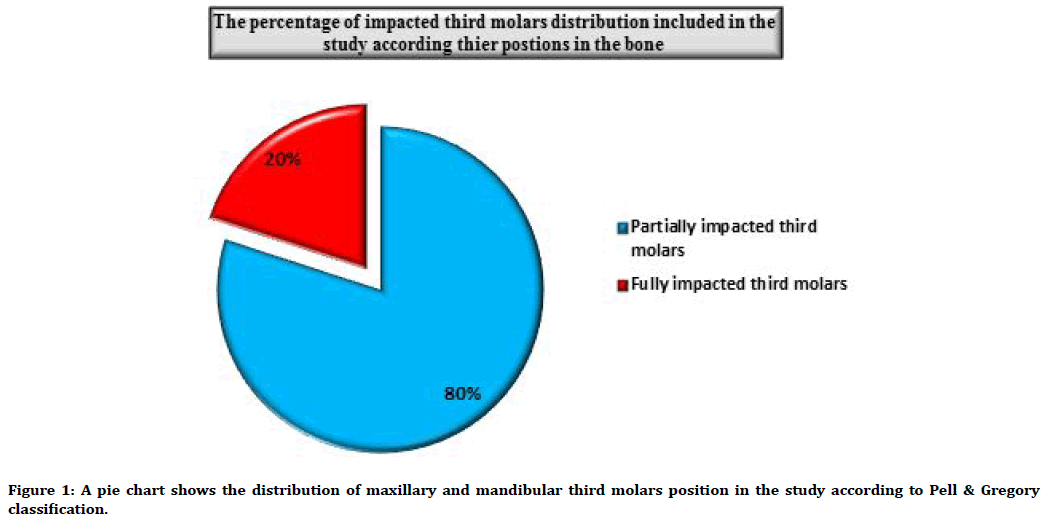
The radiographical examinations showed that 160 patients have bilateral maxillary and mandibular partially impacted third molars and forty patients have bilateral maxillary and mandibular fully impacted third molars.
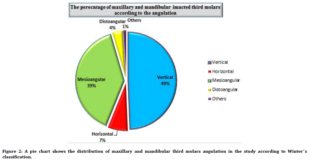
The impacted third molars included in this study were distributed according to the classification of Pell et al. [33]. According to their positions in the bone; whether not impacted, partially impacted and fully impacted in the bone (the non-impacted third molars not included in this study) and Winter’s classification [34] according to their angulation in the bone; whether the impactions were a vertical, horizontal, mesioangular, distoangular and others) Figures 1 and 2.

Figure 1. A pie chart shows the distribution of maxillary and mandibular third molars position in the study according to Pell & Gregory classification.

Figure 2. A pie chart shows the distribution of maxillary and mandibular third molars angulation in the study according to Winter’s classification.
Inclusion criteria
The following conditions were included in the study
• Patients with bilateral maxillary and mandibular partially and fully impacted third molars.
• Patient with normal occlusion.
• Patient with good oral hygiene.
Exclusion criteria
The following conditions were excluded from the study
• Patient without third molar impactions or with normally erupted third molars.
• Extensive pathology in the jaws and dentition which may affect the occlusion.
• Patients with fractured anterior teeth or class IV restorative filling.
• Posterior or anterior missing teeth.
• Patient with moderate to severe mobility of teeth.
• Any craniofacial anomaly.
• Patients with orthodontic treatment or dental appliance.
• Patients with malocclusion or crowding.
• Patients with dental attrition.
• Patients with presence of parafunctional habits.
Measurement of overjet
The measurements of overjet were done by a graduated metal ruler on each patient with impacted third molars. The metal ruler was covered by a disposable dental sleeve after each measurement. Overjet is estimated as the extent between the upper and lower incisors at the point of utmost severity. It is estimated from the labial surface of the more lingual positioned mandibular incisor and the incisal edge of the more labial positioned maxillary incisor. A normal overjet range considered in the study was measured between 2-4 mm [35].
Statistical analyses
Data were analyzed using the Statistical Package for the Social Sciences (SPSS for Windows, version 19). The relations between the groups were analyzed using the Pearson Chi-square test. The level of significance was 0.05 and p-value considered significant if it is <0.05.
Results
The study showed that among two hundred patients, there were only twenty cases of patients with partially impacted third molars had increased overjet and only five cases of patients with fully impacted third molars had found with an increased overjet.
The relationship between the presence of impacted third molars and overjet extent was tested by using Pearson’s chi-squared test and the p-value was accepted as significant if it is ˂0.05. The statistical analysis showed that there was no significant relationship between the presence of impacted third molars and overjet extent.
The results also showed that there was no any significance between males and females regarding the effect of impacted third molars on the overjet distance. The chi-square statistic is (0) and the p-value is (1). This result is not significant at p<0.05 Table 1.
| Patients samples | Increased overjet | Normal overjet | Total |
|---|---|---|---|
| Patients with partially impacted third molars | 20 [11 males 9 females] | 140 [73 males 67 females] | 160 |
| Patients with completely impacted third molars | 5 [3 males 2 females] | 35 [19 males 16 females] | 40 |
| Totals | 25 | 175 | 200 |
Table 1: Distribution of patients samples with impacted third molars position and overjet status.
Discussion
Overjet measurements are very important because of its value in analysis and treatment plan in case of overjet discrepancies where orthodontic or surgery is necessary.
Harrison et al. stated that it is particularly important to obtain an accurate measurement of overjet to detect the malocclusion [36].
So, many studies discussed the importance of overjet parameter in the development of malocclusion while some studies have been observed different factors which have a major or minor effect on the overjet; Kaisa et al. used the overjet parameter to find whether there is a relationship between the changes in the overjet and arch width between the ages (7-32) years [37].
Ingrid et al. observed the association between the overjet and overbite linear measurements and the inter-incisal space restricted by the upper and lower incisors morphology [38].
Terie et al. studied the effect of sever malocclusions (as in sever overjet) on the alveolar bone height [39].
Livia et al. found that the increased exposure to the risk of dental trauma was associated to increased overjet [40].
This study is unique because it is observed whether the presence of impacted third molars may be affected the overjet extent and it found there was no significant relationship between the presence of impacted third molars and overjet extent.
This conclusion is consistent with the explanation stated that the increase in overjet was due to defect in the growth of jaw rather than it was caused by the malposition of the teeth, but till now, no significant data has been published [41].
But on the other hand, and in regard to the increase in overjet in some patients with partially impacted third molars, it has been observed that the most cases associated with this increase in overjet were mesioangulated maxillary third molars rather than other cases, so this type of impaction may cause impact on the adjacent teeth in causing an increase in the mesial shifting, in turn to the maxillary anterior teeth causing some discrepancy and an increase in overjet. So this explanation may lead to the assumption that if we increase the sample size of maxillary mesio-angulated impacted third molars, we may get a significant effect regarding the overjet extent.
Conclusions
The study concluded that there was no relationship between the presence of impacted third molars and overjet extent.
References
- Moyers RE. Handbook of orthodontics. 4th Edn. Chicago: Year Book Medical Publisher; 1988.
- Broadbent BH. A new X-ray technique and its application in orthodontica. Angle Orthod 1931; 1:45–46.
- Williams R. The diagnostic line. Am J Orthod 1969; 5:458–476.
- Proffit WR, Phillips C, Tulloch JFC, et al. Surgical versus orthodontic correction of skeletal class II malocclusion in adolescents: Effects and indication. Int J Adult Orthod Orthog Surg 1992; 209-220.
- Bishara SE. Impacted maxillary canines:a review. Am J Orthod Dentofacial Orthop 1992; 101:159–71.
- Becker A. The orthodontic treatment of impacted teeth 2nd Edn London: Informa Healthcare. 2007.
- Jacoby H. The etiology of maxillary canine impactions. Am J Orthod. 1983; 84:125–32.
- Ngan P, Hornbrook R, Weaver B. Early timely management of ectopically erupting maxillary canines. Semin Orthod 2005; 11:152–63.
- Grover PS, Lorton L. The incidence of unerupted permanent teeth and related clinical cases. Oral Surg Oral Med Oral Pathol 1985; 59: 420−425.
- Aktan AM, Kara S, Akgünlü F, et al. The incidence of canine transmigration and tooth impaction in a Turkish subpopulation. Eur J Orthod 2010; 32:575−581.
- Sukh R, Singh GP, Tandon P. Interdisciplinary approach for the management of bilaterally impacted maxillary canines. Contemp Clin Dent 2014; 5:539–544.
- Kaya GS, Aslan M, Ömezli MM, et al. Some morphological features related to mandibular third molar impaction. J Clin Exp Dent 2010; 2:12–17.
- Hattab FN, Rawashdeh MA, Fahmy MS. Impaction status of third molars in Jordanian students. Oral Surg Oral Med Oral Pathol Radiol Endod. 1995; 79:24–29.
- Scherstén E, Lysell L, Rohlin M. Prevalence of impacted third molars in dental students. Swed Dent J 1989; 13:7–13.
- Brown LH, Berkman S, Cohen D, et al. A radiological study of the frequency and distribution of impacted teeth. J Dent Assoc S Afr 1982; 37:627–30.
- Fanning EA, Moorrees CF. A comparison of permanent mandibular molar formation in Australian aborigines and Caucasoids. Arch Oral Biol 1969; 14:999–1006.
- Haidar Z, Shalhoub SY. The incidence of impacted wisdom teeth in a Saudi community. Int J Oral Maxillofac Surg. 1986; 15:569–571.
- Kramer RM, Williams AC. The incidence of impacted teeth. A survey at harlem hospital. Oral Surg Oral Med Oral Pathol 1970; 29:237–241.
- Quek SL, Tay CK, Tay KH, et al. Pattern of third molar impaction in a Singapore Chinese population: A retrospective radiographic survey. Int J Oral Maxillafac Surg 2003; 32:548–552.
- Lima CJ, Silva LC, Melo MR, et al. Evaluation of the agreement by examiners according to classifications of third molars. Med Oral Patol Oral Cir Bucal 2012; 17:216–281.
- Dachi SF, Howell FV. A survey of 3,874 routine full-mouth radiographs. II. A study of impacted teeth. Oral Surg Oral Med Oral Pathol 1961; 14:1165–1169.
- Bishara SE, Andreasen G. Third molars: A review. Am J Orthod 1983; 83:131–141.
- Grover PS, Lorton L. The incidence of unerupted permanent teeth and related clinical cases. Oral Surg Oral Med Oral Pathol 1985; 59:420–425.
- Elsey MJ, Rock WP. Influence of orthodontic treatment on development of third molars. Br J Oral Maxillofac Surg 2000; 38:350-353.
- Khan NB, Chohan AN, AlMograbi B, et al. Eruption time of permanent first molars and incisors among a sample of saudi male school children. Saudi Dent J 2006; 18:18-24.
- Pahkala R, Pahkala A, Laine T. Eruption pattern of permanent teeth in a rural community in northeastern Finland. Acta Odontol Scand. 1991; 49:341-349.
- Kruger E, Thomson WM, Konthasinghe P. Third molar outcomes from age 18 to 26: Findings from a population-based New Zealand longitudinal study. Oral Surg Oral Med Oral Pathol Oral Radiol Endod 2001; 92:150-155.
- Odusanya SA, Abayomi IO. Third molar eruption among rural Nigerians. Oral Surg Oral Med Oral Pathol 1991; 71:151-154.
- Hugoson A, Kugelberg CF. The prevalence of third molars in a Swedish population. An epidemiological study. Community Dent Health 1988; 5:121–138.
- Yuasa H, Sugiura M. Clinical postoperative findings after removal of impacted mandibular third molars: Prediction of postoperative facial swelling and painbased on preoperative variables. Br J Oral Maxillofac Surg 2004; 42:209-214.
- Hattab FN, Alhaija ES. Radiographic evaluation of mandibular third molar eruption space. Oral Surg Oral Med Oral Pathol Oral Radiol Endod 1999; 88:285-291.
- Alling CC, Alling RD. Indications for management of impacted teeth. In: Alling CC, Helfrick JF, Alling RD. Impacted Teeth Philadelphia: WB Saunders. 1993; 49-54.
- Pell GJ, Gregory BT. Impacted mandibular third molars: classification and modified techniques for removal. Dent Digest 1933; 39: 330–338.
- Winter GB. Impacted mandibular third molar. St Louis, American Medical Book Co.1926.
- Ackerman JL, Proffit WR. The characteristics of malocclusion: A modern approach to classification and diagnosis. Am J Orthod 1969; 56:443-454.
- Harrison JE, O’Brien KD, Worthington HV. Orthodontic treatment for prominent upper front teeth in children. Cochrane Database Syst Rev 2007; 18: 3452.
- Heikinheimo K, Nyström M, Heikinheimo T, et al. Dental arch width, overbite, and overjet in a Finnish population with normal occlusion between the ages of 7 and 32 years. Eur J Orthod 2012; 34:418-26.
- Tonni I, Pregarz M, Ciampalini G, et al. Overjet and overbite influence on cyclic masticatory movements: A CT Study. ISRN Radiol 2013; 2013:932805.
- Bjørnaas T, Rygh P, Bøe OE. Severe overjet and overbite reduced alveolar bone height in 19-year-old men. Am J Orthod Dentofacial Orthop 1994; 106:139-45.
- Antunes LAA, Gomes IF, Almeida MH, et al. Increased overjet is a risk factor for dental trauma in preschool children. Indian J Dent Res. 2015; 26:356-60.
- Proffit WR. Contemporary orthodontics. 2nd Edn Chapter 1. St. Louis, USA: Mosby, 1993.
