Research - (2022) Volume 10, Issue 3
Effect of Different Marginal Cement Space Thickness on the Retention of Monolithic Zirconium Crown Using Different Luting Agents
Huda Rasheed Hameed Alazzawi* and Adel Farhan Ibraheem
*Correspondence: Huda Rasheed Hameed Alazzawi, Department of Restorative and Esthetic Dentistry, College of Dentistry, Baghdad University, Iraq, Email:
Abstract
Aim: The objective of this study done in vitro was to compare and assess the effects of various marginal cement gap thicknesses on retention of zirconium with using various luting agents. Materials and Method: A 48 freshly first premolar teeth of comparable shape and size. Teeth were prepared by one operator with the aid of modified dental surveyor to receive a monolithic zirconia crown restoration according to the guidelines recommended for KATANATM zirconia. The prepared teeth then based on the setting of marginal cement space thickness, they were separated into two main groups (n=24), Group Aa: zero μm cement space around the margin, Group : 25μm cement space around the margin and additional space 80 μm starting 1 mm above the margin for both groups. Each group then divided into 3 subgroup (N=8) according to luting used: (A1, B1) using RelyX U200 self-adhesive cement ,(A2,B2) using RelyX Ultimate adhesive cement and (A3,B3) using Riva Luting Plus RMGIC . Samples were subjected to tensile test, dislodgment force recorded in MPa. Statistical analyses: Two-way ANOVA test and Post hoc Bonferroni test significance at level of 0.05 was used. Result: showed that the highest mean retentive stress value was recorded with subgroup B3 (9.208),followed by B2(6.683),A3(6.057), A2(5.200),B1(5.046) ,A1(4.049). Concerning the failure-mode the majority of samples revealed adhesive failure between teeth and cement. Conclusion: With in limitation of this study, using 25 um marginal cement space thickness result in statically significant increase in retention value for all types of luting agent used.
Keywords
Monolithic zirconium crown, Luting agents, Plaque accumulation, Crown cementation
Introduction
Because of its esthetic, biocompatibility, and mechanical features, a noticeable shift towards high-strength, all-ceramic crowns in recent years. In particular, those manufactured from zirconium oxide ceramics [1]. However, a usual cause of failure of crowns and bridges was reported as a loss of retention [2]. The resistance of restoration to being dislodged by pressures parallel to its insertion path is known as retention. Several factors can affect the retention of a single crown, including the tooth preparation geometry, the quantity of dislodging pressures, the roughness of the restoration internal surface, and the crown itself materials to be cemented, film thickness, and other luting attributes cement [3]. Furthermore, the preservation of a crown restoration is influenced by restoration fitness [4]. A well-adapted marginal fit is thought to reduce the dental plaque accumulation , secondary caries, and pulpitis, all of which can harm tooth structures and their supporting systems. Crown restorations have a shorter lifespan when periodontal disease develops [5,6]. These are common reasons for the failure of crown restorations [7].The preparation design, fabrication technique, material type of all ceramic crowns, quality and precision of impression in traditional workflow, accuracy of scanning device, software design, spacer settings, and accuracy of milling machine in digital workflow are all factors that may affect crown restoration fitness [8]. According to many researchers that the cement spaces presence during crown fabrication help in improve its fitness during crown cementation, also retention of cemented crown will be improve [9]. It provides more space for the luting agent between the tooth preparation surface and the crown fitting surface, thus decreasing stress areas created during cementation and improving the definitive restoration's fitness and retention ,As a result, fitness and the final restoration's retention will be boosted [8]. When the cement space is too large, the seated crown becomes loose on the tooth, and its resistance to dislodgment decreases. In addition, the risk of, crown loosening ,during function will, considerably increase , which effect ,on its longevity [3]. Definitive cementation considered as an essential step through the restorative procedure. For adequate marginal sealing and retention of the restoration in time, effective luting agent selection and standardized application of cementation protocol are critical [10]. However, there are other aspects to consider when deciding which luting agent to utilize.
Materials and Method
A total 48 sound maxillary premolar was selected of similar in size and shape. To prevent dehydration of teeth, they kept in deionized distill water at all stage of the study [11]. All teeth mounted in auto-polymerizing acrylic 2-cm below the CEJ to simulate the supporting bone.
All sample were prepared according to manufacturing guidelines for KATANA zirconium crown with aid of dental surveyor that holded turbine hand-piece in such a way that the bur kept in parallel to long axis of tooth for axial preparation and perpendicular to the long axis during planner occlusal reduction for standarazation purpose.
Occlusion and axial reduction 1 mm-1.5 mm, chamfer finishing line (0.8 mm) and one mm above the cemento-enamel junction, 4 mm occluso-gingival height with tapering angle for all teeth (6º total convergence angle).
The sample were then isolated into two primary bunches agreeing to the minimal cement space thickness(n=24): A: zero μm minimal cement space and B: 25 μm minimal cement space and additional 80 μm cement thickness starting 1 mm above the margin for both group. Each sub group (n=8) depend on the luting cement used: (A1, B1) using RelyX U200 self- adhesive resin cement (3M ESPE, Germany), (A2, B2) using RelyX Ultimate adhesive resin cement(3M ESPE, Germany) and (A3, B3) using Riva Luting Plus resin modified glass ionomer cement (SDI, Australia).
Medit i500 intra-oral scanner (Medit, Korea) was used for scanned all samples. Using Auto CAD architecture software for measuring the surface area (mm2) of the scanned preparation crown [12]. Surface area of prepared teeth was measure using Auto CAD software.
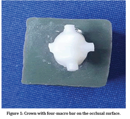
In-lab Sirona CAD SW 16.1 was used to design the crown restoration. One crown was made first and then used as reference for all crown [13]. Using the same milling machine MC X5 for all crowns (Sirona Dental System, Germany) and sintered into in-fire HTC speed furnace to have their original size, strength and final color. Four outer macro-retention bars (one bar on each side) on the occlusal surface was made to assist in removal of the crown after cementation procedure (Figure 1) [12,14].

Figure 1: Crown with four-macro bar on the occlusal surface.
Cementation procedure for each group was done according to manufacturer’s instructions ,using custom made holding and cementation device to maintain a constant seating force perpendicular to the occlusal surface of the crown during the cementation process [15] .A 5 Kg load (approximately 50 N) was applied in order to simulate the clinical condition during the cementation procedure “biting force” [16].
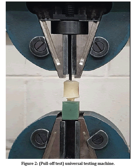
A second block of cold cure clear acrylic with screw incorporated at the top was cover the crown, preparing for tensile pull-off test , by a computer controlled UTM in a speed 0.5 mm/ min until failure. The dislodgment force was recorded and divided by the total surface area of each preparation to yield the retentive stress value in (mpa) (Figure 2).

Figure 2:(Pull-off test) universal testing machine.
The surfaces of detached teeth and crowns were checked using a magnifying lens. Failure was classified as show in Table 1 [17]. Retentive stresses were evaluated using two-way analysis of variance (ANOVA) and Bonferroni test at 0.05 level of significance.
| Mode of failure | Â Nature |
|---|---|
| Type I | Cohesive failure in teeth |
| Type II | Adhesive failure between teeth and cement |
| Type III | Cohesive failure in the cement |
| Type IV | Adhesive failure between cement and zirconia surface |
| Type V | Cohesive failure in ceramic |
Table 1: Classification of failure criteria.
Results
The statistics results of this study were listed in (Table 2)show that the highest mean value of stress failure was recorded by Subgroup B3(25μm cement space thickness and cemented with the Riva plus resin cement) (9.208MPa), while the lowest mean value was recorded by SubgroupA1 (0 μm cement space thickness using Relyx U200 cement) (4.049 MPa). Twoway ANOVA test show a significant difference in subgroups (P<0.001) (Table 3).
| Groups | Minimum | Maximum | Mean | ± SD | ± SE |
|---|---|---|---|---|---|
| A1 | 2.23 | 5.601 | 4.049 | 1.191 | 0.421 |
| A2 | 3.268 | 6.454 | 5.2 | 1.088 | 0.385 |
| A3 | 4.577 | 7.03 | 6.057 | 0.743 | 0.263 |
| B1 | 4.32 | 5.973 | 5.046 | 0.542 | 0.191 |
| B2 | 5.057 | 8.705 | 6.683 | 1.295 | 0.458 |
| B3 | 7.993 | 10.121 | 9.208 | 0.787 | 0.278 |
Table 2: Descriptive, statistics of, the stress failures, of the, different subgroups, within each, group measured, in MPa.
| Subgroup | Mean difference | P value | ||
|---|---|---|---|---|
| A1 | A2 | -1.152 | 0.023 | Sig. |
| A3 | -2.008 | 0 | ||
| A2 | A3 | -0.856 | 0.087 | NS |
| B1 | B2 | -1.637 | 0.002 | Sig. |
| B3 | -4.162 | 0 | ||
| B2 | B3 | -2.525 | 0 | |
Table 3: Bonferroni test for comparison of retentive stress among the different subgroups.
Discussion
The chosen for zirconium oxide crown for this study due to their superior physical properties that withstand during the tensile pull-off test [18]. Designed the crown in this study used modern art technique (CAD/CAM), in provision of, cement space to, set up a high, precision procedure [19] ,Two different marginal cement spaces were selected for this study (0 μm ,25 μm)in order to evaluate their effect on retentive stress of zirconia crowns [20,21]. while the remaining radial cement space set on the 80 μm starting 1mm above the finishing lines in accordance with the manufacturer’s setting of Ceric In-Lab sw 16.1, and as approved by Taha et al. [12] the 80 μm cement space had a significant increase in retention value than 100 μm and 120 μm space.
The result of this study showed that all tested zirconium crowns have mean retentive stress values exceeding, the clinically acceptable, limit needed, to hold crown, successful [12,22,23],and the statically highest mean retentive stress value was recorded with 25 μm subgroup B3(9.208) while lowest one was recorded with 0 μm subgroup A1 (4.049).
As finding of this study utilizing marginal cement space thickness 25 μm significantly higher than that when 0 um marginal cement space ,this explained by the fact that using of 25 μm, space at the marginal area could decrease the frictional resistance formed during seating of crown restoration [24]. Furthermore, 25 μm marginal, cement space decreased the internal and marginal gaps as reported by Hammood et l. [20] and Increase in retention may be a result of the improved adaptation (fitness) So increasing the cement space significantly improved the fitness of zirconia crowns as confirmed by Ahmed et al. [25].
At the 0 μm marginal cement space ,as the crown approaches the final position, there is no way for exceeded cement to escape through the marginal area. this result in incomplete seating of crown due to shortage of space for cement film , which result to the formation of hydraulic pressure under the crown, which increases until it meets the seating force, making further seating impossible [15].
So increasing space between the prepared tooth surface and the intaglio surface of the restoration, reducing stress areas formed during cementation and thereby resulting in improved fitness and retention of the definitive restoration, This was in accordance with previous studies concerning the increase in cement spacer thickness [26].
Zirconia crowns can be bonded either nonadhesively using ordinary cements or adhesively with resin-cement due to its great flexural strength [23]. Therefore, Riva lutes plus RMGIC, Relyx U200 self-adhesive and Relyx Ultimate resin cement ware chosen to act as three different luting agent. So there are other finding concerns the type of luting agent utilized. The result showed statically significant differences, The highest mean retentive value was recorded with Riva luting plus RMGIC.
This finding disagree with , Olivera which they found a significant better retention for resin cement in comparing to conventional GIC, Also finding doesn’t agree with [14] which found there are no statically significant differences in mean value of retention when using RMGIC and adhesive resin cement. This might be due to chemical adhesion of RMGIC to the dentin improving the retention of RIVA luting plus RMGIC [27], Also It has high flexural strength and high shear bond strength to dentin and zirconia enhances longevity of a glass-ionomer luting cement against force of tensile test.
Adhesive failure (type II) was the most mode of failure found in this study. This suggests that the mechanical interlock to air-abraded roughened zirconia surface was greater than mechanical interlock to the tooth [28], and might be also attributed to the application of Z-prime plus which enhances the strength bond of the resincement to zirconium crowns [16].
Conclusion
Marginal cement space thickness 25 μm improve the retention of zircon crowns for all types of luting agents, statically there was significant differences in retentive properties obtained from all cements type with the highest mean retentive stress for Riva luting plus RMGIC.
References
- Pilo R, Harel N, Nissan J, et al. The retentive strength of cemented zirconium oxide crowns after dentin pretreatment with desensitizing paste containing 8% arginine and calcium carbonate. Int J Mol Sci 2016; 17:426.
- Sreeramulu B, Suman P, Ajay P. A comparison between different luting cements on the retention of complete cast crowns-an in vitro study. Int J Healthc Biomed Res 2015; 3:29-35.
- Rosenstiel SF, Land MF. Contemporary fixed prosthodontics-e-book. Elsevier Health Sciences 2015.
- Heintze SD. Crown pull-off test (crown retention test) to evaluate the bonding effectiveness of luting agents. Dent Materials 2010; 26:193-206.
- Valderhaug J, Ellingsen JE, Jokstad A. Oral hygiene, periodontal conditions and carious lesions in patients treated with dental bridges: A 15-year clinical and radiographic follow-up study. J Clin Periodontol 1993; 20:482-489.
- Syrek A, Reich G, Ranftl D, et al. Clinical evaluation of all-ceramic crowns fabricated from intraoral digital impressions based on the principle of active wavefront sampling. J Dent 2010; 38:553-559.
- Sailer I, Fehér A, Filser F, et al. Five-year clinical results of zirconia frameworks for posterior fixed partial dentures. Int J Prosthod 2007; 20.
- de Paula Silveira AC, Chaves SB, Hilgert LA, et al. Marginal and internal fit of CAD-CAM-fabricated composite resin and ceramic crowns scanned by 2 intraoral cameras. J Prosthet Dent 2017; 117:386-392.
- Eames WB, O'Neal SJ, Monteiro J, et al. Techniques to improve the seating of castings. J Am Dent Assoc 1978; 96:432-437.
- Mobilio N, Fasiol A, Mollica F, et al. Effect of different luting agents on the retention of lithium disilicate ceramic crowns. Materials 2015; 8:1604-1611.
- Abdo SB, Masudi SA, Luddin N, et al. Fracture resistance of over-flared root canals filled with MTA and resin-based material: An in vitro study. Brazilian J Oral Sci 2012; 11:451-457.
- Taha FA, Ibraheem AF. The effect of die spacer thickness on retentive strength of all zirconium crowns (an in vitro study). Int J Med Res Health Sci 2019; 8:22-27.
- Hamza TA, Sherif RM. In vitro evaluation of marginal discrepancy of monolithic zirconia restorations fabricated with different CAD-CAM systems. J Prosthet Dent 2017; 117:762-766.
- Aleisa K, Alwazzan K, Al-Dwairi ZN, et al. Retention of zirconium oxide copings using different types of luting agents. J Dent Sci 2013; 8:392-398.
- Abdullah LS, Ibraheem AF. The effect of finishing line designs and occlusal surface reduction schemes on vertical marginal fit of full contour CAD/CAM zirconia crown restorations (a comparative in vitro study). Int J Dent Oral Health 2017; 4:1-6.
- Kim SM, Yoon JY, Lee MH, et al. The effect of resin cements and primer on retentive force of zirconia copings bonded to zirconia abutments with insufficient retention. J Adv Prosthod 2013; 5:198-203.
- Akkus E, Begum TS. Evaluation of surface treatment methods on the bond strength of zirconia ceramics systems, resin cements and tooth surface. Balkan J Dent Med 2015; 19:101-12.
- Ernst CP, Cohnen U, Stender E, et al. In vitro retentive strength of zirconium oxide ceramic crowns using different luting agents. J Prost Dent 2005; 93:551-558.
- Mehl C, Harder S, Steiner M, et al. Influence of cement film thickness on the retention of implant-retained crowns. J Prosthod 2013; 22:618-25.
- Hammood ED. The effect of marginal cement space thickness on the marginal and internal fitness of monolithic zirconia crowns using different types of luting agents (Doctoral dissertation, University of Baghdad).
- Nayyef MI, Ibraheem AF. Marginal microleakage of monolithic zirconia crowns using different marginal cement space thickness and luting cements. J Res Med Dent Sci 2021; 9:17.
- Palacios RP, Johnson GH, Phillips KM, et al. Retention of zirconium oxide ceramic crowns with three types of cement. J Prosthetic Dent 2006; 96:104-14.
- Ali RH, Kassim M. The Influence of cement spacer thickness on retentive strength of monolithic zirconia crowns cemented with different luting agents (a comparative in-vitro study). Indian J Forensic Med Toxicol 2021; 15.
- Hoang L. Accuracy and precision of die spacer thickness with combined computer-aided design and 3-D printing technology. Dissertation Thesis 2014.
- Ahmed WM, Shariati B, Gazzaz AZ, et al. Fit of tooth-supported zirconia single crowns-A systematic review of the literature. Clin Exp Dent Res 2020; 6:700-716.
- El-Anwar MI, Tamam RA, Fawzy UM, et al. The effect of luting cement type and thickness on stress distribution in upper premolar implant restored with metal ceramic crowns. Tanta Dent J 2015; 12:48-55.
- Czarnecka B, Deregowska-Nosowicz P, Limanowska-Shaw H, et al. Shear bond strengths of glass-ionomer cements to sound and to prepared carious dentine. J Mater Sci 2007; 18:845-849.
- Shahin R, Kern M. Effect of air-abrasion on the retention of zirconia ceramic crowns luted with different cements before and after artificial aging. Dent Materials 2010; 26:922-928.
Indexed at, Google Scholar, Cross Ref
Indexed at, Google Scholar, Cross Ref
Indexed at, Google Scholar, Cross Ref
Indexed at, Google Scholar, Cross Ref
Indexed at, Google Scholar, Cross Ref
Indexed at, Google Scholar, Cross Ref
Indexed at, Google Scholar, Cross Ref
Indexed at, Google Scholar, Cross Ref
Indexed at, Google Scholar, Cross Ref
Indexed at, Google Scholar, Cross Ref
Indexed at, Google Scholar, Cross Ref
Indexed at, Google Scholar, Cross Ref
Indexed at, Google Scholar, Cross Ref
Indexed at, Google Scholar, Cross Ref
Indexed at, Google Scholar, Cross Ref
Indexed at, Google Scholar, Cross Ref
Author Info
Huda Rasheed Hameed Alazzawi* and Adel Farhan Ibraheem
Department of Restorative and Esthetic Dentistry, College of Dentistry, Baghdad University, IraqCitation: Huda Rasheed Hameed Alazzawi, Adel Farhan Ibraheem, Effect of Different Marginal Cement Space Thickness on the Retention of Monolithic Zirconium Crown Using Different Luting Agents, J Res Med Dent Sci, 2022, 10 (3):175-179.
Received: 04-Feb-2022, Manuscript No. JRMDS-22-52364; , Pre QC No. JRMDS-22-52364 (PQ); Editor assigned: 07-Feb-2022, Pre QC No. JRMDS-22-52364 (PQ); Reviewed: 21-Feb-2022, QC No. JRMDS-22-52364; Revised: 25-Feb-2022, Manuscript No. JRMDS-22-52364 (R); Published: 04-Mar-2022
