Research - (2020) Advances in Dental Surgery
Duration of Mobility Reduction Followed by Management of Luxation Injuries
Meera Theenathayalan, Bhagya Lakshmi T* and Deepak S
*Correspondence: Bhagya Lakshmi T, Department of Pediatric and Preventive Dentistry, Saveetha Dental College and Hospitals,Saveetha Institute of Medical and Technical Sciences Saveetha University, 162, Poonamallee High Road, Chennai, India, Email:
Abstract
Traumatic injuries emerge as an increasingly significant threat to the dental health of children and adolescents. A dental injury should always be considered an emergency and thus be treated immediately to relieve pain, facilitate reduction of displaced teeth, reconstruct lost hard tissue and improve prognosis. The aim of the study was to evaluate the duration of mobility reduction after the management of the luxation injuries. In this study around 56 pedo patient’s data was collected from patient’s dental records who reported with luxation injuries, between june 2019 to March 2020, Saveetha Dental College and Hospital, Chennai. The values obtained were tabulated and documented. The data were then entered into SPSS software for statistical analysis. The association between study variables was calculated using the chi-square test. Age groups were split into 0-9 years, 10-14 years, and 15-18 years. Among the selected data most commonly affected were males (75%) (around the age group 10-14 years). 53.5% of the study showed the most affected were the 10-14 years age group with luxated teeth, followed by 15-18 years (35.7%). The most common time duration taken for reduction of mobility after management of the luxated teeth was found to be less than 3 months (37.5%). The chi square test showed negative correlation between teeth no. and duration (P value >0.05). Luxation injuries are a rare condition/accident, mostly affecting children between 10-14 years of age. Mostly the follow up and home care contributes to better healing followed by trauma dental injuries. In growing individuals, careful and delegated monitoring is essential to diagnose the injury.
Keywords
Dental Trauma, Luxation injuries, Mobility, Reduction
Introduction
Traumatic injuries emerge as an increasingly significant threat to the dental health of children and adolescents. A dental injury should always be considered an emergency and thus be treated immediately to re1ici.e pain, facilitate reduction of displaced teeth, reconstruct lost hard tissue, and improve prognosis. According to Andreas et al. [1], approximately 50% of children are exposed to dental trauma before school leaving age. Trauma to supporting tissues are among the most common dental injuries, occasionally leading to pulp canal obliteration, necrosis, and root resorption [1]. Tooth maturity at the time of injury has been identified as one of the most decisive factors affecting post traumatic recovery of pulp [2].
Tooth luxation as a clinical diagnosis covers a broad spectrum of injuries. Most luxations represent combined injury to the pulp and periodontium. The emergency treatment of displaced permanent teeth seen soon after injury consists of careful repositioning, and as the repositioned tooth often tends to migrate incisally, a flexible splint should be applied for 2-3 weeks. Teeth which are concussed or subluxated are normally not splinted. After emergency treatment, the patient is scheduled for follow-up controls. Pulp survival following luxation depends on the extent of the injury. The prevalence of subsequent permanent necrosis is increased in displaced teeth with complete root formation, where a constricted apical foramen apparently prevents revascularization of severed pulp tissue [3]. Pulp canal obliteration has been observed predominantly in displaced teeth with wide apical aperture [4]. Trauma to the periodontal ligament during the luxation injury can induce external root resorption. Small areas of cell damage are repaired from adjacent sound tissue while larger injuries to the periodontal ligament elicit replacement resorption with concomitant pulp injection, bacterial toxins through exposed dentinal tubuli potential the activity of the resorptive process, resulting in rapid replacement of tooth and surrounding bone by granulation tissue [5].
Presence of injection in pulp has been related to internal root resorption [6], which appears to be a relatively rare finding in permanent dentition [2]. In recent years, many aspects of the relationship between type of injury and management and healing complication have been clarified, however knowledge concerning factors influencing long term prognosis remains fragmentary. It has been demonstrated that most luxation injuries heal spontaneously [7], avoiding the traumatic experience of tooth extraction. The clinician’s skills and experience with pediatric patients is of utmost importance for managing the patients and parents or care’s behaviors in the emergency solution.
Acute dental injury can be a very costly problem for a society, in the form of direct costs (including labor and material) and indirect costs to the patient (including lost school time, loss of income for parents who miss work, long observation periods, renewed treatments). This highlights the need to collect data dealing not only the causes and types of tooth injuries, but also with treatment outcomes. A better understanding of healing processes following injury might enable the practitioner to create a biologically sound (and possibly more economical) treatment strategy based on principles of wound healing and which may provide a more conservative, less aggressive treatment in selected cases [8]. Previously our team had conducted numerous clinical trials [9-21], and questionnaire surveys [22,23] over the past 5 years. Now we are focusing on retrospective studies. The idea for this study stemmed from the current interest in our community. The aim of this study was to evaluate the duration of mobility reduction after the management of the luxation injuries.
Materials and Methods
This study was approved by the research ethics committee of saveetha dental college. The data was collected from patients records between June 2019 and March 2020, in Saveetha Dental College and Hospital, Chennai. The dental records of 56 patients who reported to the clinic for management of Luxated teeth were investigated by collecting the data and records were entered in the excel sheet. The male and female distribution among the study population was evaluated. The collection of data was divided on 4 parameters, the age of the patient, the gender of the patient, tooth number and duration of mobility management of the luxated tooth. After grouping the parameters, data copied to the software and statistical analysis was carried out. Statistical analysis was done using IBM SPSS software. The significance level was at <0.05. Descriptive analysis and chi-square tests were done. Graphs were tabulated.
Inclusion criteria
Patients of age group 0-18 years only included, and both male and females with avulsion were included.
Exclusion criteria
Other than luxation cases, other injuries or dental trauma were excluded.
Results and Discussion
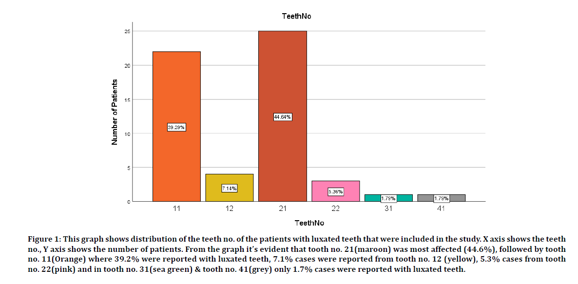
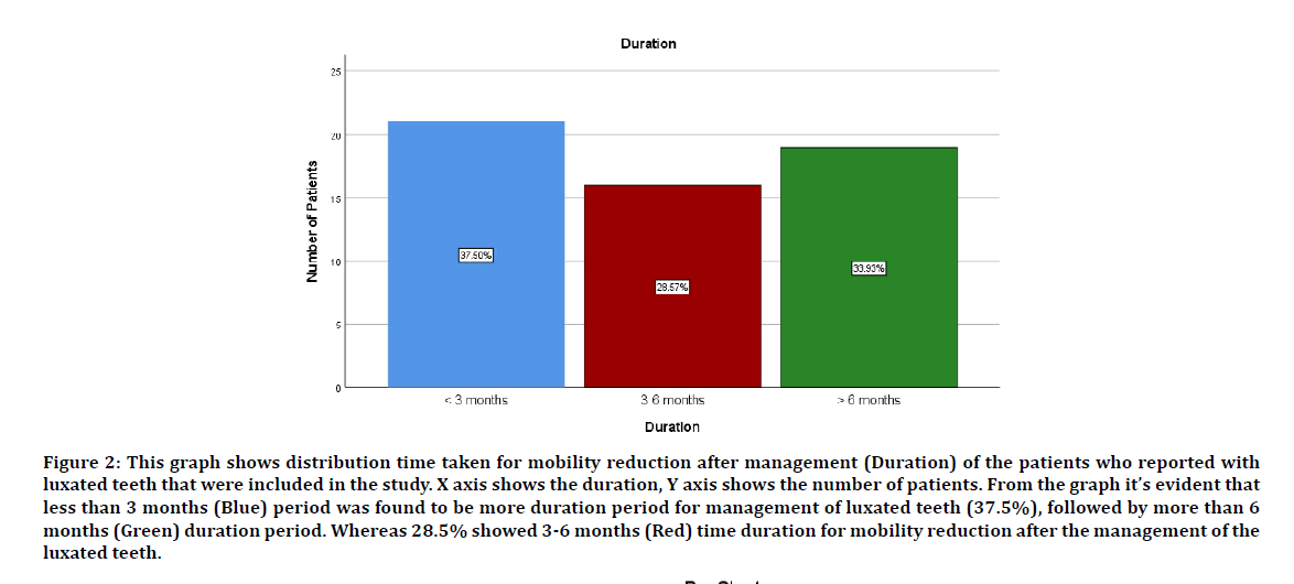
Luxation was most seen in the maxillary and mandibular, central and lateral incisors. The most affected tooth no. was 21 where twenty five patients were reported with luxation (44.6%), followed by tooth no. 11 where twenty two cases were reported (39.3%), tooth no. 12 where four cases were reported with luxation (7.1%), tooth no. 22 where three cases were reported with luxation (5.4%), tooth no. 31 and tooth no. 41 where one case each was reported with luxation (1.8%) (Figure 1). The incidence of duration was associated with the mobility reduction after the management of luxated teeth. The most duration taken to reduce was less than 3 months were 21 cases showed mobility reduction after the management of luxation (37.5%), followed by more than 6 months period where 19 cases showed mobility reduction after management of luxation (33.9%) and in 3-6 months period 16 cases showed mobility reduction after management (28.6%) (Figure 2).

Figure 1: This graph shows distribution of the teeth no. of the patients with luxated teeth that were included in the study. X axis shows the teeth no., Y axis shows the number of patients. From the graph it’s evident that tooth no. 21(maroon) was most affected (44.6%), followed by tooth no. 11(Orange) where 39.2% were reported with luxated teeth, 7.1% cases were reported from tooth no. 12 (yellow), 5.3% cases from tooth no. 22(pink) and in tooth no. 31(sea green) & tooth no. 41(grey) only 1.7% cases were reported with luxated teeth.

Figure 2: This graph shows distribution time taken for mobility reduction after management (Duration) of the patients who reported with luxated teeth that were included in the study. X axis shows the duration, Y axis shows the number of patients. From the graph it’s evident that less than 3 months (Blue) period was found to be more duration period for management of luxated teeth (37.5%), followed by more than 6 months (Green) duration period. Whereas 28.5% showed 3-6 months (Red) time duration for mobility reduction after the management of the luxated teeth.
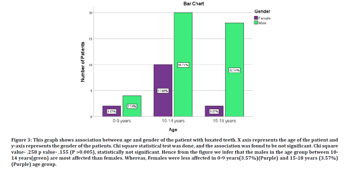
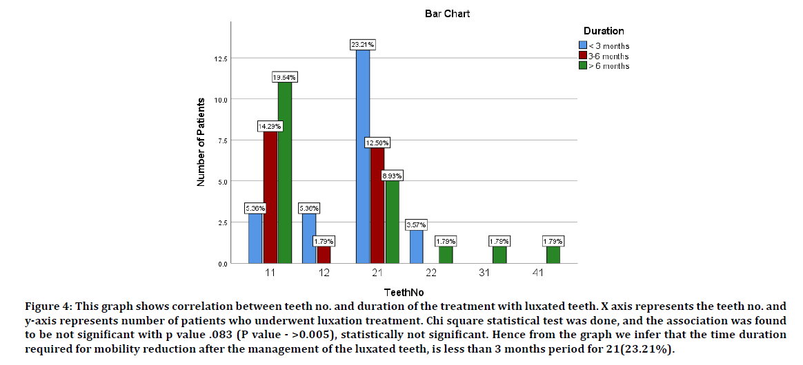
Among the 56 pedo patient is, in the age group 0-9 years, two female patients, four males were reported with luxation, in the 10-14 years age group, ten female patients, and twenty male patients reported with luxation. In the 15- 18 years age group, two female patients, and eighteen male patients reported with luxation (Figure 3). Each tooth had its own time for mobility reduction after management of luxation. In less than 3 months period three cases with luxation in tooth no. 11, three cases in tooth no. 12, thirteen cases in tooth no. 21 and two cases with luxation in tooth no. 22 showed mobility reduction , whereas in the 3-6 months period, eight cases with luxation in tooth no. 11, one case in tooth no. 12, seven cases with luxation in tooth no. 21 showed mobility reduction. In more than 6 months period, eleven cases with luxation in tooth no. 11, five cases with luxation in tooth no. 21, and one case with luxation in tooth no. 22, tooth no. 31 & tooth no. 41 showed mobility reduction after the management (Figure 4). Correlations between the age and gender was determined which showed p value to be 0.155 which showed a negative correlation and correlation between the teeth number and duration was seen , p value was found to be .083 which also showed a negative correlation between the two parameters.

Figure 3: This graph shows association between age and gender of the patient with luxated teeth. X axis represents the age of the patient and y-axis represents the gender of the patients. Chi square statistical test was done, and the association was found to be not significant. Chi square value- .258 p value- .155 (P >0.005), statistically not significant. Hence from the figure we infer that the males in the age group between 10- 14 years(green) are most affected than females. Whereas, Females were less affected in 0-9 years(3.57%)(Purple) and 15-18 years (3.57%) (Purple) age group.

Figure 4: This graph shows correlation between teeth no. and duration of the treatment with luxated teeth. X axis represents the teeth no. and y-axis represents number of patients who underwent luxation treatment. Chi square statistical test was done, and the association was found to be not significant with p value .083 (P value - >0.005), statistically not significant. Hence from the graph we infer that the time duration required for mobility reduction after the management of the luxated teeth, is less than 3 months period for 21(23.21%).
The minimum time for a follow-up examination (6 months) should be considered relevant because most of the late complications as compared to some earlier studies [24]. The higher frequency of late complications as compared to earlier studies [2]. May be since tooth injuries with severe periodontal trauma were observed more often than by other authors. Another explanation may be that only these patients with complaints reported for the follow up. The optimal treatment for a luxated tooth has yet to be determined.
Shapira et al. [25] have suggested that these are three options available to the dental practitioner: a) Await spontaneous re eruption in intrusive luxation especially in immature cases. b) Immediate surgical reduction and fixation (or) c) Orthodontic repositioning. Andreason et al. [26,27] has stated that immature will re erupt spontaneously, while surgical repositioning of mature teeth is not advisable as this procedure may lead to extensive marginal bone loss. Tusley et al. [28] have investigated spontaneous re eruption and orthodontic extrusion as options for intrusive luxation injuries options for experimenting. Less severity intruded and mobile teeth responded well to orthodontic treatment extrusion while the deeply embedded teeth became ankylosed and failed to respond.
The more conservative option of observation was divided. On the case reported, in endodontic intervention study were the tooth were carefully monitored for containing signs of mobility and its re eruption in three months period post trauma, if endodontics had been required earlier, gingival surgery or orthodontic repositioning to provide access to the pulp may be employed. Calcium hydroxide was deemed the medication of choice in the endodontic treatment, as andreasan [29,30] has reported that external root resorption is a common sequel to pulp necrosis associated with intrusive luxation injuries. In luxation injuries the tooth position is altered, resulting in possible interference with occlusion and articulation. Based on these the management is done, and the reduction of mobility is seen [31-34].
Conclusion
In this study,the most time duration taken for reduction in management of luxation injuries in less than 3 months was 37.5% , whereas in more than 6 months’ time duration 33.9% were seen. The management of the luxation injuries can be done easily. Early results can be obtained, duration of management will be reduced by this. Mostly the follow up and home care contributes to better healing followed by trauma dental injuries. In growing individuals, careful and diligent monitoring Is essential for diagnosing the injury. Timely and appropriate management of luxation will offer the best prognosis for the patient.
Acknowledgement
The authors are thankful to the Director of Saveetha Dental College and Hospital, Chennai.
Author Contribution
Meera has contributed to data collection, study design, data analysis, results, tables and manuscript preparation.
Dr. Bhagyalakshmi has contributed to the manuscript preparation, proof heading of the manuscript and reviewing the manuscript.
Dr. Deepak has contributed to reviewing the manuscript.
Conflict of Interests
There is no conflict of interest.
References
- Andreasen JO. Traumatic injuries of the teeth. WB Saunders Company; 1981.
- Andreasen JO. Luxation of permanent teeth due to trauma A clinical and radiographic follow-up study of 189 injured teeth. Eur J Oral Sci 1970; 78:273–86.
- Andreasen FM. Pulpal healing after luxation injuries and root fracture in the permanent dentition. Dent Traumatol 1989; 5:111–131.
- Andreasen FM, Zhijie Y, Thomsen BL, et al. Occurrence of pulp canal obliteration after luxation injuries in the permanent dentition. Dent Traumatol 1987; 3:103–115.
- https://www.scienceopen.com/document?vid=e496e959-8a0e-4d67-ba19-c10888617739
- Wedenberg C, Lindskog S. Experimental internal resorption in monkey teeth. Dent Traumatol 1985; 1:221–227.
- Colak I, Markovic D, Petrovic B, et al. A retrospective study of intrusive injuries in primary dentition. Dent Traumatol 2009; 25:605–610.
- Andreasen FM. Pulpal healing after luxation injuries and root fracture in the permanent dentition. Dent Traumatol 1989; 5:111–31.
- JeevananDan G. Kedo-S paediatric rotary files for root canal preparation in primary teeth-Case report. J Clin Diagn Res 2017; 11:ZR03.
- Gurunathan D, Shanmugavel AK. Dental neglect among children in Chennai. J Indian Soc Pedod Prev Dent 2016; 34:364.
- Govindaraju L, Jeevanandam G, Subramanian EMG. Comparison of quality of obturation and instrumentation time using hand files and two rotary file systems in primary molars: A single-blinded randomized controlled trial. Eur J Dent 2017; 11:376–379.
- Somasundaram S, Ravi K, Rajapandian K, et al. Fluoride content of bottled drinking water in Chennai, Tamilnadu. J Clin Diagn Res 2015; 9:ZC32.
- Jeevanandam G, Govindaraju L. Clinical comparison of Kedo-S paediatric rotary files vs manual instrumentation for root canal preparation in primary molars: a double blinded randomised clinical trial. Eur Arch Paediatr Dent 2018; 19:273–278.
- Govindaraju L, Jeevanandam G, Subramanian EMG. Clinical evaluation of quality of obturation and instrumentation time using two modified rotary file systems with manual instrumentation in primary teeth. J Clin Diagn Res 2017; 11:ZC55.
- Panchal V, Jeevanandam G, Subramanian EMG, Others. Comparison of instrumentation time and obturation quality between hand K-file, H-files, and rotary Kedo-S in root canal treatment of primary teeth: A randomized controlled trial. J Indian Soc Pedod Prev Dent 2019; 37:75.
- Christabel SL, Gurunathan D. Prevalence of type of frenal attachment and morphology of frenum in children, Chennai, Tamil Nadu. World J Dent 2015; 6:203–207.
- Packiri S, Gurunathan D, Selvarasu K. Management of paediatric oral ranula: a systematic review. J Clin Diagn Res 2017; 11:ZE06.
- GovinDaraju L, Gurunathan D. Effectiveness of chewable tooth brush in children-A prospective clinical study. J Clin Diagn Res 2017; 11:ZC31.
- Subramanyam D, Gurunathan D, Gaayathri R, et al. Comparative evaluation of salivary malondialdehyde levels as a marker of lipid peroxidation in early childhood caries. Eur J Dent 2019; 12:67–70.
- http://www.ijpronline.com/ViewArticleDetail.aspx?ID=7041
- Lakshmanan L, Mani G, Jeevanandam G, et al. Assessing the quality of root canal filling and instrumentation time using kedo-s files, reciprocating files and k-files. Br Dent Sci 2020; 23:7.
- Govindaraju L, Jeevanandam G, Subramanian EMG. Knowledge and practice of rotary instrumentation in primary teeth among Indian dentists: A questionnaire survey. J Int Oral Health 2017; 9:45.
- Ravikumar D, Jeevanandam G, Subramanian EMG. Evaluation of knowledge among general dentists in treatment of traumatic injuries in primary teeth: A cross-sectional questionnaire study. Eur J Dent 2017; 11:232–237.
- Andreasen FM, Pedersen BV. Prognosis of luxated permanent teeth—the development of pulp necrosis. Dent Traumatol 1985; 1:207–220.
- Shapira J, Regev L, Liebfeld H. Re-eruption of completely intruded immature permanent incisors. Dent Traumatol 1986; 2:113–116.
- Andreasen JQ, Ravn JJ. Epidemiology of traumatic dental injuries to primary and permanent teeth in a Danish population sample. Int J Oral Surg 1972; 1:235–239.
- Andreasen JO, Andreasen FM. Dental trauma: Quo vadis. Tandlaege Bladet 1989; 93:381–384.
- Turley PK, Joiner MW, Hellstrom S. The effect of orthodontic extrusion on traumatically intruded teeth. Am J Orthod 1984; 85:47–56.
- Lieger O, Graf C, El-Maaytah M, et al. Impact of educational posters on the lay knowledge of schoolteachers regarding emergency management of dental injuries. Dent Traumatol 2009; 25:406–412.
- DiAngelis AJ, Andreasen JO, Ebeleseder KA, et al. International association of dental traumatology guidelines for the management of traumatic dental injuries: Fractures and luxations of permanent teeth. Dent Traumatol 2012; 28:2–12.
- Malmgren B, Andreasen JO, Flores MT, et al. International association of dental traumatology guidelines for the management of traumatic dental injuries: Injuries in the primary dentition. Dent Traumatol 2012; 28:174–182.
- Flores MT, Malmgren B, Andersson L, et al. Guidelines for the management of traumatic dental injuries. III. Primary teeth. Dent Traumatol 2007; 23:196–202.
- Flores MT, Andreasen JO, Bakland LK. Guidelines for the evaluation and Management of traumatic dental injuries editors note. Dent Traumatol 2001; 17:193–1966.
- Andersson L. Epidemiology of traumatic dental injuries. J Endod 2013; 39:2–5.
Author Info
Meera Theenathayalan, Bhagya Lakshmi T* and Deepak S
1Department of Pediatric and Preventive Dentistry, Saveetha Dental College and Hospitals,Saveetha Institute of Medical and Technical Sciences Saveetha University, 162, Poonamallee High Road, Chennai, India2Department of Conservative Dentistry and Endodontics, Saveetha Dental College and Hospitals, Saveetha Institute of Medical and Technical Sciences Saveetha University, 162, Poonamallee High Road, Chennai, India
Citation: Meera Theenathayalan, Bhagya Lakshmi T, Deepak S, Duration of Mobility Reduction Followed by Management of Luxation Injuries, J Res Med Dent Sci, 2020, 8 (7): 280-285.
Received: 29-Sep-2020 Accepted: 02-Nov-2020 Published: 09-Nov-2020
