Research - (2020) Advances in Dental Surgery
Determination of the Anteroposterior Maxillary Central Incisors Position Relative to the Forehead Iraqi Study
Abeer Moayad Jead1* and Mohammed Nahidh2
*Correspondence: Abeer Moayad Jead, Dentist, Ministry of Health, Iraq, Email:
Abstract
Background: Evaluating the anteroposterior position of the maxillary incisors is an important step in the diagnosis and treatment planning in order to get better facial and dental esthetics. This study aimed to evaluate and compare the anteroposterior position of the maxillary central incisors in both genders and to find out whether there is a relation between this position relative to the forehead inclination.
Samples and methods: Eighty dental students (40 males and 40 females) having normal dental and skeletal relationships and pleasing profile agreed to participate in this study. Standardized profile photograph on smiling was taken for each student and analyzed by AutoCAD program to assess the anteroposterior position of the maxillary central incisors and the inclination of forehead. Independent sample t-test and Pearson's Chi-square were used to assess the gender difference, while Pearson's correlation coefficient was used to assess the relationship.
Results: Males showed maxillary central incisors positioned significantly more anteriorly relative to the forehead in comparison with females. In most of the studied sample, the maxillary central incisors were located anterior to the point glabella. Moderate to strong, direct, high significant correlations were found between maxillary central incisors position and forehead inclination in both genders.
Conclusions: The forehead is considered as a helpful landmark for assessing the facial profile as it correlated significantly with the anteroposterior position of the maxillary central incisors.
Keywords
Facial aesthetics, forehead inclination, Maxillary central incisors position
Introduction
One of the most important elements of facial aesthetics is the maxillary central incisors. Treatment with orthodontic approach only or combined with orthognathic surgeries can changed the anteroposterior position (AP) and inclination of these teeth. These changes had great effects on the smiling profile esthetics percept by orthodontists and lay persons [1-3].
Many facial landmarks including nose, lips and chin had a major role in assessing the anteroposterior position of displayed maxillary central incisors in profile. After many studies and observations, Andrews et al. [4] favored the forehead to be a reference landmark used to determine the anteroposterior position of maxillary central incisors. His observations on persons with facial harmony led him to reach a conclusion about the presence of a direct relation between the inclination and prominence of forehead and the anteroposterior positions of the teeth and jaws. Moreover, he considered forehead as a stable landmark in contrast to the internal radiographic landmarks and its relationship with the maxillary incisors can be predictable and repeatable. Schlosser et al. [1] concluded that trained or untrained people are sensitive to the erroneous anteroposterior relationship of the maxillary incisors to the forehead and this is the way that society instinctively utilizes for determining profile acceptance. Many researches had been done world-wide assessing the anteroposterior position of the maxillary central incisors relative to the forehead on American white and African samples, Chinese and Indians [3-12].
To the best of author's knowledge, there is no Iraqi study performed in this field, so this study aimed to evaluate the position of the maxillary central incisors relative to the forehead by measuring the anteroposterior position of the maxillary central incisors and their location to the forehead in both genders, and to find out whether there is a relation between this position and the forehead inclination in a sample of Iraqi adults with normal dental and skeletal relationships and pleasing facial profile.
Samples and Methods
Study design
This prospective study was approved by the ethical and scientific committees in the Department of Orthodontics, University of Baghdad School of Dentistry, Iraq.
Samples
Eighty participants (40 males and 40 females) were recruited according to specific criteria from the students in the College of Dentistry, University of Baghdad between December 2018 and April 2019. The inclusion criteria included:
Were Iraqi Arabs in origin.
Age ranged between 20 to 23 years.
All had balanced facial profile with normal dental and skeletal relationships [13,14].
No one had a history of bad oral habits, orthodontic treatment, dentofacial deformities, plastic and orthognathic surgeries.
Methods
Explanation the purpose of the study was demonstrated by the researcher for each participant and in case of agreement, consent form was signed. History taking and clinical examination were performed on the dental chair and standardized right side profile photographs were captured during smiling in natural head position using mobile I-phone 6 camera with the aid of Planmeca ProMax Dimax3 X-ray unit in the department of Orthodontics, College of Dentistry/ University of Baghdad.
The photographic technique involved establishing the subjects in natural head position like in preparation for image exposure. Standing subjects were initially asked to assume their arms by their sides to establish orthoposition, then instructed to close their eyes and perform a series of neck bending exercises by tilting the head upward and downward until comfortable position of natural balance was achieved. After that, subjects reopened their eyes and looked into their eyes reflection on the mirror mounted on the stand 137 cm in front of the patient's nose [15].
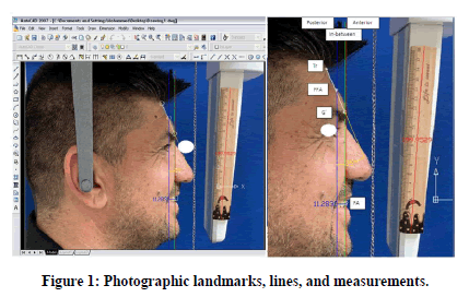
Subjects were then asked to stay still, with teeth lightly together and lips relaxed, then the ear rods of the cephalostat were gently inserted into the external auditory meati and profile facial photographs were then taken with a one meter distance between the camera and patient's head [16]. The most important point was that the maxillary central incisors and forehead must be fully exposed in photographic image; otherwise the photo will be neglected. Every profile photograph was imported and analyzed using AutoCAD computer program to calculate the angular and linear measurements. First of all, the magnification was corrected for each photo using a wooden ruler as a caliper. According to the definitions of Andrew (5), the photographic landmarks were located and lines drawn, then forehead inclination angle and the anteroposterior position of the maxillary incisor were measured directly on the photographs (Figure 1).

Figure 1. Photographic landmarks, lines, and measurements.
Photographic landmarks
The following landmarks were utilized:
Glabella (G'): It is the most inferior aspect of the forehead.
Superion: It is the most superior aspect of the forehead when the forehead is either rounded or angular in contour.
Trichion (Tr'): It is defined as the hairline, and it is the most superior aspect of the forehead when the forehead is of relatively flat contour.
The forehead facial axis (FFA) point: It is the midpoint between trichion and glabella for foreheads with flat contour or the midpoint between superion and glabella for foreheads with rounded or angular contour.
The facial axis (FA) point: It is the point on the facial axis of the maxillary central incisor that separates the gingival half of the clinical crown from the occlusal half.
Photographic lines
The reference lines were constructed
Line 1: Perpendicular through the FFA point.
Line 2: Perpendicular through glabella.
Line 3: Perpendicular through the maxillary central incisor’s FA point.
Line 4: for assessing forehead inclination connected glabella to the uppermost point of the clinical forehead i.e. superion or trichion.
Photographic measurements
The anteroposterior position of the maxillary central incisor and the forehead inclination were determined as followed:
The anteroposterior relationship of the maxillary central incisors to the forehead was measured as the distance between line 1 and line 3. A positive value was assigned when the maxillary central incisors (line 3) were anterior to the FFA point (line 1) and when the incisors were posterior to the FFA point, a negative value was given.
Forehead inclination was measured as the angle between line 4 and line 1.
The position of the maxillary central incisors relative to points FFA and glabella was determined. It is allocated as anterior when they were positioned anterior to glabella, posterior when they were positioned posterior to FFA and in-between when they were positioned between FFA and glabella.
Statistical analyses
Data were analyzed using SPSS software version 24. In this study, the following statistics were used:
Descriptive statistics: including means, standard deviations, minimum and maximum values, frequency (No.), percentages, and statistical tables and figures.
Inferential statistics: including:
Intra-class correlation coefficient test and Cohen's Kappa test: for intra- and inter-examiner reliability.
Independent sample t-test: to compare the measured variables between both genders.
Pearson's Chi-square test: to test any statistically significant genders differences for the position of the maxillary incisors.
Pearson's correlation coefficient test (r): to test the relation between the measured variables in both genders.
In the statistical evaluation, the following levels of significance are used:
Non-significant NS P>0.05
Significant S 0.05 ≥ P>0.01
Highly significant HS P ≤ 0.01
Results
Inter-and intra-examiner reliability
After good training on using the software, five photographs were selected, and measurements were performed by the professional orthodontist and the researcher to check the inter-examiner reliability.
After seven days, the researcher re-measured the same five photographs again to evaluate the inter-examiner reliability. Intra-class correlation coefficient test and Cohen's Kappa test were used to test the reliability for the position of the maxillary central incisors.
The results of intra-class correlation coefficient test indicated excellent reliability both for intra- and inter-examiner. On the other hand, Cohen's Kappa test showed perfect inter- and intra-examiner reliability for determination the position of the maxillary central incisor.
Descriptive statistics and genders difference
The descriptive statistics and gender difference for the measured variables were shown in Table 1. The results revealed that male group had significantly higher mean values for forehead inclination and maxillary central incisors position in comparison with female group. The higher standard deviation values were related to the higher range between the minimum and maximum values.
| Variables | Genders | Descriptive statistics | Gender difference (d.f.=78) | ||||
|---|---|---|---|---|---|---|---|
| Mean | S.D. | Min | Max | t-test | P-value | ||
| Forehead | Males | 22.05 | 5.965 | 11 | 33 | 4.451 | 0 (HS) |
| inclination (º) | Females | 16.8 | 4.479 | 5 | 29 | ||
| AP Incisors | Males | 19.245 | 14.009 | -7.49 | 43.59 | 3.196 | 0.002 (HS) |
| position (mm.) | Females | 9.308 | 13.8 | -29.8 | 33.99 | ||
Table 1: Descriptive statistics and genders difference for the measured variables.
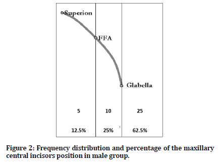
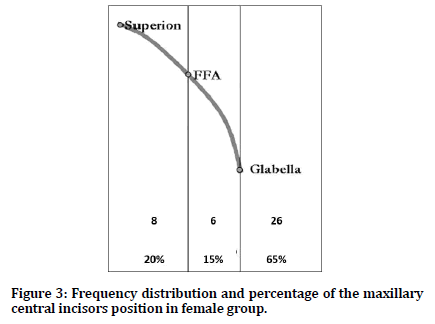
Regarding the position of maxillary central incisors relative to the forehead; Table 2 and Figures 2 and 3 demonstrated the frequency distribution and percentages of the maxillary central incisors position relative to the points FFA and glabella. In males group, 62.5% of the cases had their maxillary central incisors positioned anterior to glabella and 25% between the glabella and FFA, while 12.5% were posterior to FFA.
| Genders | Posterior | In between | Anterior | Total | |
|---|---|---|---|---|---|
| Males | N | 5 | 10 | 25 | 40 |
| % | 12.5 | 25 | 62.5 | 100 | |
| Females | N | 8 | 6 | 26 | 40 |
| % | 20 | 15 | 65 | 100 | |
| X2=1.712, d.f.=2, P-value=0.425 (NS) | |||||
Table 2: Frequency distribution, percentages, and gender difference regarding the position of the maxillary central incisors.

Figure 2. Frequency distribution and percentage of the maxillary central incisors position in male group.

Figure 3. Frequency distribution and percentage of the maxillary central incisors position in female group.
Regarding females group, in the highest percentage of the cases, the maxillary central incisors were located anterior to glabella (65%), followed by 20% posterior to FFA and in only 15% of the cases, they lie between FFA and glabella. Chi-square test indicated nonsignificant gender difference.
The relationship between forehead inclination and anteroposterior position of the maxillary central incisors
The relationship between the forehead inclination and the position of the maxillary central incisors was determined in Table 3 for both genders. The relationship was strong, direct, and highly significant in males and moderate, direct, and highly significant in females.
| Relation | Males | Females |
|---|---|---|
| r | 0.877 | 0.658 |
| P-value | 0.000 (HS) | 0.000 (HS) |
Table 3: Relationship between the forehead inclination and maxillary central incisors' position in both genders.
To reach a primary and then definitive orthodontic diagnosis, orthodontists must rely on many diagnostic aids which are the frontal photographic with lip relaxed and while smiling and profile photographic with lips relaxed only. As previously mentioned, there is a correlation between the prominence and the inclination of forehead and the AP positions of the teeth and jaws, so profile photographic examination in smile with bared maxillary incisors is far important just like the frontal photograph. This is the first Iraqi study that deals with this subject.
The samples selected for the present study included young adult subjects to minimize the effect of any remaining skeletal growth [17,18]; as most of the facial growth is nearly completed by 16-17 years of age [19]. Those students were selected with normal dental and skeletal relationships to exclude the abnormal dental or skeletal relationships that may affect the maxillary central incisors position. AutoCAD software is used in the current study. This program is accurate, reliable, and easy to manipulate with a simple method for correction of magnification [20]. Previous studies [5,8,11] tried to resize the photos, taken from magazines and journals, to the real size and use the ruler and protractor for measuring. This could not be so accurate like measuring with AutoCAD.
The Inclination of the forehead
In the present study, the inclination of the forehead in male groups was higher significantly than females; this comes in agreement with Zou et al. study [10] on Chinese sample. The gender difference could be attributed to the greater variations in the inclinations and amount of frontal bossing in males as the frontal bones in males are thick, less rounded and slope backwards, while females had thin, smooth, more vertical frontal bone, as a results, the glabella tends to be more anterior in men [8].
Dumont et al. [21] and Al-Mashhadany et al. [22] found that the soft tissue thickness is higher in males than females because of the effect of testosterone hormone in facilitating the synthesis of collagen that provide males with a thick skin, on the other hand, the estrogen hormone in females facilitates the synthesis of hyaluronic acid in addition to the decreasing in the synthesis of collagen making their skin thinner.
Previous Iraqi studies [15,23-25] indicated that Iraqi males had large and more protrusive noses than females. Enlow et al. [26] stated that, "because of the larger, more protuberant character of the male nose, the part of the forehead contiguous with it also necessarily remodels into a more protrusive position. Therefore, the male forehead tends to be more sloping, in contrast to a more bulbous, upright female forehead. The supraorbital and glabellar parts of the male forehead tend to be quite protrusive, as compared with the much less Neanderthal-like character of the female forehead". This confirms the findings of the current study.
The mean values of the forehead inclination in Iraqi sample were near to that of Chinese but far from the American (White and African) and Indian samples. This can be attributed to difference in sample selection, number, and ethnic variation. Moreover, many studies depended on photographs from magazines and resized them to the real size and this may inherent a difference.
The anteroposterior maxillary central incisors position
The anteroposterior position of the maxillary central incisors relative to the forehead was significantly higher in males than females. This partially agreed with Singh et al. [9] and Zou et al. [10] in the mean values but not in the statistical difference. Singh et al. [9] used soft tissue nasion not FFA as a reference point.
The anteroposterior position of the maxillary central incisors is influenced by many factors like the lips and tongue forces, dentoalveolar compensation, presence of bad oral habits, skeletal relation, problems in the nasal passages, their morphology, inclination and their state in the dental arch whether crowding or spacing [13,14].
On reviewing Table 4, the Iraqi sample showed more prominent incisors than other studied samples. This difference could be due to prominent maxilla of the Iraqis or protrusive maxillary anterior teeth [15].
| Studies | Andrews et al. [5] | Ajmera, et al. [7] | Adams et al. [8] | Zou et al. [10] | Abrol et al. [11] | Gidaly et al. [12] | Present study | |
|---|---|---|---|---|---|---|---|---|
| Year | 2008 | 2012 | 2013 | 2015 | 2018 | 2019 | 2019 | |
| Genders | Females | Females | Males | Both | Females | Females | Both | |
| Country | USA | India | USA | China | India | USA | Iraq | |
| Sample size | 94 | 100 | 101 | 65 | 50 | 48 | 80 | |
| Forehead inclination (º) | 13.7 | 12.963 ± 3.244 | 19.04 ± 4.58 | M=21.01 ± 5.21 | 12.7 ± 3.797 | 26.7 ± 6.95 | M=22.05 ± 5.965 | |
| F= 13.63 ± 3.88 | F= 16.8 ± 4.479 | |||||||
| AP maxillary central incisor position (mm) | 2.5 ± 1.9 | 2.348 ± 2.391 | 3.22 ± 3.17 | M=1.38 ± 2.02 | 2.85 ± 1.877 | 8.58 ± 3.96 | M=19.245 ± 14.009 | |
| F= 0.66 ± 1.87 | F= 9.308 ± 13.800 | |||||||
| Maxillary incisor position | Anterior | 3% | 5% | 1% | M=0% | 12% | - | M=62.5% |
| F=0% | F=65% | |||||||
| In between | 93% | 87% | 91% | M=86.2% | 82% | M=25% | ||
| F= 82.9% | F= 15% | |||||||
| Posterior | 4% | 8% | 8% | M=13.3% | 6% | M=12.5% | ||
| F=17.1% | F=20% | |||||||
| M=Males; F=Females | ||||||||
Table 4: Summary of the studies published regarding the forehead inclination and incisors position.
The position of maxillary central incisors
Reviewing Table 4 revealed that in all previous studies, the maxillary central incisors lie mostly between the glabella and FFA.
Iraqi sample differed greatly from others as in the majority of the cases, the maxillary central incisors located in front of glabella. This confirms findings of higher mean values of the anteroposterior position of the maxillary central incisors and comes in accordance with Hernández-Alfaro et al. [6] and Singh et al. [9] although they used soft tissue nasion as a reference point.
The relationship between forehead inclination and anteroposterior position of the maxillary central incisors
The relationship between the forehead inclination and anteroposterior position of the maxillary central incisors was moderate to strong, direct highly significant (Table 3). This comes in agreement with the findings of other studies [4,5,7,8,10-12] hence the forehead is used as a useful landmark for assessing anteroposterior maxillary central incisors position.
Limitations of the study
This study did not address the inclination of the central incisors that may have a great effect on the anteroposterior position of the maxillary central incisors.
Conclusion
The conclusions that can be drawn from this study were:
Maxillary central incisors were significantly positioned more anteriorly relative to the forehead in males compared to females.
The maxillary central incisors were located anterior to the point glabella in most of the sample studied.
This study confirmed that forehead is a valuable landmark for assessing the facial profile as it correlated significantly with the anteroposterior position of the maxillary central incisors.
Treatment ambitions should comprise a harmonious anteroposterior relationship between the maxillary central incisors and the forehead for patients with a specified malocclusion.
Including a lateral smiling photograph is especially useful for the diagnostic purposes.
Further studies are needed to assess the anteroposterior maxillary central incisors position in relative to forehead in different age groups, malocclusion types, facial types, and ethnic groups (Kurds in North of Iraq and Negroids in Basra city).
Moreover, further study is recommended to compare the anteroposterior maxillary central incisors position relative to forehead after orthodontic treatment with different appliance prescriptions like Roth, MBT, Damon and Insignia also after orthodontic treatment accompanied by orthognathic surgeries.
Conflict of Interest
The author has no conflict of interest to declare.
Financial Disclosure
The author declared that this study has received no financial support.
References
- Schlosser JB, Preston CB, Lampasso J. The effects of computer-aided anteroposterior maxillary incisor movement on ratings of facial attractiveness. Am J Orthod Dentofacial Orthop 2005; 127:17-24.
- Ghaleb N, Bouserhal J, Bassil-Nassif N. Aesthetic evaluation of profile incisor inclination. Eur J Orthod. 2011; 33:228-235.
- Cao L, Zhang K, Bai D, et al. Effects of maxillary incisor labiolingual inclination and anteroposterior position on smiling profile esthetics. Angle Orthod 2011; 81:121-129.
- Andrews LF. The six elements of orofacial harmony. The andrews. J Orthod Orofac Harmony. 2000; 1:1-9.
- Andrews WA. AP relationship of the maxillary central incisors to the forehead in adult white females. Angle Orthod. 2008; 78:662-669.
- Hernández-Alfaro A. Upper incisor to soft tissue plane (UI-STP): A new reference for diagnosis and planning in dentofacial deformities. Med Oral Patol Oral Cir Bucal 2010; 15:e779-e781.
- Ajmera AJ, Toshniwal NG. Assessing the AP position of maxillary central incisor using forehead: A smiling profile photographic study. J Ind Orthod Soc 2012; 46:188-192.
- Adams M, Andrews W, Tremont T, et al. Anteroposterior relationship of the maxillary central incisors to the forehead in adult white males. Art Practice Dentofac Enhancement. 2013; 14:e2–e9.
- Singh V, Sharma P, Kumar P, et al.. Evaluation of anteroposterior relationship of maxillary central incisors to a soft tissue plane in profile analysis. J Ind Orthod Soc 2014; 48:180-183.
- Zou B, Zhou Y, Lowe AA, et al. Changes in anteroposterior position and inclination of the maxillary incisors after surgical-orthodontic treatment of skeletal class III malocclusions. J Cranio-Maxillofac Surg 2015; 43:1986-1993.
- Abrol V, Abrol K. Anteroposterior relationship of maxillary central incisor to forehead: A photographic study. J World Fed Orthod 2018; 7:150-155.
- Gidaly MP, Tremont T, Lin CP, et al. Optimal antero-posterior position of the maxillary central incisors and its relationship to the forehead in adult African American females. Angle Orthod. 2019; 89:123-128.
- Cobourne MT, DiBiase AT. Handbook of orthodontics. 2nd Edn Edinburgh: Elsevier; 2016.
- Littlewood SJ, Mitchell L. An introduction to orthodontics. 5th Edn Oxford: Oxford university press; 2019.
- Kadhom ZM, Al-Janabi MF. Soft-tissue cephalometric norms for a sample of Iraqi adults with class I normal occlusion in natural head position. J Bagh Coll Dent 2011; 23:160-166.
- Ahmed HMA, Ali FA. Dental arches dimensions, forms and the relation to facial types in a sample of Iraqi adults with skeletal and dental class I normal occlusion. J Bagh Coll Dent 2012; 24:99-107.
- Sinclair PM, Little RM. Dentofacial maturation of untreated normals. Am J Orthod 1985; 88:146–156.
- Little RM, Riedel RA. Postretention evaluation of stability and relapse– mandibular arches with generalized spacing. Am J Orthod Dentofacial Orthop 1989; 95:37-41.
- Jones ML, Oliver RG. W and H Orthodontic notes. 6th Edn Oxford: Wright; 2000.
- Nahidh M, Al-Jarad AF, Aziz ZH. The reliability of AutoCAD program in cephalometric analysis in comparison with pre¬programmed cephalometric analysis software. Iraqi Dent J 2012; 34:35-40.
- Dumont RE. Mid-facial tissue depths of white children: An aid in facial feature reconstruction. J Forensic Sci 1986; 31:1463-1469.
- Al-Mashhadany SM, Al-Chalabi HMH, Nahidh M. Evaluation of facial soft tissue thickness in normal adults with different vertical discrepancies. Inter J Sci Res 2017; 6:938-942.
- Nahidh M, Yassir YA. The relationship between the nasal length and projection with the cranial base and jaws morphologies. Iraqi Orthod J 2008; 4:24-28.
- Nahidh M. Nose and skeletal patterns, is there a relationship? J Bagh Coll Dent 2009; 21:113-117.
- Al-Janabi SM, Ali FA. Photogrammetric analysis of facial soft tissue profile of Iraqi adults’ sample with Class I normal occlusion: (A cross sectional study). J Bagh Coll Denti 2013; 25:164-172.
- Enlow DH, Hans MG. Essentials of facial growth. 1st Edn Philadelphia: WB. Saunders Co 1996.
Author Info
Abeer Moayad Jead1* and Mohammed Nahidh2
1Dentist, Ministry of Health, Baghdad, Iraq2Department of Orthodontics, College of Dentistry, University of Baghdad, Iraq
Citation: Abeer Moayad Jead, Mohammed Nahidh, Determination of the Anteroposterior Maxillary Central Incisors Position Relative to the Forehead Iraqi Study, J Res Med Dent Sci, 2020, 8 (7): 9-15.
Received: 17-Sep-2020 Accepted: 09-Oct-2020
