Case Report - (2019) Volume 7, Issue 5
Determination of Expressiveness of the Compensation Sagittal Curve in People with Various Types of Structure of Jaws
Olga P Ivanova1*, Maria V Vologina1, Viktor I Shemonaev1, Sergey V Chernenko2 and Victoria V Bavlakova1
*Correspondence: Olga P Ivanova, Department of Orthodontics, The Volgograd State Medical Universit,The Ministry of Health of the Russian Federation, Volgograd, Russia, Email:
Abstract
This article is devoted to the problem of determining the severity of the sagittal curvature in people with dolichognatic, mesognathic and brachygnathic structure of the jaws. Determination of sagittal curve is an important element of diagnosis and treatment planning in orthodontic and orthopedic dental clinics. It is given by measuring the distance between the occlusal surface of the teeth and the aesthetic plane in people with different types of jaw structure. As a result of the study it was revealed that each stoma type has a different degree of the severity of sagittal curvature. For a dolichognathic structure of jaws the sagittal curve has the highest degree, for people with a mesognathic structure of jaws-average degree, for people with brachygnathic type of a structure of jaws the sagittal curve has the smallest degree of expressiveness in difference from previous. In the present study, it is aimed to investigate the severity of the sagittal curve in people with dolichognathic, mesognathic, and brachygnatic types of jaw structure based on the survey conducted among 80 people.
Keywords
Sagittal curve, Creation of a sagittal curve, Statement of teeth, Different type of jaws
Introduction
The study of the sagittal curve devoted a lot of work, both foreign and domestic researchers [1,2]. The first information about the surfaces of teeth is found in the works of John Hunter in 1780 [3]. The anteroposterior curvature of the occlusal surfaces, starting from the top of the lower canine, continuing along the tops of the buccal cusps of premolars and molars and ending at the anterior border of the mandible branch, in 1890 was named the Spee curve in honor of the author, who gave it a clear description [4]. The ideal Spee curve is positioned in such a way that the continuation of its curve extends through the condylar processes.
The lower jaw performs complex movements that depend on the anatomical occlusal curve formed by the teeth in the sagittal and transversal direction. To reproduce the movements of the lower jaw, Gysi et al. developed an articulator and anatomical method for masticatory teeth, which is used for complete adentia [5]. To reconstruct the shape of the dentition, Monson proposed a spherical surface technique that has been improved by modern authors [6]. However, in literary sources there are averaged data on the sagittal curve and there is no information about determining its severity in people with a different type of jaw structure, which was the purpose of our study. The purpose of the study is to determine the severity of the sagittal curve in people with dolichognathic, mesognathic, and brachygnatic types of jaw structure.
Materials and Methods
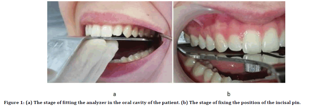
We conducted a survey of 85 people of the first period of mature age and physiological occlusion. Of these, 29 people with mesognathic, 34 with brahygnatichesky and 22 with dolichognathic jaw structure. The distance from the cusp tips, the cutting edges of the teeth of the upper jaw to the occlusal plane was measured. The role of the occlusal plane was performed by the metal surface of the analyzer of the HIP-plane. The analyzer was installed in the pterygoids. After the cusp tips of the canines contact with the surface of the analyzer, they fixed the position of the incisal pin. Thus, we obtained five control points: Pterygium pits, tearing tubercles of canines and incisal pin, which was located in the projection of the palatine suture in the region of the third palatal folds (Figure 1).

Figure 1: (a) The stage of fitting the analyzer in the oral cavity of the patient. (b) The stage of fixing the position of the incisal pin.
After fitting, a hard silicone mass was applied to the surface of the analyzer and an impression was obtained, which was a negative display of the position of the teeth of the upper jaw relative to the occlusal plane. The position of each tooth was measured using a micrometer installed in the imprint of each cusp. The thickness of the mass indicated the vertical position of the tooth relative to the occlusal plane in millimeters (Figure 2).
Figure 2: (a) Analyzer of the HIP-plane. (b) Silicone impression on the surface of the analyzer. (C) Measurement of the thickness of the silicone impression using a micrometer.
Results
As a result of the study, we found that the vertical position of the teeth of the upper jaw relative to the occlusal plane depends on the type of the jaw structure. Obtained results based on the survey are reported in Table 1.
| Tooth number | Point of occlusion | The distance from the point of occlusion of the teeth of the upper jaw to the occlusal plane in mm | ||
|---|---|---|---|---|
| Brachygnathic Type |
Mesognathic Type |
Dolichognathic type |
||
| 1 | Cutting edge | 0 | 0 | 0 |
| 2 | Cutting edge | 0.2 ± 0.05 | 0.3 ± 0.07 | 0.8 ± 0.18 |
| 3 | Cutting cup | 0 | 0 | 0 |
| 4 | Buccal cusp | 0 | 0 | 0 |
| Palatal cusp | 0.05 ± 0.01 | 0.3 ± 0.07 | 0 | |
| 5 | Buccal cusp | 0 | 0 | 0.85 ± 0.1 |
| Palatal cusp | 0.05 ± 0.01 | 0.2 ± 0.05 | 0.15 ± 0.02 | |
| 6 | Mesiobuccal cusp | 0.1 ± 0.02 | 0.25 ± 0.05 | 1.7 ± 0.15 |
| Mesiopalatal cusp | 0.15 ± 0.02 | 0.2 ± 0.02 | 0.45 ± 0.1 | |
| Distobuccal cusp | 0.2 ± 0.05 | 0.25 ± 0.05 | 1.95 ± 0.019 | |
| Distopalatal cusp | 0.25 ± 0.05 | 0.3 ± 0.07 | 1.35 ± 0.1 | |
| 7 | Mesiobuccal cusp | 0.75 ± 0.07 | 0.95 ± 0.09 | 2.05 ± 0.25 |
| Mesiopalatal cusp | 0.4 ± 0.1 | 0.5 ± 0.08 | 1.65 ± 0.15 | |
| Distobuccal cusp | 1.05 ± 0.07 | 1.35 ± 0.09 | 2.75 ± 0.15 | |
| Distopalatal cusp | 0.9 ± 0.09 | 1.6 ± 0.09 | 2.35 ± 0.12 | |
Table 1: The vertical position of the teeth of the upper jaw relative to the occlusal plane.
Obtained results, reported in Table 1 to be discussed in following section.
Discussion
Based on the results we noted that, the vertical position of the masticatory teeth of the upper jaw in patients with a dolichognathic jaw structure occupied the highest position relative to the occlusal plane. This fact determined the steep form of the compensation sagittal curve, starting from the second premolar, ending with the molar region, while the sagittal curve in patients with brachygnatic jaw structure in the same region was the most gentle, as it shown in Figure 3.
Figure 3: The compensation sagittal curve in the form of a diagram in a comparative aspect.
For the mesognathic structure of the jaws, the sagittal curve had an average degree of manifestation in comparison with the two previous types [7]. When assessing the vertical position of the upper jaw incisors relative to the occlusal plane, it was established that the lateral incisors of patients with a dolichognathic jaw structure had the highest position in contrast to the position of the teeth of the same name of patients with meso and brachygnathic jaw structure [8].
Conclusion
The formation of the correct shape of the occlusal surface is the key to the quality treatment of patients in the clinic of orthodontics and prosthetic dentistry. Errors and mistakes at the stages of manufacturing prosthetic structures lead to repeated prosthetics and volumetric correction of the finished work. Ignoring additional bends to form a sagittal curve when using the straight wire technique inevitably leads to a relapse of the pathology after treating patients with dental anomalies and deformities. The difference in the expression of the sagittal curve in people with different types of jaw structure affects the position of the articular heads and the nature of the movement of the mandible. Since the most pronounced sagittal curve corresponds to the dolichognathic structure of the jaws, the mesognathic structure of the jaws is moderate, and the brachygnatic type is the least pronounced, an individual approach to each clinical situation is required. Thus, taking into account the individual structure of the jaws and the characteristic severity of the sagittal curve, it is possible to reduce the percentage of errors at the stages of the reconstruction of the form of dentition. The results of the study can be applied in the clinic of orthodontics, therapeutic and orthopedic dentistry.
References
- https://www.webmd.com/oral-health/guide/children-and-orthodontics#1
- Carlson JE. Physiological occlusion. Midwest press 2009; 218.
- Homann AV. Hilsher Uchebnik of the dentoprosthetic equipment Part 2. 2010; 904.
- Yu LI. Orthopedic stomatology. LLC meditsinskoye news agency 2012; 640.
- Rampulla D, Pelosi A. From 2D to 3D Modern orthopedic stomatology.2017; 27:6–13.
- Dawson PE. Functional occlusion: From a temporal and mandibular joint before planning smile/St. Petersburg E. Dawson. The lane with English under the editorship of Konev DB. App Med 2016; 529.
- Noh J, Neumann U. A survey of facial modeling and animation techniques. USC Technical Report 1998; 99–705.
- Samuel S, Bender IB, Hacke G. The dental pulp: Biologic considerations in dental procedures. Philadelphia: Lippincott 1984.
Author Info
Olga P Ivanova1*, Maria V Vologina1, Viktor I Shemonaev1, Sergey V Chernenko2 and Victoria V Bavlakova1
1Department of Orthodontics, The Volgograd State Medical Universit, The Ministry of Health of the Russian Federation, Volgograd, Russia2State Institute Post Graduate Education, Branch of Russian Medical Academy of Continuing Vocational Education, Novokuznetsk, Russia
Citation: Olga P Ivanova, Maria V Vologina, Viktor I Shemonaev, Sergey V Chernenko, Victoria V Bavlakova, Determination of Expressiveness of the Compensation Sagittal Curve in People with Various Types of Structure of Jaws, J Res Med Dent Sci, 2019, 7(5):21-24.
Received: 14-Aug-2019 Accepted: 11-Sep-2019

