Research - (2021) Volume 9, Issue 9
Correlation between Apical Microleakage and Penetration of Various Root Canal Sealers: An In Vitro Study
Nur A Alalaf1* and Emad Farhan Alkhalidi2
*Correspondence: Nur A Alalaf, Ministry of Health, Nineveh Health Directorate, Iraq, Email:
Abstract
Introduction: This study aimed comparatively evaluate the relationship between penetration in simulated lateral canals and apical microleakage of AH Plus Jet, TotalFill BC and GuttaFlow Bioseal root canal sealers. Materials and methods: Thirty single-rooted extracted human mandibular premolars were decoronated and the root canal length was standardized at15mm, simulated lateral canals were made. After chemo-mechanical preparation, the specimens were randomized into three experimental groups according to the root canal sealers: TotalFill BC, AH Plus Jet, and GuttaFlow Bioseal root canal sealers. In all groups, root canal filling was performed with a single cone technique. The specimens were immersed in 2% methylene blue dye solution and then, subjected to the clearing technique. A stereomicroscope was used to observe the specimens. The penetration depth of root canal sealers in simulated lateral canals and apical microleakage were assessed by a four-grade scoring system (0.3). Data were statistically analyzed by Spearman correlation test at 5% significance. Results: Statistically, there was no significant correlation between penetration depth and apical microleakage of AH Plus Jet sealer (P>0.05). Instead, there was a significant correlation between penetration depth and apical microleakage of TotalFill BC sealer at apical simulated lateral canals whereas GuttaFlow Bioseal sealer showed a significant correlation at coronal simulated lateral canals (P<0.05). Conclusions: GuttaFlow Bioseal and TotalFill BC sealers showed an association between their penetration depth and apical microleakage whereas AH Plus Jet sealer showed no association between its penetration depth and apical microleakage.
Keywords
Root canal sealers, Clearing technique, Apical microleakage technique, Penetration depth
Introduction
Root canal filling is clinically a challenge to be succeeded since it is the final operative phase of root canal treatment which completes the clinical procedures of accurate diagnosis, and chemo-mechanical preparation [1]. Moreover, Gutmann et al. [2] stated that along with the removal of debris and microorganism, good adaptation of filling material to the dentinal wall is essential to achieve hermetic sealing of root canal filling through the penetration of the filling material into the dentinal tubules thus, complete cleaning and sealing of the anatomical ramifications aid in achieving tridimensional sealing of the entire root canal cavity, and consequently root canal filling success [3]. Also, it had been suggested by Ingle et al. [4] in the so-called “Washington study” that apically infected periapical exudate into the incompletely filled root canal space represented about 60% of root canal treatment failure therefore, tridimensional sealing of root canal system is of utmost important factor influencing root canal treatment success.
Root canal sealers should have good sealing ability in static and dynamic conditions since; sealing ability reduces any space that permits penetration of oral fluid between the gutta-percha, the dentin wall, and resisting filling dislodgment following treatment [5]. The sealing ability of the root canal sealer is frequently assessed by microleakage, and bond strength tests [6]. Hence, the good sealing ability of root canal sealer is related to improved adhesion and reduced microleakage values [7].
Theoretically, root canal sealer penetration into dentinal tubules could improve the sealing ability of a root canal filling due to micro-mechanical interlocking [8]. Subsequently, the concept of the root canal sealer penetrating dentinal tubules as a requirement to improve the sealing ability of root canal fillings has been created [9]. Hence, this in vitro study aimed to estimate the association between penetration depth and apical microleakage among experimental root canal sealers in a single experimental group.
Materials and Methods
A total of 30 human single-rooted mandibular premolars were used as study specimens, the teeth were kept in a 0.1% thymol solution at room temperature till the time of the experiment [10].
A preoperative periapical radiograph (Carestream, USA) was taken for teeth in bucco-lingual directions and mesio-distal direction to ensure the presence of a single straight root canal and exclude teeth with prior root canal treatment, calcification, internal or external resorption [11].
The teeth were horizontally decoronated at/below the cementoâ??enamel junction (CEJ) by a high speed fissure bur under continuous water cooling [12] to obtain a standardized root canal length of 15 mm [13].
Simulated lateral canals preparation
The simulated lateral canals were made by parallel milling machine (bio-art 1000MAX, Brazil), measured and prepared at 3 mm (apical third), 6 mm (middle third), and 10 mm (coronal third) from the root apex by a 0.3 mm active end cylinder-shaped bur [14]. The connection between the outer root surface and the main root canal by simulated lateral canals was identified by a 21mm, size 08 K-file (Dentsply Maillefer, Switzerland) and a periapical radiograph was taken if the K-file did not enter the main root canal, and the specimen was excluded from the experimental procedure [15].
Preparation of specimens
The specimens were accessed then, a size 10 K-file was introduced into root canal until it was just visible at the apical foramen, and the length of K-file was measured, working length was established by subtracting 1 mm from this length [16]. The instrumentation procedure had been done by the crown down technique by Protaper Next rotary system nickel-titanium files (Dentsply Maillefer ,Switzerland) in sequential order from (X1-X2) [11]. The irrigation procedure had been done using 2 ml of 5% sodium hypochlorite (NaOCl) (CHLORAXID, ul. Kwiatkowskiego) was used as an irrigant, and 17% Ethylene diamine tetra acetic acid gel (EDTA) (Dentsply Maillefer, Switzerland) was used as a lubricant during the instrumentation procedure [17].
After end of the instrumentation, all specimens were washed with 5ml of 5% NaOCl solution for 1 min subsequently 5ml of distilled water, 5ml of 17% EDTA solution for 1 min, and finally with 5ml of distilled water [18].
Specimens grouping and root canal filling
The specimens were randomly allocated into three experimental groups (N=10) according to the type of the root canal sealers: AH Plus Jet, TotalFill BC, and Gutta Flow Bioseal sealers. All experimental root canal sealers were handled along with manufacturer’s instructions and root canal filling was completed by a single cone filling technique.
Apical dye leakage technique
A coat of nail polish (FloDerm, P.R.C) was carried out on the external surface of specimens except the apical part about 1mm free from nail paint then, after one hour another coat was applied upon the coat of nail paint had completely dried, the specimens were immersed in a 2% methylene blue dye solution for 48 hours [19].
Clearing technique
This technique was accomplished by following the phases: First phase was decalcification process by immersing the specimens into 5% nitric acid for 4 days that had been changed every day, shaken three times in a day and on the 4th day the specimens were tested by trying to thrust a thin needle through the coronal third. If the needle went easily through, the specimens were soft and ready for the next phase [17]. The next phase was dehydration process by immersing the specimens in 70% ethyl alcohol solution for 12 hours followed by 80% ethyl alcohol solution for 12 hours, 90% ethyl alcohol solution for 6 hours and finally in 100% ethyl alcohol solution. The final phase was transparency by immersing the specimens in 100% methyl salicylate for 2 hours till the specimens made transparent [20].
Stereomicroscopic evaluation
All the specimens were examined under a stereomicroscope (OPTIKA, Italy) at 10X magnification, linear apical leakage from the apex of the root to the most coronal extent of dye penetration was evaluated [21] and measured in millimetres on digital images of specimens that had been captured by an attached camera (OptikamB5, Italy) on a stereomicroscope [22].
Apical microleakage values were scored as: Score 0: If no leakage. Score 1: If leakage less than or equal 0.5 mm. Score 2: If leakage from 0.51mm to less than 1 mm .Score 3: If leakage more than 1 mm [17].
The penetration of root canal sealer in simulated lateral canals was examined under a stereomicroscope (OPTIKA, Italy) at 10X magnification and evaluated on digital images of specimens that had been captured by an attached camera (OptikamB5, Italy) on a stereomicroscope [23].
The depth of penetration into the simulated lateral canals thirds (apical, middle and coronal) was scored as: Score 0: No sealer penetration. Score1: If sealer was present in less than half of the lateral canal. Score 2: If sealer covered more than half of the lateral canal. Score 3: Lateral canal was completely filled with sealer [13].
Statistical analysis
Data were analyzed by the Statistical Package for Social Sciences (SPSS, version 25). Spearman correlation coefficient was estimated to measure the strength of correlation between the penetration depth scores and the apical microleakage scores at a 5% significance level (p <.05).
Results
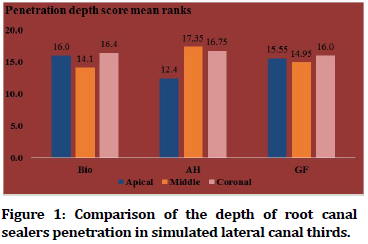
All experimental root canal sealers revealed the ability to penetrate simulated lateral canals, the measured mean of depth of penetration scores of root canal sealers in the apical, middle and coronal thirds simulated lateral canal, number of specimens(N), and percentage(%) were assessed (Table 1 and Figure 1).
| Bio | AH | GF | ||||
|---|---|---|---|---|---|---|
| Penetration score | N | (%) | N | (%) | N | (%) |
| Apical | ||||||
| 0 | 5 | (50.0) | 5 | (50.0) | 4 | (40.0) |
| 1 | 0 | 0 | 1 | (10.0) | 0 | 0 |
| 2 | 0 | 0 | 3 | (30.0) | 3 | (30.0) |
| 3 | 5 | (50.0) | 1 | (10.0) | 3 | (30.0) |
| Middle | ||||||
| 0 | 4 | (40.0) | 3 | (30.0) | 4 | (40.0) |
| 1 | 2 | (20.0) | 0 | 0 | 1 | (10.0) |
| 2 | 2 | (20.0) | 4 | (30.0) | 2 | (20.0) |
| 3 | 2 | (20.0) | 3 | (30.0) | 3 | (30.0) |
| Coronal | ||||||
| 0 | 3 | (30.0) | 2 | (20.0) | 2 | (20.0) |
| 1 | 1 | (10.0) | 3 | (30.0) | 2 | (20.0) |
| 2 | 3 | (30.0) | 2 | (20.0) | 4 | (40.0) |
| 3 | 3 | (30.0) | 3 | (30.0) | 2 | (20.0) |
Table 1: The penetration depth scoring distribution of experimental root canal sealers.
Figure 1.Comparison of the depth of root canal sealers penetration in simulated lateral canal thirds.
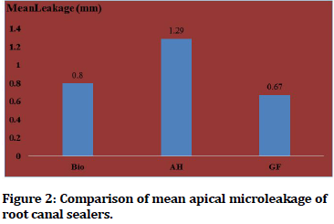
The highest mean apical microleakage was observed on AH Plus Jet sealer (1.290) mm whereas the least mean apical micro leakage was observed on GuttaFlow Bioseal sealer (0.670)mm as shown in Figure 2.
Figure 2.Comparison of mean apical microleakage of root canal sealers.
All experimental root canal sealers showed no apical microleakage in various distributions. The highest distribution of score 0 in which no leakage was observed on GuttaFlow Bioseal sealer which was 30.0% whereas TotalFill BC sealer, and AH Plus Jet showed 20.0% as shown in Table 2.
| Bio | AH | GF | ||||
|---|---|---|---|---|---|---|
| Leakage score | N | (%) | N | (%) | N | (%) |
| 0 | 2 | (20.0) | 2 | (20.0) | 3 | (30.0) |
| 1 | 1 | (10.0) | 1 | (10.0) | 2 | (20.0) |
| 2 | 3 | (30.0) | 0 | 0 | 2 | (20.0) |
| 3 | 4 | (40.0) | 7 | (70) | 3 | (30.0) |
| Total | 10 | (100.0) | 10 | (100.0) | 10 | (100.0) |
Table 2: Apical microleakage scoring distribution of experimental root canal sealers.
Spearman correlation test indicated no significant correlation between the penetration depth scores and the apical microleakage scores of AH Plus Jet sealer. Furthermore, there was a significant positive Spearman correlation in TotalFill BC sealer between penetration depth scores and the apical microleakage scores in the apical third. Regarding the coronal third, there was a significant positive Spearman correlation in GuttaFlow Bioseal sealer between penetration depth scores and the apical microleakage scores (Table 3).
| Leakage Score | ||||||
|---|---|---|---|---|---|---|
| Bio | AH | GF | ||||
| Penetration Score | Spearmen correlation coefficient (rho) | p-value | Spearmen correlation coefficient (rho) | p-value | Spearmen correlation coefficient (rho) | p-value |
| Apical | 0.84 | *0.002 | -0.162 | 0.655 | -0.238 | 0.507 |
| Middle | 0.06 | 0.87 | -0.222 | 0.537 | -0.338 | 0.34 |
| Coronal | 0.37 | 0.293 | 0.386 | 0.27 | 0.808 | *0.005 |
Table 3: Analysis of the Spearman correlation among experimental root canal sealers.
Discussion
In this study, there is no significant correlation between the penetration depth scores and the leakage scores of AH plus jet sealer indicated that there was no significant monotonically increased association. This could be caused by AH Plus Jet sealer contains epoxide component [24] that has to soften the effect on gutta-percha being as a partial solvent [25], bond with root dentin by adamantine, and good flow ability [26]. In addition to low solubility on complete setting altogether improve the penetration ability into root canal complexities through micro-mechanical interlocking hence, AH Plus Jet sealer only bonds mechanically with the root dentin [27]. On the other hand, fast polymerization reaction and subsequently sealer shrinkage was occurred during an early stage of polymerization reaction resulted in micro gap formation, also this sealer is naturally acidic and hydrophobic while the root dentin is hydrophilic in nature and the dentin-sealer interface is relatively hydrophilic [28]. Hence, adhere poorly to humid root dentin and decrease the ability of complete adaptation and filling of the hydrophilic root canal [29]. Moreover, the sealing ability of AH Plus Jet sealer could be affected by silicone oils resulted in poor wettability and high surface tension [30]. Also, this sealer consists of largesized particles (1.5-8) μm which could not easily penetrate small dentinal tubules, particularly at the apical area hence, high risk of apical microleakage that enhances dye penetration [31].
There is no significant association in TotalFill BC sealer between the penetration depth scores and the leakage scores except in the apical third of the simulated lateral canal (p<0.05) which was p = 0.002 whereas the Spearman correlation coefficient was positive (rho = 0.84) indicated that there was a significant monotonically increased association at the apical third of the simulated lateral canal. This could be caused by the ability of the nanoparticles sized less than 2 μm in diameter of this sealer to penetrate apical dentinal tubules to form micro-mechanical interlocking then, hydroxyapatite formation along the mineral infiltration zone hence, this sealer bonds mechanically and chemically [32].
There was no significant correlation in GuttaFlow Bioseal sealer except between the penetration depth scores and the leakage scores in the coronal third of the specimens (p<0.05) which was p = 0.005 whereas the Spearman correlation coefficient was positive (rho= 0.808) indicated that there was a significant monotonically increased association between the penetration scores and the leakage scores at the coronal third of the simulated lateral canal. This could be caused by the ability of nanoparticles sized less than 30μm of guttapercha, thixotropic property, and physically, it is comparable to the gutta-percha core material. In addition to the ability of this sealer to expand as much as 0.2% after setting hence, filling the irregular spaces of the root canal wall and into dentinal tubules hence, GuttaFlow Bioseal bonds mechanically, physically and chemically with the root dentin [32].
TotalFill BC and GuttaFlow Bioseal sealers showed a significant monotonically increased association between the penetration depth scores and the leakage scores at the apical, coronal thirds of the simulated lateral canal respectively. Hence, if sealer penetration into simulated lateral canal had been increased, apical microleakage had not been decreased which could be due to the advantageous biological properties of calcium silicatebased TotalFill BC and GuttaFlow Bioseal sealers primarily resulted from their bioactivity ability [33] which considered a significant property for chemical bonding resulted from water sorption and solubility even after complete setting resulted in sealer mass loss, decrease dimensional stability, and high risk of apical microleakage [28]. Hence, a negative impact on the sealing ability of root canal sealer [34]. Moreover, the bio mineralization ability has not been absolutely confirmed that the mineral infiltration zone influences the outcome of root canal filling treatment [35], either positively, calcium ions will react with the carbon dioxide forming calcite crystals [36]. Hence, decrease micro gaps and porosity as well as increase the retention of the root canal sealer [37] or negatively, hydroxyapatite formation did not decrease microleakage values due to its porous shape [38].
In this study a single experimental group had been standardized, designed and aimed to evaluate the potential correlation between root canal sealer penetration and apical microleakage in which the same specimens had been made two different experiments to examine two independent variables; hence, it could be possible to evaluate the cause and result in correlation thus, increase the power of statistical analysis since typical correlation analysis is assumed that any random variable factor affects only one subject and this requirement could not possible when two or more different experiments [39].
The results of this study are in conformance with De- Deus et al. [39] showed that no significant correlation between tubular penetration and leakage of AH Plus sealer. In contrast, Attur et al. [40] revealed that there was a positive correlation between microleakage and tubular penetration of AH26 sealer.
Conclusion
The penetration ability of AH Plus jet sealer was not a dependent factor influencing its apical microleakage values as no significant association was accepted between penetration depth scores and microleakage scores of AH Plus jet sealer. The penetration ability of TotalFill BC sealer in apical root thirds was a dependent factor influencing its apical microleakage values as a positive association was accepted between penetration depth scores and microleakage scores. The penetration ability of GuttaFlow Bioseal sealer in coronal root thirds was a dependent factor influencing its apical microleakage values as a positive association was accepted between penetration depth scores and microleakage scores.
References
- Schilder H. Filling root canals in three dimensions. J Endod 2006; 32:281â??290.
- Gutmann JL. Adaptation of injected thermoplasticized gutta-percha in the absence of the dentinal smear layer. Int Endod J 1993; 26:87â??92.
- Alkaabi W, Alshwaimi E, Farooq I, et al. A micro-computed tomography study of the root canal morphology of mandibular first premolars in an Emirati population. Med Princ Pract 2017; 26:118â??124.
- Ingle JI, Simon JH, Machtou P, et al. Outcome of endodontic treatment and re-treatment. In: Ingle JI, and Bakland LK. Textbook of endodontics. 5th Edn. BC Decker Inc, Hamilton, London 2002; 748.
- Schwartz RS. Adhesive dentistry and endodontics. Part 2: Bonding in the root canal system—the promise and the problems: A review. J Endod 2006; 32:1125â??1134.
- Wennberg A, Orstavik D. Adhesion of root canal sealers to bovine dentine and gutta-percha. Int Endod J 1990; 23:13â??19.
- Brainstetter J, Von Fraunhofer A. The physical properties and sealing action of endodontic sealer cements: A review of the literature. J Endod 1982; 8:312â??316.
- Chandra SS, Shankar P, Indira R. Depth of penetration of four resin sealers into radicular dentinal tubules: A confocal microscopic study. J Endod 2012; 38:1412–1416.
- Moon YM, Shon WJ, Baek SH, et al. Effect of final irrigation regimen on sealer penetration in curved root canals. J Endod 2010; 36:732â??736.
- Tomer AK, Gupta R, Ramachandran M, et al. Comparison of the apical sealing ability of calcium hydroxide, MTA, and silicone based sealers. IJADS 2018; 4:3â??5.
- Candeiro GTM, Lavor AB, Lima ITF, et al. Penetration of bioceramic and epoxy-resin endodontic cements into lateral canals. Braz Oral Res 2019; 33:1â??6.
- Devarajan M, Ahamed SH. Comparative evaluation of dentinal penetration of three different endodontic sealers-Scanning electron microscopic study. IJCR 2018; 10:70600â??70605.
- Heda DU, Kubde R, Shenoi P, et al. To assess and compare depth of penetration of an epoxy amine based resin sealer in simulated lateral canals after manual, sonic & ultrasonic agitation-An in vitro stereomicroscopic study. GJRA 2019; 8:1â??3.
- Tanomaru-Filho M, Sant'anna-Junior A, Bosso R, et al. Effectiveness of gutta-percha and Resilon in filling lateral root canals using the obtura II system. Braz Oral Res 2011; 25:205â??214.
- Fernandez R, Restrepo JS, Aristizaba LDC, et al. Evaluation of the ï¬lling ability of artiï¬cial lateral canals using calcium silicate-based and epoxy resin-based endodontic sealers and two guttapercha ï¬lling techniques. Int Endod J 2015; 49:365â??373.
- Akcay H, Arslan H, Akcay M, et al. Evaluation of the bond strength of root-end placed mineral trioxide aggregate and biodentine in the absence/presence of blood contamination. Eur J Dent 2016; 10:370–375.
- Amanda B, Suprastiwi E, Usman M. Comparison of apical leakage in root canal obturation using bioceramic and polydimethylsiloxane sealer (In Vitro). OJST 2018; 8:24â??34.
- Kumari M, Taneja S, Bansal S. Comparison of apical sealing ability of lateral compaction and single cone gutta percha techniques using different sealers: An in vitro study. J Pierre Fauchard Acad 2017; 31:67â??72.
- Elshinawya MI, Abdelazizb KM, Khawshhalc AA, et al. Sealing ability of two adhesive sealers in root canals prepared with different rotary file systems. Tanta Dent J 2019; 16:21â??24.
- De Gregorio C, Estevez R, Cisneros R, et al. Efficacy of different irrigation and activation systems on the penetration of sodium hypochlorite into simulated lateral canals and up to working length: An in vitro study. J Endod 2010; 36:1216–1221.
- Lone MM, Khan FR. Evaluation of micro leakage of root canals filled with different obturation techniques: An in vitro study. J Ayub Med Coll Abbottabad 2018; 30:34â??43.
- Teixeira CK, Da Silva SS, Waltrick SBG, et al. Effectiveness of lateral and secondary canal filling with different endodontic sealers and obturation techniques. RFO Passo Fundo 2017; 22:182â??186.
- Lankar A, Mian RI, Mirza AJ, et al. A comparative evaluation of apical sealability of various root canal sealers used in endodontics. IMJ 2018; 25:39â??41.
- Tedesco M, Felippe MC, Felippe WT, et al. Adhesive interface and bond strength of endodontic sealers to root canal dentine after immersion in phosphate buffered saline. Microsc Res Tech 2014; 77:1015–1022.
- Tagger M, Greenberg B, Sela G. Interaction between sealers and gutta-percha cones. J Endod 2003; 29:835–837.
- Santini A, Miletic V. Comparison of the hybrid layer formed by silorane adhesive, one-step self-etch, etch, and rinse systems using confocal micro-raman spectroscopy and SEM. J Dent 2008; 36:683–691.
- Khader AM. SEM penetration depths evaluation of three root canal sealers. J Int Oral Health 2016; 8:191–194.
- Gandolfi MG, Siboni F, Prati C. Properties of a novel poly siloxane- guttapercha calcium silicate-bio glass-containing root canal sealer. Dent Mater 2016; 32:113â??126.
- Roggendorf MJ, Ebert J, Petschelt A, et al. Influence of moisture on the apical seal of root canal fillings with five different types of sealer. J Endod 2007; 33:31â??33.
- Drukteinis S, Peciuliene V, Maneliene R, et al. In vitro study of microbial leakage in roots filled with EndoREZ sealer/EndoREZ points and AH Plus sealer/conventional gutta-percha points. Stomatol 2009; 11:21â??25.
- Primus CM. Products and distinctions. In: Camilleri J. Mineral trioxide aggregate in dentistry: From preparation to application. Springer, Verlag, Berlin, Heidelberg 2014; 151â??156.
- Al-Haddad A, Che Ab Aziz ZA. Bioceramic-based root canal sealers: A review. Int J Biomater 2016; 2016:1â??10.
- Huang Y, Orhan K, Celikten B, et al. Evaluation of the sealing ability of different root canal sealers: a combined SEM and micro-CT study. J Appl Oral Sci 2018; 26:1–8.
- Donnermeyer D, Bürklein S, Dammaschke T, et al. Endodontic sealers based on calcium silicates: A systematic review. Odontology 2019; 107:421â??436.
- Jeong JW, DeGraft-Johnson A, Dorn SO, et al. Dentinal tubule penetration of a calcium silicate-based root canal sealer with different obturation methods. J Endod 2017; 43:633â?? 637.
- Holland R, de SouzaV, Nery MJ, Otoboni Filho JA, et al. Reaction of rat connective tissue to implanted dentin tubes filled with mineral trioxide aggregate or calcium hydroxide. J Endod 1999; 25:161â?? 166.
- Iacono F, Gandolfi MG, Huffman B, et al. Push-out strength of modified portland cements and resins. Am J Dent 2010; 23:43â?? 46.
- Weller RN, Tay KC, Garrett LV, et al. Microscopic appearance and apical seal of root canals filled with gutta-percha and proroot endo sealer after immersion in a phosphate-containing fluid. Int Endod J 2008; 41:977â?? 986.
- De-Deus G, Brandão MC, Leal F, et al. Lack of correlation between sealer penetration into dentinal tubules and sealability in nonbonded root fillings. Int Endod J 2012; 45:642–651.
- Attur KM, Kamat S, Shylaja KA, et al. A comparison of apical seal and tubular penetration of mineral trioxide aggregate, zinc oxide eugenol, and AH26 as root canal sealers in laterally condensed gutta-percha obturation: An in vitro study. Endodontology 2017; 29:20â??25.
Author Info
Nur A Alalaf1* and Emad Farhan Alkhalidi2
1Ministry of Health, Nineveh Health Directorate, Iraq2Department of Conservative Dentistry, College of Dentistry, University of Mosul, Iraq
Citation: Nur A Alalaf, Emad Farhan Alkhalidi, Correlation between Apical Microleakage and Penetration of Various Root Canal Sealers: An In Vitro Study, J Res Med Dent Sci, 2021, 9(9): 10-15
Received: 06-Jul-2021 Accepted: 09-Mar-2021


