Research - (2021) Volume 9, Issue 10
Computed Tomography Imaging in Evaluating Mediastinal MassesâA Diagnostic Approach with Histopathological Correlation
*Correspondence: P Naveen, Department of Radiodiagnosis, Sree Balaji Medical College and Hospital, India, Email:
Abstract
Computed tomography imaging techniques have contributed significantly to the detection, characterization and staging of mediastinal masses. In this study the anterior mediastinum was the most commonly involved compartment (n=17, 56.7%), followed by superior mediastinum (n=16, 53.3%). Cough was the most common clinical symptom constituting 80 % followed by dyspnea 53.3%, chest pain 23.3% and fever 16.6%. In our study, isolated compartmental involvement is common in posterior mediastinum (n=7, 23.4%) followed by superior (n=4, 13.3%) middle (n=4, 13.3%) and anterior mediastinum (n=3, 10%) and Lymph nodal masses formed majority of the cases with 30% and Lymphoma (66.7%) being most common lymph nodal mass.
Keywords
Computed tomography, Mediastinum, Lymph node
Introduction
The aim of this study is to determine the accuracy of the diagnosis of mediastinal masses by Computed Tomography and to correlate its findings with histopathology. This study reviews the variety of the disease processes involving the mediastinum. Emphasis is based on illustrating the specific diagnostic CT features and its correlation with histopathology findings that allow one to distinguish between the different types of mediastinal masses. The multitude of diseases affecting the mediastinum varies considerably, ranging from tumors (benign to extremely malignant), cysts, vascular anomalies, lymph node masses and mediastinal fibrosis. Hence every possible effort has to be made to arrive at a specific diagnosis at the earliest. Computed Tomography has revolutionized in the diagnosis of mediastinal lesions. It is one of the finest non-invasive imaging modalities available for imaging of the thorax. Computed Tomography has good spatial resolution and shorter imaging time, besides being less expensive and being more widely available. It is capable of defining the precise anatomical details and characterizing the nature, site and extent of the disease. Co-existing lung abnormalities and calcification within the lesions are better appreciated on CT. CT gives much more detail of extent and involvement of disease. This also underlines the importance of close cooperation with the histo-pathologist and the clinicians in diagnosis and management [1-7]. The additional role of CT in performing CT guided biopsies of lesions cannot be over emphasized. The purpose of this study is to study the characteristics and determine the differential diagnosis of Mediastinal masses by Computed Tomography and to study the distribution of mediastinal masses also to determine the accurate delineation and extensions of the tumors. Then to correlate the histo-pathological diagnosis to the findings of CT scan where possible.
Methodology
Source of data
All cases referred to the department of Radio-Diagnosis for clinically suspected Mediastinal masses at Sree Balaji Medical College and Hospital over a period of two years were included in the study.
Sample size
30 cases.
Type of study
Prospective study.
Inclusion criteria
Patients with symptoms of clinically suspected Mediastinal Masses investigated by CT scan and subsequently proved by histopathology.
Exclusion criteria
• Patients with prior treatment elsewhere on presentation.
• Recurrent mediastinal masses after treatment.
• Patient with abnormal renal function test and contrast sensitivity.
All the cases were studied on a 32 Slice Siemens Somatom computed tomography machine.
Preparation of patient
Patients were kept nil orally 4 hrs prior to the CT scan to avoid complications while administrating contrast medium. Risks of contrast administration were explained to the patient and consent was obtained prior to the contrast study.
Routine anteroposterior topogram of the thorax was initially taken in all patients in the supine position. An axial section of 5 mm thickness was taken from the level of thoracic inlet to the level of suprarenal. In all cases pre-contrast study was followed by post-contrast study, image acquisition was done with intermittent suspended inspiration. For post-contrast study, 80-100ml of dynamic intravenous injection of Diatrizoate-meglumine (Trazograf 76%; Urograffin 76%, 60%) at a dose of 300mg of Iodine / Kg body weight (in children) was given and axial section were taken from thoracic inlet to the level of suprarenals.
Sagittal and coronal reconstructions were made wherever necessary. The magnification mode was commonly employed, and the scans were reviewed on a direct display console at multiple window settings (i.e. soft tissue (mediastinal) window, Lung window and Bone window to examine the wide variation of tissue density and also to look for osseous involvement.The pre and post contrast attenuation values, the size, location of the mass, presence of calcification, mass effect on adjoining structures and others associated findings were studied.
Results
Results are explained in the figures (Figures 1 to Figure 5).
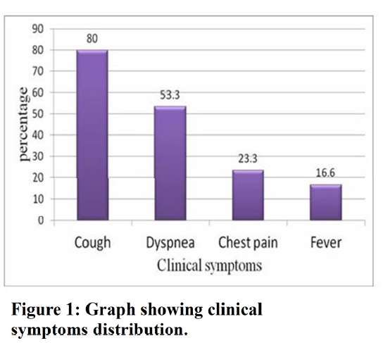
Figure 1. Graph showing clinical symptoms distribution.
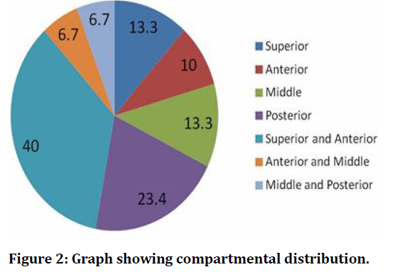
Figure 2. Graph showing compartmental distribution.
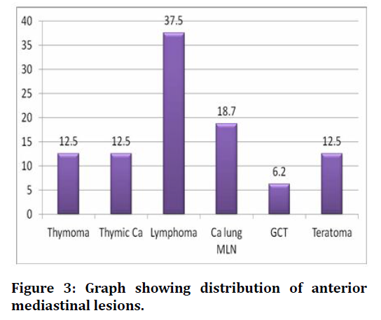
Figure 3. Graph showing distribution of anterior mediastinal lesions.
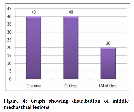
Figure 4. Graph showing distribution of middle mediastinal lesions.
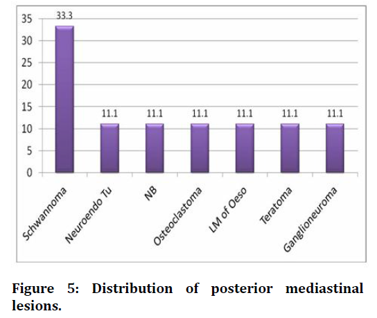
Figure 5. Distribution of posterior mediastinal lesions.
Radiological images
Axial CT scan of thorax of different patients complaining of cough shows, lobulated, soft tissue attenuating mass at superior and anterior mediastinal compartments appearing hypodense on non- contrast study (a) and (b). On post contrast study (c) it shows homogenous enhancement. Mediastinal vessels encased (Figure 6).
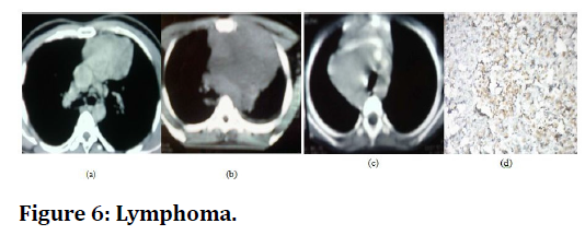
Figure 6. Lymphoma.
Discussion
The initial detection of mediastinal masses can be achieved mainly by chest radiograph (Frontal and Lateral views) and once found, they can be localized, further characterized and staged by CT. However the main objective is to determine if the lesion is malignant or benign, as accordingly the further management depends. Our study comprises a total of 30 patients from in-patient department and is conducted for a period of 2 years in the department of Radio diagnosis, SBMCH.
According to the Davis et al. [8] study in 400 consecutive patients with mediastinal masses, chest pain constituted the most common symptom i.e. 30%, followed by fever 20% whereas in this study, cough was the most common clinical symptom. Felson in 1978 in a series of 550 cases reported, there is no predilection for the masses to occur in the anterior mediastinum. Isolated compartmental involvement is common in posterior mediastinum (n=7, 23.4%) followed by superior (n=4, 13.3%) middle (n=4, 13.3%) and anterior mediastinum (n=3, 10%). However the anterior mediastinal is most commonly involved in trans-compartmental lesions (n=12, 40% and n=2, 6.7%). Therefore anterior mediastinum (n=17, 56.7%) was collectively the most common compartment involved, followed by superior mediastinum (n=16, 53.3%), posterior mediastinum (n=9, 30.1%) and middle mediastinum (n=6, 20.0%). In the similar studies conducted by [7-10] found thymic lesions as common mediastinal lesions. In a study by Chen etalon 34 patients with CT diagnosis of thymic mass, thymoma constituted 91 %, thymic cyst 2.9%. Whereas in our study, of the 5 patients with thymic mass, thymoma constituted 60% and thymic carcinoma constitute 40%. According to previous studies Thymoma is most commonly seen between 50-60 years which is comparable to our study in which the 3 patients with thymoma where of age 44, 62 and 63 years respectively.
References
- Wychulis AR, Payne WS, Clagett OT, et al. Surgical treatment of mediastinal tumors: A 40 year experience. J Thoracic Cardiovas Surg 1971; 62:379-92.
- Benjamin SP, McCormack LJ, Effler DB. Primary tumors of the mediastinum. Chest 1972; 62: 297-303.
- Mcloud TC, Kalisher L, Stark P, et al. Intrathoracic lymph node metastasis from extrathoracic neoplasms. Am J Roentgenol 1978; 131: 403-407.
- Baron RL, Levitt RG, Sagel SS, et al. Computed tomography in the evaluation of mediastinal widening. Radiology 1981; 138:107-113.
- Baron RL, Lee JKT, Sagel SS, et al. Computed tomography of the abnormal thymus. Radiology 1982; 142:127-34.
- Webb WR. Advances in computed tomography of the thorax. Radiol Clin North Am 1983; 21:723-729.
- Sones PJ et al. Effectiveness of CT in evaluating intrathoracic masses. Am J Roentgenol 1982; 139:469-475.
- Davis et al. Primary cysts and neoplasms of the mediastinum: Recent changes in clinical presentations, methods of diagnosis, management and results. Ann Thorac Surg 1987; 44:229-237.
- Chen J, Weisbrod GL, Herman SJ. Computed tomography and pathologic correlations of thymic lesion. J Thoracic Imaging 1988; 3: 61-65.
- Cohen AJ, Thompson LN, Edwards FH, et al. Primary cysts and tumors of the mediastinum. Ann Thorac Surg 1991; 51: 378-386.
Author Info
Department of Radiodiagnosis, Sree Balaji Medical College and Hospital, Chennai, IndiaCitation: Eldho Sajeev, P Naveen, Computed Tomography Imaging in Evaluating Mediastinal Masses–A Diagnostic Approach with Histopathological Correlation , J Res Med Dent Sci, 2021, 9(10): 268-270
Received: 28-Sep-2021 Accepted: 12-Oct-2021
