Research - (2020) Volume 8, Issue 3
Computed Tomography Evaluation of Sella Turcica Dimension in Skeletal Class III Malocclusion Among Adult Ukrainian Peoples
Al-cablany Ebrahem Hezam1,2* and Makarova Alexendra N2
*Correspondence: Al-cablany Ebrahem Hezam, Department of Orthodontics, Faculty of Dentistry, IBB University, IBB, Yemen, Email:
Abstract
Introduction: The sella Turcica is a midline depression in the sphenoid bone which contains the pituitary gland and distal portion of the pituitary stalk. The Sella in orthodontics is most an important skeletal constant unilateral landmark in all cephalometric analysis of the neurocranial and craniofacial complex which located at the center of sella turcica. It's used to measurement the positions of maxilla and mandible in relation to the cranium and to themselves which help in investigation, diagnosis and treatment plan.
Aims : The purpose of this study was to evaluation of Sella Turcica dimension(area and perimeter) in skeletal class III malocclusion (as study group) in comparative with skeletal class I normal occlusion (as control group) among adult Ukrainian peoples by using 3D Cone-beam computed tomography (CBCT).
Materials and Methods: Pre-treatment 3D Cone-beam computed tomography radiographs for 62 subjects of adult patients (18-27 years old ) , 31 subjects (10 male, 21 female) with class I normal occlusion as control group and 31 subjects (12 male ,19 female ) with class III skeletal malocclusion according to ANB (The angle formed at A point, Nasion point and B point of skull) and FMA (The angle formed between the mandibular plane and Frankfort Horizontal plane) angle. The digital tracing of sella Turcica was done by the area and perimeter measures icon tool for all subjects in window of the advanced 3d-imaginag software Ez3d2009. The independent samples T test was used to assessed the relationship between skeletal type and sella Turcica dimension, also was used to determine if skeletal type showed the significant differences. Standard devotions, standard errors, mean values and normality of data were generated for all parameters.
Result and discussion: When comparing the area of sella Turcica for two group, there were significant differences between the study group and the control group (P=0.0116) while the sella perimeter were non-significant differences (P=0.0662). Furthermore, when our result was compared with those in other global data, disparities dimensions among different populations were observed.
Conclusion: When sella size was compared with skeletal type, a significant difference was found in diameter size between Class I and Class III subjects. Larger diameter values were present in skeletal Class III subjects, while smaller diameter sizes were apparent in Class I subjects, so the size of Sella Turcica and pituitary gland play important role to determine the skeletal developmental, classification and occlusion of jaws. The results of the present study of sella area and perimeter may be used as reference standards for Ukrainian subjects in relation to sella Turcica size.
Keywords
Sella Turcica, Area, Perimeter, 3D Computed Tomography, Digital
Introduction
The sella Turcica is a midline depression in the sphenoid bone which contains the pituitary gland and distal portion of the pituitary stalk. The sphenoid bone is divided into a central portion, characterized by two great and two lesser wings extending outward from the sides of the body, and two pterygoid processes. The superior surface of the body is the saddle-like sella Turcica [1]. Its border superiorly by the diaphragma sella, inferiorly by thin floor of cortical bone below which lies the sphenoid sinus, laterally by cavernous sinus, anteriorly by the tuberculum sellae, anterolaterally by the tow anterior clinoid processes. Anteroinferiorly, the foramen rotundum and Posterior to the sella are the tow posterior clinoid processes, dorsum sellae. The sella turcica name derived from Latin Sella that means sedes or saddle while turcica refers to the Turks [2].
Sella is most an important skeletal constant unilateral landmark in cephalometric analysis of the neurocranial and craniofacial complex which located at the center of sella turcica it used to measurement the positions of maxilla and mandible in relation to the cranium and to themselves. The most of cephalometric analysis and studies of the skull depend on the sella point to make liner and angular measurements, like Downs analysis (1948) [3], where he was used the sella landmark to make Y-axis plane that extend from sella to Gonthon (S-Gn) as one of skeletal reference plane, which indicate the degree of downward, reward or forward position of chin in relation to upper face. The Y-axis angle (growth axis angle) that formed from S-Gn plane and Frankfort plane was used to measure the cranium growth it ranges (53º-66º). Steiner analysis (cephalometric analysis of the dental and skeletal relationships of a human skull was presented by Steiner at 1953) [4] in which SN plane (represents the anterior cranial base and is formed by projecting a plane from the Sella-Nasion line) substituted FH plane(Frankfort horizontal plan formed by projection a plane from Infraorbital- Porion line ) because the variation in the Porion point, also the S-N points located at the med-sagital plane of the head and move minimally with any deviation of the head from true profile position. The SN plane used to make SNA (82º), SNB (80º) as skeletal sagital angular measurement and SN-mandibular plane,Y-axis as skeletal vertical angular measurement. Sella landmark point was used also, in another's analysis like, Sassouni analysis (1955) [4], Harvold analysis (1974) and Rickett analysis (1960) and others.
In relation to the sella size, a variety of conditions can lead to sellar enlargement, including tumors of the pituitary or functional hypertrophy of the pituitary, which may occur in primary hypothyroidism or primary hypogonadism, tumors, hypertrophy syndrome, normal variants anatomy and empty sella. As enlarged sella Turcica is a significant finding, suggesting the presence of a pituitary neoplasm. An empty sella can be completely asymptomatic. Also, the sella Turcica size may be smaller in primary hypopituitarism, growth hormones and others. According to Taveras and Wood [5], 17 mm is the upper limit of normal for the maximum anteroposterior diameter of the sella. The depth measured perpendicular to the sella floor, from a line drawn between dorsum and tuberculum, should not exceed 13 mm in most cases. The normal width varies between 10 and 15 mm. These are only guidelines and sella Turcica enlargement can only be used as a suggestion of pituitary abnormality and is certainly not sufficient for diagnosis.
Investigators have also attempted to use the area and the volume of the sella Turcica to serve as better predictors of pituitary disease, analysis and diagnosis. According to Silverman [6]. The size of sella Turcica at age from 1 month to 18 years of age reported that sella Turcica was larger in males than in females except during puberty as this occurred about 2 years earlier and more pronounced in females than in males.
The sella Turcica is best visualized on lateral views of the skull. The sellar floor can be studied on frontal radiographs angled tangentially to the plane of the floor [7].
The shape of sella Turcica with three basic shape ovals, round and flat. Focal erosion or sclerotic of floor in some cases effect in size and shape of ST [8]. Deposition of bone was seen on the tuberculum sellae and resorption at the posterior boundary of Sella Turcica up to 16-18 years of age. The sella point is displaced backward and downward during growth and development.
The morphology is very important for evaluating, diagnostic growth changes and orthodontic treatment results are to be evaluated. There is an increasing interest in the study of human craniofacial dysmorphology, but there are few cephalometric standards available in growth and development [9]. Origin of the pituitary gland is a result of interaction between oral ectoderm which gives rise to anterior pituitary and neural ectoderm gives rise to posterior pituitary. The pituitary fossa differentiates directly from the hypophyseal cartilage which in turn is derived from the cranial neural crest cells of the early chondrocranium.
During embryological development, Sella Turcica area is the key point for the migration of the neural crest cells to the frontonasal and maxillary developmental fields. Formation and development of the anterior part of the pituitary gland, sella turcica, and teeth share in common, the involvement of neural crest cells, and dental epithelial progenitor cells differentiate through sequential and reciprocal interaction with neural crest-derived mesenchyme [10]. Posterior part of the pituitary gland develops from the paraxial mesoderm which is closely related to notochordal induction [11].
A close interrelationship exists between the development of brain tissue and the bones surrounding the Brain-neurocranium [12].
During the last few years 3D cephalometric analysis able to describe anatomical landmarks both on hard and soft tissues has been introduced [13]. Cone-beam computed tomography (CBCT) is currently being used with orthodontic patients since it offers threedimensional (3D) craniofacial imaging as an alternative to conventional radiography and computed tomography (CT). Moreover, The images produced with CBCT are not magnified, CBCT may replace some of the diagnostic tools used in orthodontics, such as two-dimensional (2D) cephalometry. Today, most clinicians are replacing conventional radiographic records with CBCT, since it can provide a series of slides which are then reconstructed in 3D, giving, thus, much more information of the structures studied [14]. The reference angles was used to determine the skeletal type are ANB (A point, Nasion, B point) angle which indicates whether the skeletal relationship between the maxilla and mandible is a normal skeletal class I (+2 degrees), a skeletal Class II (+4 degrees or more), or skeletal class III (0 or negative) relationship and FMA (Frankfortmandibular plane angle) which formed by the intersection of the Frankfort horizontal plane and the mandibular plane.
Materials and Methods
62 Subjects with three dimensional cone beam tomography X-ray for adult patients between 18-27 years old age, 31 subjects (10 male, 21 female) with class I normal occlusion as control group and 31 subjects (12 male, 19 female ) with class III skeletal malocclusion according to ANB and FMA angle as study group were screened for the following criteria.
All control group with class I skeletal relation.
All study group with class III skeletal relation.
Good facial symmetry/proportion shown.
None of the subjects should have undergone any orthodontic or maxillofacial/plastic surgery in the past.
Digital measuring method for all cases by the area and perimeter determinations icon in the software program.
Ethical Approval and acquisition of Informed Consent was obtained for all cases.
The study protocol has been submitted for approval by the Ethical Committee of our medical academy, which complies with the Declaration of Helsinki.
Subjects with cleft lip or palate and other craniofacial deformities.
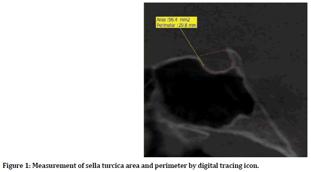
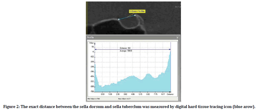
All cases had randomly selected and not undergone previous orthodontic treatment, they were come to orthodontic department of Academy, for orthodontics consultation. Between April 2019 and January 2020. All the research related work in this study was done in orthodontic department of Ukrainian medical Academy, in Poltava city. 3D CBCT (advanced 3d-imaginag software Ez3d2009) high quality was used for all cases, it was recorded by the same trained radiographic technicians were taken in Natural Head Position (NHP) and simultaneously in centric occlusion and lips in repose. The radiographs were distributed according to skeletal Class and gender. All measurements were rehashed 3 times. After the primary measurements were finished, the normal of three readings of every measurement was considered for the last factual examination in request to minimize the intra-analyst variety. The digital tracing of sella turciac was done by the area and perimeter measures icon tool for all subjects in window of the advanced 3d-imaginag software Ez3d2009 as shown in Figure 1. Also, for more assurance the anterior and posterior skeletal limit of sella turcica was measured by bone density profile icon tool as shown in Figure 2.

Figure 1. Measurement of sella turcica area and perimeter by digital tracing icon.

Figure 2. The exact distance between the sella dorsum and sella tuberclum was measured by digital hard tissue tracing icon (blue arow).
The independent samples T test was used to assess the relationship between skeletal type and sella turcica dimension, also was used to determine if skeletal type showed the significant differences. Standard devotions, standard errors, mean values and normality of data were generated for all parameters.
Results
The digital measurements of the sella turcica are presented in Table 1. The average area and perimeter of sella turcica for both class III as study group and class I as control group are shown. When comparing the area of sella Turcica for two groups, there were significant differences between the study group and the control group (P=0.0116) while the sella perimeter were nonsignificant differences (P=0.0662). The mean dimension area of sella turcica for study group was 63.3 mm2, while it was in control group 58.3 mm2 with 5 mm2 differences. The sella turcica perimeter mean for calculated 31.4 mm for study group and 30.4 mm for control group were differ only 1 mm.
| Variable | Class | Mean | Std.D | Std.E | P-value |
|---|---|---|---|---|---|
| Sella perimeter mm | III | 31.4 | 1.936 | 0.3478 | 0.0662 |
| I | 30.4 | 1.932 | 0.3472 | ||
| Sella area mm2 | III | 63.3 | 8.303 | 1.4914 | 0.0116* |
| I | 58.3 | 6.563 | 1.1789 |
Table 1: Independent T test comparing the sella area and perimeter for two different skeletal types.
The sella turcica area and perimeter in this study were compared with the results of previous studies of Malay [15], Bangladesh [16], Brazil [17], Iraq [18] and Greece [19]. In different societies as showed in the Table 2. Furthermore, when our result was compared with those in other global data, disparities in all dimensions among different populations were observed.
| Study | Sella area mm2 | Sella perimeter mm | Data collection instrument |
|---|---|---|---|
| Present study | 60.8 | 30.9 | CBCT |
| Malay | 65.29 | 32.8 | CT |
| Bangladesh | 54.93 | 29.99 | CT |
| Brazil | 41.2 | 28.6 | CBCT |
| Iraq | 65.29 | 32.35 | CT |
| Greece | 46.1 | 30.7 | Cephalometric |
Table 2: Global and present study data of sella turcica measurements.
Discussion
This prospective study describes the perimeter and area dimensions of the sella turcica in Ukrainian subjects with two ( Class I, and Class III ) different skeletal types for adult age group were Statistically significant correlations were found(sella area) between skeletal type as the class III was showed higher mean value in sella area and perimeter 63.3 mm2, 31.3 mm respectively. Were it’s in the class I 58.3 mm2, 30.4 mm respectively?
In related to gender of subjects, most of studies don’t show significant big variation. According to Tetradis and Kantor there was are a tendency of increased size with age, but this was not consistent between all successive age groups [20]. Axelsson et al. [9] had also showed a stable increase in size for both genders during growth. Quakinine, et al. performed a microsurgical anatomical study on 250 sphenoidal blocks obtained from cadavers of different ages. They found that the average transverse width of the sella turcica was 12 mm, the length (anteroposterior diameter) 8 mm, and the average height (vertical diameter) 6 mm [21]. According to Preston a skeletal/facial type Class I, Class II, and Class III. His findings showed no statistically significant correlation between facial type and the mean sella area of the pituitary fossa, were pituitary fossa increased in size with age and found a positive correlation of the area of the sella to age. After 26 years of age, no significant increase was observed on the size [22].
Alkofide EA, et al. performed a cephalometric radiographs study of 180 patients of different ages in Saudi subject, when skeletal type and linear dimensions of sella turcica were evaluated, a significant difference was found. When comparing skeletal Class II and Class III subjects, a significant difference was observed between the diameter of the sella turcica in both Classes. An increase in diameter size appears to be more common in Class III subjects, while a reduced diameter size is more prevalent in Class II individuals, also the non-significant differences of sella turcica length, width, diameter and three different heights of the sella turcica (anterior, posterior, and median) between genders. It was found that there were no statistically significant differences between males and females in all the three linear dimensions [23].
The results of linear and area dimensions and morphological shape of sella Turcica in Malay population were showed No statistically significant differences in gender for all linear and area measurements except at sella height anterior [18]. The parameters for conventional measurements were three dissimilar sella height (anterior, posterior and median), sella length, diameter and width, where all of them deliberated in relation with Frankfort reference line (FH). Total area of sella turcica also considered. No important contrasts in size of the sella were found between sexes [16]. Haritha P performed a lateral cephalometric study on 180 subjects in the age group 9 to 27 years were grouped into Class I, Class II, and Class III (60 subjects in each group). When he was compared the sella size with skeletal type, there was a significant difference as the class III was larger [24].
The differences between various measurements studies of sella turcica are may be due to the use of different landmarks, protocol and techniques of radiographic and degree of radiographic enlargement. Also, the variation in the results probably due to anatomy of population, skeletal characteristic, accuracy of measurements and radiographic, measurements techniques and protocol.
The importance of sella turcica it contain pituitary gland that regulate the growth and functions of the body by its secretions, also its effect on the type growth of mandible bone and maxilla, and thus it contribute to determination the type of occlusion. The pituitary gland is the brain of the endocrine organs, therefore natural to take all this interest in living human. According to many authors and studies the height of the pituitary gland was usually 2 mm shorter than the actual depth of the sella, so during the measurements, analysis and diagnosis should be taken into consideration.
Conclusion
When sella size was compared with skeletal type, a significant difference was found in diameter size between Class I and Class III subjects. Larger diameter values were present in skeletal Class III subjects, while smaller diameter sizes were apparent in Class I subjects, so the size of Sella Turcica and pituitary gland play important role to determine the skeletal developmental, classification and occlusion of jaws. The results of the present study of sella area and perimeter may be used as reference standards for Ukrainian subjects in relation to sella Turcica size also can be used for discovering Pathological enlargement of the pituitary fossa, providing reference data in Comparison with others populations, as the present study were showed differences with previous studies in area and perimeter dimensions.
Source of Support
None.
Conflict of Interest
None declared.
References
- Moore KL, Dalley AF, Agur AM. Clinically oriented anatomy. Lippincott Williams & Wilkins 2013.
- Valpy FE. A Manual of Latin Etymology, as Ultimately Derived... from the Greek Language. Longman 1852.
- Bishara SE. Textbook of orthodontics (1 bs): Saunders Company. Philadelphia 2001; 119.
- Proffit WR, Fields HW, Sarver DM. Contemporary orthodontics. Elsevier Health Sciences 2006.
- Friedland B, Meazzini MC. Incidental finding of an enlarged sella turcica on a lateral cephalogram. Am J Orthodont Dentofac Orthop 1996; 110:508-512.
- Silverman,FN, Roentgen standards fo-size of the pituitary fossa from infancy through adolescence. Am J Roentgenol Radium Therapy Nuclear Med 1957; 78:451-460.
- Maya M, Pressman BD. Pituitary imaging, in the pituitary. Elsevier 2017; 645-669.
- Du Boulay GH. Principles of X-ray diagnosis of the skull. Butterworth Heinemann 2016.
- Axelsson S, Storhaug K, Kjær I. Post-natal size and morphology of the sella turcica. Longitudinal cephalometric standards for Norwegians between 6 and 21 years of age. Eur J Orthodont 2004; 26:597-604.
- Miletich I, Sharpe PT. Neural crest contribution to mammalian tooth formation. Birth Defects Res Part C: Embryo Today: Reviews 2004; 72:200-212.
- Kjær I, Fischer-Hansen B. The adenohypophysis and the cranial base in early human development. J Craniofacial Genetics Developmental Biol 1995; 15:157-161.
- Kjaer, I. Prenatal traces of aberrant neurofacial growth. Acta Odontologica Scandinavica 1998; 56:326-330.
- Harrell WE, Scarfe WC, Pinheiro LR, et al. Applications of CBCT in Orthodontics. In Maxillofacial Cone Beam Computed Tomography Springer 2018; 645-714.
- Dindaroğlu F, Yetkiner E. Cone beam computed tomography in orthodontics. Turkish J Orthodont 2016; 29:16.
- Hasan HA, Alam MK, Yusof A, et al. Size and morphology of sella turcica in Malay populations: A 3D CT study. J Hard Tissue Biol 2016; 25:313-320.
- Islam M, Alam MK, Yusof A, et al. 3D CT study of morphological shape and size of sella turcica in Bangladeshi population. J Hard Tissue Biol 2017; 26:1-6.
- Ruiz CR, Wafae N, Wafae GC. Sella turcica morphometry using computed tomography. Eur J Anatomy 2020; 12:47-50.
- Hasan HA, Alam MK, Abdullah YJ, et al. 3DCT morphometric analysis of sella turcica in Iraqi population. J Hard Tissue Biol 2016; 25: 227-232.
- Andredaki M, Koumantanou A, Dorotheou D, et al. A cephalometric morphometric study of the sella turcica. Eur J Orthodont 2007; 29:449-456.
- Argyropoulou M, Perignon F, Brunelle F, et al. Height of normal pituitary gland as a function of age evaluated by magnetic resonance imaging in children. Pediat Radiol 1991; 21:247-249.
- Ouaknine G, Hardy J. Microsurgical anatomy of the pituitary gland and the sellar region 1 the pituitary gland. Am Surgeon 1987; 53:285-290.
- Choi WJ, Hwang EH, Lee SR. The study of shape and size of normal sella turcica in cephalometric radiographs. Imaging Sci Dent 2001; 31:43-49.
- Alkofide EA. The shape and size of the sella turcica in skeletal Class I, Class II, and Class III Saudi subjects. Eur J Orthodont 2007; 29:457-463.
- Sathyanarayana HP, Kailasam V, Chitharanjan AB. The size and morphology of sella turcica in different skeletal patterns among South Indian population: A lateral cephalometric study. J Indian Orthodont Society 2013; 47:266-271.
Author Info
Al-cablany Ebrahem Hezam1,2* and Makarova Alexendra N2
1Department of Orthodontics, Faculty of Dentistry, IBB University, Yemen2Department of Orthodontics, Ukrainian Medical Stomatological Academy, Ukraine
Citation: Al-cablany Ebrahem Hezam, Makarova Alexendra N, Computed Tomography Evaluation of Sella Turcica Dimension in Skeletal Class III Malocclusion Among Adult Ukrainian Peoples, J Res Med Dent Sci, 2020, 8 (3):203-208.
Received: 21-May-2020 Accepted: 11-Jun-2020
