Research - (2019) Volume 7, Issue 5
Computed Tomographic Analysis of Mental Foramen for Age and Sex Determination in Iraqi Population
Hawraa Noori Atallah1*, Sarmad A Hameed2, Zahraa M Al-Fadhily3 and Abdulrazzaq Ibrahim4
*Correspondence: Hawraa Noori Atallah, Department of Oral and Maxillofacial Surgery, University of Kufa, Iraq, Email:
Abstract
Objectives: A study conducted to investigate the location of the mental foramen using computed tomography to evaluate whether there is a correlation between this radiographic parameter with age and gender of the Al-Najaf population and evaluate it relates to forensic odontology.
Materials and methods: 72 computed tomographic images were studied to measure the distance from the lower border of the mental foramen to the lower border of the mandible. The study sample was divided into two groups, group I age (14-35) and group II age (36-60) years (each group consisted of 36 subjects), and also grouped according to sex (each group consisted of 36 subjects).
Statistical analysis: Statistical analysis was done by using SPSS (statistical package for social sciences) version 20. In which we use mean and standard deviation as descriptive statistics. For analysis of data, we use independent sample t-test and Pearson correlation coefficient. p-value ≤ 0.05 regarded as significant.
Results: There is a significant difference in the location of the mental foramen to the lower border of the mandible which was higher in males than females while there is no significant difference regarding the age of the subject.
Conclusion: There could be sexual dimorphism regarding the location of the mental foramen to the lower border of the mandible. This difference does not seem to be found regarding the age of the subject in Al-Najaf population.
Keywords
Mental foramen, Inferior border of the mandible, Forensic dentistry, Computed tomography, Age and sex
Introduction
Assessment of chronological age plays a fundamental role in both medical and legal domains. It can be considered a complex procedure involving many factors. Alteration associated with chronological age are noticed with both soft and hard tissues. Dental hard tissues and bone are among the tissues which are considered refractory to fire and could remain for a prolonged period post burial [1,2]. Hence, forensic odontology has received a significant role to be a tool in the skeletal and dental remains identification. Because of the limited precision in technique applied for estimation of the existing age at death, dental hard tissues have been considered to demonstrate age-related changes [3]. Mandible has been sought to be a reliable source for agerelated changes as the mandible is a hard and a durable bone showing a high rate of dimorphism [4]. In the absence of sufficient ante mortem records, radiographs can play a valuable role in the identification of human remains. Mental foramen appears on radiographs as either round, slit-like or an irregular radiolucent area that is completely or partially corticated. Numerous studies have investigated mental foramen in terms of; position concerning sex, and the change in location of mental foramen related to age [5].
As computed tomography (CT) has been utilized widely in different medical fields including clinical dentistry (as maxillofacial surgery and dental implants), it has been considered to evaluate the age and sex of the 72-human sample from Al-Najaf city, Iraq depending on the mental foramen position concerning the inferior margin of the mandible.
Materials and Methods
The sample of study composed of 72 computed tomographic images for subjects attending the department of computed tomography in Al- Sadr Medical City in Najaf for various medical purposes after taking some information from them. The study starts from October 2017 to June 2018. The study subjects were informed about the objectives of the study and consent form was obtained before using their data. The study protocol was approved by the Medical Ethics Committee in University of Kufa.
The study sample was divided into two groups: group I age (14-35) and group II age (36-60) years, each group consisted of 36 subjects (18 males and 18 females).
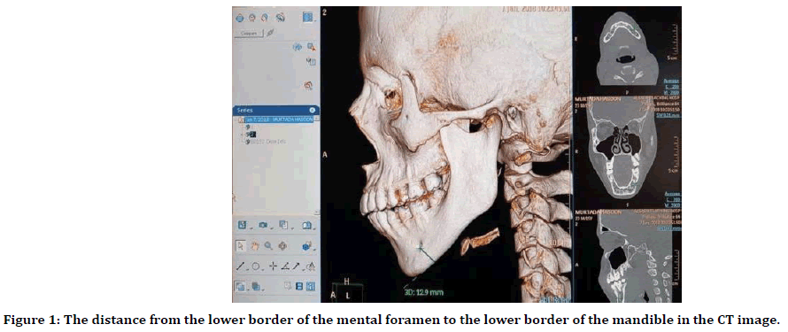
The distance from the lower border of the mental foramen to the lower border of the mandible was measured in 3D (volume) view of CT by using the tool that measures the distance between two points, (Figure 1).

Figure 1. The distance from the lower border of the mental foramen to the lower border of the mandible in the CT image.
Exclusion criteria
→ Patients with surgical intervention to the mandible or undergone orthognathic surgery.
→ Patients who have either radiopaque or radiolucent mass in the mandible.
→ Patients with severe alveolar bone resorption that affect the mental foramen location.
→ Patients who have malignant tumors in the mandible or the tongue.
→ Unidentified mental foramen.
→ Patients who have congenital or developmental disturbances in the mandible.
Statistical analysis
Statistical analysis was done by using SPSS (statistical package for social sciences) version 20. In which we use mean and standard deviation as descriptive statistics. For analysis of data we use independent sample t-test and pearson correlation coefficient. The analysis of data was done firstly to compare between the two age groups as a whole independent of sex, then further analysis done, so each age group divided into males and females, then the comparison was conducted between the males and females of the two age groups separately.
Results
According to an independent t-test, there were no statistically significant difference in the distance from the lower border of the mental foramen to lower border of the mandible between those 14- 35 years (group I) and those 36-60 years (group II) (Table 1), both males and females (Table 2).
| Age/years | N | Mean | Std. Deviation | p-value | |
|---|---|---|---|---|---|
| Mental foramen to lower border of mandible (mm) | 14-35 | 36 | 13.672 | 1.2134 | 0.153 |
| 36-60 | 36 | 14.144 | 1.54077 |
Table 1: Age difference between two groups.
| Age/years | N | Mean | Std. Deviation | p-value | ||
|---|---|---|---|---|---|---|
| Mental foramen to lower border of the mandible (mm) | Males | 14-35 | 18 | 14.211 | 0.739 | 0.671 |
| 36-60 | 18 | 14.366 | 1.352 | |||
| Females | 14-35 | 18 | 13.133 | 1.3672 | 0.137 | |
| 36-60 | 18 | 13.922 | 1.7182 |
Table 2: Age difference between males in two groups and females in two groups.
The result showed that there is a statistically significant difference in the distance from the lower border of the mental foramen to lower border of the mandible between males and females which is more in males (14.2 mm) (Table 3).
| Age/years | N | Mean | Std. Deviation | p-value | |
|---|---|---|---|---|---|
| Mental foramen to lower border of mandible (mm) | Male | 36 | 14.2889 | 1.0775 | 0.02 |
| Female | 36 | 13.5278 | 1.58179 |
Table 3: Sex difference between males and females.
The futher analysis showed that this significant sex difference in mental foramen to lower border of the mandible occur within-group I (14-35 years age ) (Table 4).
| Gender | N | Mean | Std. Deviation | p-value | ||
|---|---|---|---|---|---|---|
| Mental foramen to lower border of mandible (mm) | 14-35 | males | 18 | 14.211 | 0.739 | 0.006 |
| females | 18 | 13.133 | 1.367 | |||
| 36-60 | males | 18 | 14.366 | 1.352 | 0.395 | |
| females | 18 | 13.922 | 1.718 |
Table 4: Sex difference between males and females in two age groups.
Discussion
Changes associated with the growth of human being starts from the prenatal life and continues to senescent, hard tissues are not an exclusion. Bones and teeth can have some changes in shape and/or fusion of ossification centers. These changes may remain stable after death facilitating an easy estimation of the age [6-8].
In forensic median and anthropology, the identification from human remains is of fundamental importance, particularly in criminal investigations, missed person identification in addition to replicate the life of ancient communities [8]. Sex detection depending on Morphological marks subjects to interpretation and probably inaccurate, but calculation and morphometry methods are precise and can be used to determine the sex from the skull [9-11]. Determination of sea and age of skull remains can be largely aided by a forensic dentist. Also, forensic dentistry can have a key role in the identification of the sex of victims with bodies that are mutilated to a degree that they can not be recognized because of massive damages.
The distance of the lower border of the mandible to the mental foramen was selected as a reference for this study because this distance remains relatively constant throughout life [12- 14]. The data collected from this study showed a significant difference in the distance between the lower border of the mental foramen to the lower border of the mandible which was higher in males in comparison to females, which agreed with Suragimath et al. who studied the Maharashtra population in India, study carried out in the South Indian population by Mahima et al. [15], study conducted in the North Indian population by Chandra et al. [16] and studies conducted in various parts of the world by Thomas et al. [17] and Catovic et al. [18]. The possible explanation is that males have a greater bite force due to greater muscle tone which can aid in the deposition of more bone in the lower border of the mandible [8].
In the perspective study, there was no significant difference in the position of the lower border of the mental foramen to that of the mandible. This is following Popa et al. who studied an 80 dry mandibular specimen from the charnel house of Constanţa`s Central Memorial [19].
Popa et al. [20] showed that the distance between the superior margin of the mental foramen and the inferior border of the mandible remains relatively constant throughout life.
Conclusions
Based on the data of this study, it is possible to conclude that there is sexual dimorphism in the location of the inferior border of the mental foramen to the inferior border of the mandible in Al-Najaf population of Iraq. However, the position of the mental foramen to the lower border of the mandible was not affected by age for the same sample of a population.
Conflict of Interest
The authors declare that they have no conflicts of interest.
Acknowledgments
For cooperation and help the department of computed tomography in Al-Al-Sadr Medical City is thankfully acknowledged.
References
- Mohite DP, Chaudhary MS, Mohite PM, et al. Age assessment from mandible: comparison of radiographic and histologic methods. Rom J Morphol Embryol 2011; 52:659-68.
- Prabhu S. Oral diseases in the tropics. Oxford University Press 1991.
- Dudar JC, Pfeiffer S, Saunders SR. Evaluation of morphological and histological adult skeletal age-at-death estimation techniques using ribs. J Forensic Sci 1993; 38:677-685.
- Giles E. Sex determination by discriminant function analysis of the mandible. Am J Phys Anthropol 1964; 22:129-135.
- Ghodousi, A, Sheikhi M, Zamaniet E et al. The value of panoramic radiography in gender specification of edentulous Iranian population. J Dent Mat Tech 2013; 2:45-49.
- Iyyer B, Bhalaji SI. Orthodontics, The Art and Science, 3rd Edn, Arya (Medi) Publishing House 2006; 7-8.
- Reddy NK. Identification. The essentials of forensic medicine and toxicology, 25th Edn, Hyderabad: Suguna Devi K 2006; 50-86.
- Vodanović, M, Dumančić J, Demo Z, et al. Determination of sex by discriminant function analysis of mandibles from two croatian archaeological sites. Acta Stomatologica Croatica 2006; 40:263-277.
- Humphrey LM, Dean MC, Stringer CB. Morphological variation in great ape and modern human mandibles. J Anat 1999; 195:491-513.
- Franklin D, Oxnard CE, O'Higgins P, et al. Sexual dimorphism in the subadult mandible: Quantification using geometric morphometrics. J Forensic Sci 2007; 52:6-10.
- Franklin D, O'Higgins P, Oxnard C. Sexual dimorphism in the mandible of indigenous South Africans: A geometric morphometric approach. South Af J Sci 2008; 104:101-106.
- Wical KE, Swoope CC. Studies of residual ridge resorption. Part I. Use of panoramic radiographs for evaluation and classification of mandibular resorption. J Prosthet Dent 1974; 32:7-12.
- Lindh C, Petersson A, Klinge B. Measurements of distances related to the mandibular canal in radiographs. Clin Oral Implants Res 1995; 6: 96-103.
- Güler AU, Sumer M, Sumer P, et al. The evaluation of vertical heights of maxillary and mandibular bones and the location of anatomic landmarks in panoramic radiographs of edentulous patients for implant dentistry. J Oral Rehabil 2005; 32:741-746.
- Mahima V, Patil K, Srikanth H. Mental foramen for gender determination: A panoramic radiographic study. Medico-Legal Update 2009; 9:33-35.
- Chandra A, Singh A, Badni M, et al. Determination of sex by radiographic analysis of mental foramen in North Indian population. J Forensic Dent Sci 2013; 5:52-55.
- Thomas C. A radiological survey of the edentulous mandible relevant to forensic dentistry. J Den Res 2003; 82:941-943.
- Ćatović A, Bergman V, Ćatić A, et al. Influence of sex, age and presence of functional units on optical density and bone height of the mandible in the elderly. Acta Stomatologica Croatica 2002; 36:327-328.
- Asrani VK, Shah JS. Mental foramen: A predictor of age and gender and guide for various procedures. J Forens Sci Med 2018; 4:76.
- Popa FM, Stefanescu CL, Corici PD. Forensic value of mandibular anthropometry in gender and age estimation. Rom J Legal Med 2009; 17:45-50.
Author Info
Hawraa Noori Atallah1*, Sarmad A Hameed2, Zahraa M Al-Fadhily3 and Abdulrazzaq Ibrahim4
1Department of Oral and Maxillofacial Surgery, University of Kufa, Iraq2Department of Oral Medicine and Oral Pathology, University of Kufa, Iraq
3Department of POP, University of Kufa, Iraq
4Department of Basic Sciences and Prosthodontics, Faculty of Dentistry, University of Kufa, Iraq
Citation: Hawraa Noori Atallah, Sarmad A Hameed, Zahraa M Al-Fadhily, Sana Abdulrazzaq Ibrahim, Computed Tomographic Analysis of Mental Foramen for Age and Sex Determination in Iraqi Population, J Res Med Dent Sci, 2019, 7(5):85-88.
Received: 30-Jul-2019 Accepted: 02-Oct-2019
