Research - (2021) Volume 9, Issue 9
Comparison of Yen angle in Angleâs Class 2 Division 1 and Division 2 Malocclusion in South Indian Population-A Retrospective Cephalometric Study
Preethi Rajamanickam* and Remmiya Mary Varghese
*Correspondence: Preethi Rajamanickam, Department of Orthodontics, Saveetha dental college and Hospitals, Saveetha Institute of Medical and Technical Sciences, India, Email:
Abstract
Aim: To compare the Yen angle in class 2 division 1 and class 2 division 2 in the South Indian population and to evaluate whether the position of maxillary incisor in skeletal class II patients influence the location of point M and also whether it influences the Yen angle. Methodology: 40 lateral cephalograms were obtained for the study. The samples were screened from the records of the patient who visited the Orthodontic Department of Saveetha Dental College and Hospitals. Forty pre-treatment Cephalometric radiographs were selected based on ANB, Wits appraisal, and beta angle confirming skeletal Class II. The same cephalograms were again classified into Angle’s class II division 1 and division 2 based on the position of the maxillary incisors and canines. The Yen angle is traced. Results: Statistical analysis (independent t-test) results were significantly different (P>0.001). Conclusion: There was a statistically significant difference for the values for yen angle within the two groups. The inclination of the incisor influences the position of point M and thus it influences the yen angle. Also the autorotation of the mandible to compensate for the retroclined maxillary incisors in Angle’s class II division 2 malocclusion causes anterior displacement of the G point which also influences the Yen angle. According to the results in our study, the M point is highly correlated with the inclination and position of the maxillary incisor questioning the reliability of Yen angle.
Keywords
Yen angle, Skeletal class II, Angle’s class II division 1, Angle’s class II division 2
Introduction
Several linear and angular measurements exist to assess the sagittal discrepancy between maxilla and mandible, which is of prime importance in diagnosis and treatmentplanning. However, all these measurements have their own shortcomings, which were discussed in detail by Moyers et al [1]. Earlier in 1948, evaluation of anteroposterior apical base relationship was carried out cephalometrically by forming an angle between AB and N Pog which was first devised by Downs, positive and negative variation in the values denote [2]. The relative protrusion/retrusion of the mandible was denoted by positive and negative signs Following Downs, a few years later, Riedel used the difference between [2,3] SNA and SNB angles, and ANB, to describe the apical base relationship. This method by Reidel has been widely accepted and adopted as a predominant method of evaluating anteroposterior jaw relationships. However, both Downs’ and Riedel’s methods possess their own drawbacks, as these angles are influenced by the anteroposterior and vertical variations in N. Hence, SNA and SNB do not depict the true anteroposterior position of the maxilla and mandible. Following Downs, et al. [2–4] suggested an alternative to ANB, the Wits appraisal, which is derived by drawing perpendicular lines from points A and B to the functional occlusal plane (FOP). The linear distance between the points of intersection (AO and BO) is measured to describe the anteroposterior maxillary/ mandibular relationship. According to Jacobson, in a skeletal Class I relationship, in females, AO and BO should coincide, whereas in males, BO should be 1 mm ahead of AO. Though the Wits appraisal avoids N and reduces the rotational effects of jaw growth, it uses the occlusal plane, a dental parameter, to describe a skeletal characteristic. The major drawback associated with this method is that any change in the angulation of the functional occlusal plane will obviously influence the positions of A and B and thereby the Wits appraisal reading [5]. Also, the position or the cant of the occlusal plane can be easily affected by tooth eruption and dental development [5–7]. Later, Kim, et al. [8] introduced the anteroposterior dysplasia indicator (APDI), which is a resultant obtained by addition or subtraction of the AB and palatal plane angle from the facial angle. According to Freeman, point AF is formed by dropping a perpendicular line from point A to the Frankfort horizontal plane (or X). The angle AFB is formed by a line from point AF to point B. (or AXB). Although the vertical position of A does not have an impact on AFB, the vertical displacement of B may influence. As a result, the AFB angle does not exclusively characterise the anteroposterior relationship of the body [9]. Chang, et al. [10] recommended measuring the distance between A and B projected onto the Frankfort horizontal plane. These projected points were labelled AF and BF, respectively, and the respective measurement is AF-BF. However, AF-BF will be affected by the inclination of the Frankfort horizontal plane [10,11]. Baik, et al. [12] introduced the Beta angle in 2004. While it does a good job of assessing sagittal inconsistencies, it is reliant on the points A and B, which can be sometimes not so easy to locate. In certain cases, the condyle is also not easily visible.
The development of Class II malocclusion could be attributed to several factors; hence their accurate diagnosis is important for the selection of the corresponding treatment plan [13]. Due to these existing issues, the aim of this research was to create a new cephalometric measurement to assess the sagittal relationship between the jaws, Yen angle was constructed by Neela et al [14] that involved three points namely S, midpoint of the sella turcica; M, midpoint of the premaxilla; and G, center of the largest circle that is tangent to the anterior, internal inferior, and posterior surfaces of the mandibular symphysis. Recently this angle has been gaining popularity and being used as an alternative and adjunctive to the existing skeletal dysplasia indicators due to its high reliability [15]. This study was thus carried out to check the reliability and validity of Yen angle, which was considered as one of the most reliable sagittal dysplasia indicators.
Aim
To compare the Yen angle in class 2 division 1 and class 2 division 2 in the South Indian population and to evaluate whether the position of maxillary incisor in skeletal class II patients influence the location of point M and also whether it influences the Yen angle.
The YEN angle
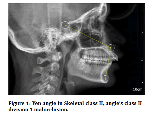
The YEN angle was developed in the Department of Orthodontics and Dentofacial Orthopaedics, YENEPOYA Dental College, Mangalore, Karnataka, India, hence its name. It uses the following three reference points: S, midpoint of the sella turcica; M, midpoint of the premaxilla; and G, centre of the largest circle that is tangent to the anterior, internal inferior and posterior surfaces of the mandibular symphysis. When S, M, and G are connected, it forms the Yen angle, measured at M (Figure 1) [14] and the following conclusions were made from the conducted study. A YEN angle between 117 and 123 degrees had a skeletal Class I pattern, angle less than 117 degrees, individuals are considered to have a skeletal Class II relationship and an angle greater than 123 degrees, the individuals had a skeletal Class III [14].
Figure 1: Yen angle in Skeletal class II, angle’s class II division 1 malocclusion.
Materials and Methodology
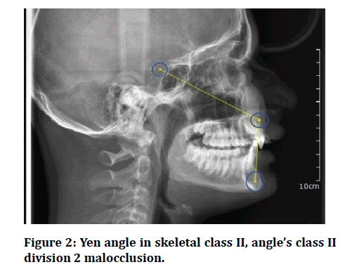
The study was conducted in the Saveetha Institute of Medical and Technical Sciences (SIMATS). This was a retrospective study; the study consisted of 40 pretreatment lateral cephalograms of 13- to 25-year-old individuals from the files of the aforementioned institution. These cephalograms were traced, and ANB, Wits appraisal, and Beta angle were measured. To determine the combined tracing, localization, and measuring error, randomly selected cephalograms were retraced 15 days after they were first evaluated. No significant difference (P >0.05) was found between the first and second measurement. To check whether the ages of the male and female groups were identical, Student’s t test was applied. This test was also used to determine whether there was a difference between the measurements of the male and female subjects. To be included in the skeletal Class II group, a patient had to have a minimum of two of the three parameters (ANB, Wits appraisal, and Beta angle), indicating a Class II relationship. A skeletal Class II relationship was indicated by an ANB of above 4 degrees, a Wits appraisal with AO ahead of BO in females or AO coinciding with or ahead of BO in males, and a Beta angle of less than 27 degrees. The center of the sella turcica, S, was eyeballed, whereas M, as proposed by Nanda, et al. as proposed by Braun et al, [16-18] were constructed using a template with concentric circles whose diameters increased in 1-mm increments and each of the two points was marked by a pinhole in the center of the template. All 40 lateral cephalograms were also classified into skeletal Class II on the basis of only the Beta angle. Then Angle’s class 2 divisions 1 and Class 2 division 2 is further classified depending upon the position of maxillary central, lateral incisors and canines. The Angle’s class 2 division 1 group consisted of 20 patients (11 males, 9 females) and the other 20 (12 females and 8 males) in the second group (Figure 2).
Figure 2: Yen angle in skeletal class II, angle’s class II division 2 malocclusion.
Eligibility criteria
Inclusion criteria
• ANB angle more than 4°.
• Skeletal class 2 pattern with Angle’s class 2 division 1 or division 2 malocclusion.
• Wits appraisal with more than average values.
• Beta angle <27 degrees.
Exclusion criteria
• Patients with skeletal malocclusion other than skeletal Class II pattern.
• Skeletal class II pattern with average inclination of incisors.
• Patients above the age of 30.
• Patients who underwent orthodontic treatment.
• Patients with TMJ disorders.
Statistical analysis: Microsoft Excel (Redmond, Washington, USA) was used to compile the data. Means and standard deviations of the YEN angle in both types of malocclusion were calculated. SPSS 23 (SPSS, Chicago, Illinois, USA) was used for statistical analysis. The data was tested to be parametric using Shapiro-Wilk test of normality. Unpaired t test was performed to compare the Yen angle values between the 2 groups. Power of the study was calculated using G-power software version 3.0 and it was found to be 90%.
Results
There was a statistically significant difference found between the two groups with a P value of 0.017 and the average Yen angle values of Group 1 and Group 2 were 109.3+/-5.4 degrees and 113.1+/-3.9 degrees respectively (Table 1).
Table 1: Unpaired t test performed between Group 1 and Group 2 showing a statistically significant difference p-value <0.05.
| Angle’s malocclusion | Mean+/-S.D(degrees) | P-value |
|---|---|---|
| Class 2 division 1 | 109.3+/-5.4 | 0.017* |
| Class 2 division 2 | 113.1+/-3.9 |
Discussion
Accurate anteroposterior analysis of jaw relationships is critically important in planning orthodontic treatment. Many linear and angular measurements are proposed in cephalometrics for this purpose. This study was thus carried out to check the reliability and validity of Yen angle, which was considered as one of the most reliable sagittal dysplasia indicators [15]. It is important that inclination of incisors do not affect such parameters as it could mislead the clinician and make interpretation very complex. Previous studies conducted, supported that Yen angle is believed to be most reliable in comparison with other skeletal dysplasia indicators. Verification of the accuracy of this parameter in maxillary incisor inclination is therefore necessary, because it involves the M point which represents the midpoint of the premaxilla.
Subjects with skeletal class II malocclusion showed good adherence to Yen angle values, but the average values differed between the Angle’s class 2 divisions 1 group and class 2 division 2 group. This lacks reliability and uniformity of the angle in skeletal class II individuals. Hence, use of Yen angle as a true indicator of sagittal dysplasia is reliable only in individuals with average inclination of incisors. This finding is however not in agreement with a previous report in patients with normal growth patterns 18 that evidenced Yen angle as most reliable, but not taking into account the various subclasses of Angle's malocclusion. This gross change in the average angulation between the Angle’s class 2 division 1 and division 2 skeletal pattern could also be attributed to the fact that Angle’s class 2 division 2 skeletal pattern is more likely to be associated with a horizontal growth pattern. The study carried out by Indukuri et al to analyze the pathognomic features associated with Angle’s class 2 division 2 malocclusion supported this finding [19].
Study carried out by Neela et al. claimed the Yen angle to be least affected by variations in facial height and jaw rotations [14] which is in contradiction with our present study results. Our study results could be justified by the fact that retroclination of upper incisors, causes retroclination of lower incisors, mandible auto rotates upward and backward to bring the lower incisors in contact with the maxillary incisors, thus increasing the values of Yen angle, compared to Angle’s class II division [1] malocclusion which most commonly is associated with vertical growth pattern. This question also creates the need for assessing the reliability of Yen angle in different growth patterns.
Conclusion
The inclination of the incisor influences the position of point M and thus it influences the yen angle. Also the autorotation of the mandible to compensate for the retroclined maxillary incisors in Angle’s class II division 2 malocclusion causes anterior displacement of the G point which also influences the Yen angle. According to the results in our study, the M point is highly correlated with the inclination and position of the maxillary incisor questioning the reliability of Yen angle.
References
- Moyers RE, Bookstein FL, Guire KE. The concept of pattern in craniofacial growth. Am J Orthod 1979; 76:136–48.
- Downs WB. Variations in facial relationships: Their significance in treatment and prognosis. Am J Orthod 1948; 34:812–40.
- Maranhão OBV. Comparison of microesthetic patterns in normal occlusion in relation to class I malocclusion treated with extractions of four premolars. Doctoral dissertation, Universidade de São Paulo 2019.
- Jacobson A. The “Wits” appraisal of jaw disharmony. Am J Orthod 1975; 67:125–38.
- Sherman SL, Woods M, Nanda RS. The longitudinal effects of growth on the Wits appraisa. Am J Orthod Dentofac Orthop 1988; 93:429–36.
- Hussels W, Nanda RS. Analysis of factors affecting angle ANB. Am J Orthod 1984; 85:411–23.
- Richardson M. Measurement of dental base relationship. Eur J Orthod 1982; 4:251–6.
- Kim YH, Vietas JJ. Anteroposterior dysplasia indicator: An adjunct to cephalometric differential diagnosis. Am J Orthod 1978; 73:619–33.
- Freeman RS. Adjusting A-N-B angles to reflect the effect of maxillary position. Angle Orthod 1981; 51:162–71.
- Chang H-P. Assessment of anteroposterior jaw relationship. Am J Orthod Dentofac Orthop 1987; 92:117–22.
- Oktay H. A comparison of ANB, WITS, AF-BF, and APDI measurements. Am J Orthod Dentofac Orthop 1991; 99:122–8.
- Baik CY, Ververidou M. A new approach of assessing sagittal discrepancies: The beta angle. Am J Orthodon Dentofac Orthop 2004; 126:100–5.
- Uribe F, Rothenberg J, Nanda R. The twin force bite corrector in the correction of class II malocclusion in adolescent patients. Orthodontic treatment of the class II noncompliant patient. St Louis: Mosby. 2006; 181-202.
- Neela PK, Mascarenhas R, Husain A. A new sagittal dysplasia indicator: The YEN angle. World J Orthod 2009; 10:147–51.
- Qamaruddin I, Alam MK, Shahid F, et al. Comparison of popular sagittal cephalometric analyses for validity and reliability. Saudi Dent J 2018;30:43–6.
- Nanda RS, Merill RM. Cephalometric assessment of sagittal relationship between maxilla and mandible. Am J Orthod Dentofac Orthop 1994; 105:328–344.
- Braun S, Kittleson R, Kim K. The G-Axis: A growth vector for the mandible. Angle Orthod 2004; 74:328–31.
- Doshi JR, Trivedi K, Shyagali T. Predictability of yen angle & appraisal of various cephalometric parameters in the assessment of sagittal relationship between maxilla and mandible in angle’s class II malocclusion. Peoples J Sci Res 2012; 5:1–8.
- Indukuri R, Narayana V, Nooney A, et al. Pathognomonic features of angle′s Class II division 2 malocclusion: A comparative cephalometric and arch width study. J Int Society Preventive Community Dent 2014; 4:105.
Author Info
Preethi Rajamanickam* and Remmiya Mary Varghese
Department of Orthodontics, Saveetha dental college and Hospitals, Saveetha Institute of Medical and Technical Sciences, Chennai, IndiaCitation: Preethi Rajamanickam, Remmiya Mary Varghese,Comparison of Yen angle in Angle’s Class 2 Division 1 and Division 2 Malocclusion in South Indian Population-A Retrospective Cephalometric Study, J Res Med Dent Sci, 2021, 9(9): 227-230
Received: 31-Aug-2021 Accepted: 14-Sep-2021


