Research Article - (2022) Volume 10, Issue 5
Comparative Evaluation of Surface Changes on Gutta-Percha Cones Treated With Different Herbal Disinfectants: A Scanning Electron Microscopic Study
Snehal Gaware*, Rishikesh Meshram, Nikhil Sathawane and Rohit Amburle
*Correspondence: Snehal Gaware, Department of Conservative Dentistry and Endodontics, Swargiya Dadasaheb Kalmegh Smruti Dental College and Hospital, Nagpur, Maharashtra, India, Email:
Abstract
Aim: The aim of this study was to analyze surface topography of gutta-percha cones after rapid chemical disinfection with 5.25% sodium hypochlorite, 96.30% aloe Vera gel and 100% lemon grass oil.
Materials and methods: From a sealed packet, 28 GP cones of size F3 protaper were selected and distributed into four groups containing 7 GP cones in each group. The following were the groups: Group 1- Control group, Group 2- Sodium hypochlorite, Group 3- Aloe Vera gel, Group 4 - Lemon grass oil. GP cones in each group were soaked in the respective disinfectant solution for 1 minute before being examined under a scanning electron microscope for the variations in the surface topography. Chi-square test was used to compare the results between the various groups in statistical analysis. P value <0.05 was considered as statistically significant.
Results: The results of the study demonstrated that aloe Vera gel caused the least surface alterations in gutta-percha cones when compared to sodium hypochlorite and lemon grass oil.
Conclusion: Within the limitation of the study, it is concluded that even though lemon grass shows high antimicrobial activity it is not as safe as aloe Vera as it causes high surface changes in gutta-percha after disinfection. Therefore,aloe Vera gel can be regarded as a safer choice for disinfecting gutta-percha cones.
Keywords
Gutta percha, Disinfection, Lemon grass, Aloe vera, Sodium hypochlorite, Surface topography, Scanning Electron microscope
Introduction
The elimination of microorganisms from the root canal and the prevention of reinfection are the two most important aspects of endodontic therapy [1]. The major goal of endodontic therapy is to keep the root canal treatment as sterile as possible, from the initial access opening until the final coronal restoration of the tooth. For endodontic therapy to be successful, it is critical to eliminate or reduce the bacteria count [2]. Utmost cares should be taken to avoid any cross contamination of the root canal system with the endodontic instruments [3].
The most commonly used obturating material in endodontics is gutta-percha points. Biocompatibility, dimensional stability, thermo plasticity and radiopacity are some of the ideal properties of gutta-percha points [4]. When the gutta-percha points are subjected to the chair-side environment, certain endogenous and exogenous bacteria like cocci, rods and yeasts may cause defilement [5]. Numerous investigations have concluded Staphylococcus to be the most prevalent microorganism infecting gutta-percha cones in their boxes and while handling with gloves [6]. Infection control is crucial for endodontic therapy success, and all stages of the treatment need to be performed under aseptic conditions, especially root canal filling. As a result, disinfecting gutta-percha cones before inserting them into the root canal is critical [4].
Gutta percha cones cannot be cleansed by the usual conventional methods wherein moist or dry heat methods are used as it may cause surface alteration to the gutta-percha cone structure due to their thermoplastic characteristics. Several compounds such as paraformaldehyde, ethyl alcohol, formocresol, poly vinyl pyrrolidone iodine, glutaraldehyde, quaternary ammonium compounds and hydrogen peroxide have been frequently utilized as disinfectants [7]. Senia et al. conducted a study and concluded that quick disinfection approach is the most successful, definitive, appropriate and cost-effective method for disinfecting gutta-percha cones. Rinsing GP cones in sodium hypochlorite (NaOCl) solution (3% and 5.25%) for a minute is considered as a standard method for disinfecting [8].
NaOCl has the ability to dissolve organic tissue and possess antibacterial properties; hence it is frequently utilized as an irrigating solution in endodontic therapy. Various preparations of NaOCl linked with surfactant molecules are commercially available as an endodontic irrigant. The inclusion of these surfactant molecules lowers the surface tension of NaOCl, making it more wettable on dentin walls. Consequently, the penetration of NaOCl solution within the root canal walls increases, and the disinfection of hard to reach areas is favored, especially within the dentinal tubules, which are inaccessible for endodontic instruments [4].
Besides chemical disinfectants, safe and effective herbal disinfectants have also been tried. Scientific literature suggests that herbs possess antimicrobial, antiseptic, antiviral, anti-fungal, and immunomodulatory properties. As a result, they can be utilized safely in the food and pharmaceutical industries with less or no adverse effects. Herbal agents are environmental friendly; however, they have rarely been tested as a pre-operative disinfectant for GP cones. In recent investigation, Shenoi et al. and Athiban et al. suggested the use of Neem bark and aloe Vera extract respectively for disinfection of gutta-percha cones. Widely used culinary plants in Asian subcontinent include Lemon grass (Cymbopogon citrates), basil (Ocimum basilicum L.), green tea (Camellia sinensis) extract which are utilized in a variety of dental and oral products [9]. Aloe Vera (AV) gel has been proven to be bacteriostatic against Staphylococcus aureus, Streptococcus pyogenic, and Salmonella paratyphi, as well as an excellent decontamination medium for GP cones [2].
In this study, an attempt was made to evaluate the surface changes on gutta-percha cones treated with different herbal disinfectants. The null hypothesis tested was there is no difference in surface topography of gutta percha after chemical disinfection with 5.25% sodium hypochlorite, 96.30% aloe Vera gel and 100 % lemon grass oil. The aim of this study was to evaluate surface topography of gutta-percha cones after chemically disinfecting with 5.25% sodium hypochlorite, 96.30% aloe Vera gel and 100% lemon grass oil [10].
Materials and Method
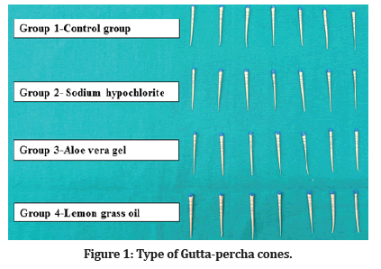
From a sealed packet, 28 GP cones of size F3 protaper were selected and distributed into four groups containing 7 GP cones in each group. The following were the groups: (Figure 1).

Figure 1: Type of Gutta-percha cones.
Group 1- Control group
Group 2- Sodium hypochlorite
Group 3- Aloe Vera gel
Group 4 - Lemongrass oil.
Group 1: Gutta-percha cones were kept without disinfection.
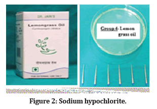
Group 2: Gutta-percha cones were disinfected with 5.25% NaOCl for 1 min (Figure 2).

Figure 2: Sodium hypochlorite.
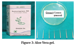
Group 3: Gutta-percha cones were disinfected with 96.30% Aloe Vera gel for 1 min (Figure 3).

Figure 3:Aloe Vera gel.
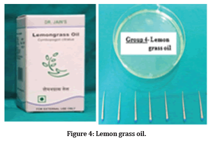
Group 4: Gutta-percha cones were disinfected with 100% Lemon grass oil for 1 min (Figure 4).

Figure 4:Lemon grass oil.
GP cones were then observed for surface topographic changes under scanning electron microscope at 100X, 300X and 1000X magnification respectively (Figure 5).

Figure 5:(A) Gutta percha cones placed for gold coating, (B) Gold coated gutta percha cones, (C) Scanning Electron Microscope.
Statistical Analysis was carried out using Chi- square test to compare the changes seen on the surface of the gutta percha cones between the groups. P value <0.05 was regarded as statistically significant. The tabulated results are given in Table 1.
Results
Seven Gutta percha cones were selected from each group and the percentage was calculated by the number of cones that showed changes divided by the total number of cones. The results were tabulated as shown in (Table 1).
| Changes seen | Total | |||||
|---|---|---|---|---|---|---|
| Mild | Moderate | Severe | No changes | |||
| Group 1 (Control Group) | Count | 0 | 0 | 0 | 7 | 7 |
| % | 0 | 0 | 0 | 100 | 100 | |
| Group 2 (Sodium hypochlorite) | Count | 0 | 4 | 3 | 0 | 7 |
| % | 0 | 57.1 | 42.9 | 0 | 100 | |
| Group 3 (Aloe Vera gel) | Count | 4 | 0 | 0 | 3 | 7 |
| % | 57.1 | 0 | 0 | 42.9 | 100 | |
| Group 4 (Lemon grass oil) | Count | 0 | 3 | 4 | 0 | 7 |
| % | 0 | 42.9 | 57.1 | 0 | 100 | |
Table 1: Distribution of the groups based on the changes seen.
In group 2 (Sodium hypochlorite), it showed severe changes with 42.9% and moderate changes with 57.1% of GP cones. The group 3 (Aloe Vera gel) groups showed no changes with 42.9% and mild changes in relation to 57.1% of GP cones. In group 4 (Lemon grass oil) groups, 57.1% of GP cones showed severe changes and 42.9% showed moderate changes (Figures 6-9).

Figure 6:SEM images of gutta-percha cones in control group under 100X, 300X and 1000X magnification respectively.

Figure 7:SEM images of gutta-percha cones treated with 5.25% sodium hypochlorite for 1min under 100X, 300X and 1000X magnification respectively.

Figure 8:SEM images of gutta-percha cones treated with 96.30% Aloe vera gel for 1 min under 100X, 300X and 1000X magnification respectively.

Figure 9:SEM images of gutta-percha cones treated with 100% lemon grass oil for 1 min under 100X, 300X and 1000X magnification respectively.
Discussion
One of the foremost goals in endodontic therapy is complete elimination of the microorganisms [5]. Thus, sterilization of endodontic instruments and materials becomes an important step [11]. Due to certain features like biocompatibility, radio opacity, dimensional stability and antibacterial activity, gutta- percha cones have been chosen as the preferred material for root canal obturation. They can also be easily retrieved from the root canals [1]. Despite the fact that the amount of microorganisms present at the time of packing was fairly low, dentists routinely use GP points ‘straight out of box’ without giving a second thought about the sterility. It was shown that the clinical use of the pack increases the number of organisms contaminating the GP cones [11].
The contamination of the gutta-percha cones in endodontic therapy consists primarily of vegetative bacterial cells rather than resistant bacterium spores [12]. Due to their thermoplastic properties, traditional sterilizing methods that employ heat cannot be used to promote disinfection of GP cones. As a result, new techniques of disinfection for gutta-percha cones are required [13]. Chemical agents that are effective at decontaminating gutta-percha cones can be used. In our study, surface topography of GP cones following rapid chemical disinfection with 5.25% sodium hypochlorite, 96.30% aloe Vera gel and 100% lemon grass oil were analyzed. The surface modifications on gutta-percha cones were examined by SEM after the disinfection process.
Since Sodium hypochlorite is a strong oxidizing agent, it induces breakdown and hydrolysis of amino acid by generating chloramines molecules. It has the ability to impair the chemical stability of chain polymer, resin, and waxes of GP cones. Such, a chemical instability would adversely affect the mechanical properties of a gutta-percha [14]. In our study sodium hypochlorite group showed severe changes with 42.9% and moderate changes with 57.1% of GP cones.
Ingle stated that dropping a cone into a good germicidal solution for a fair period of time will evidently decontaminate the cone. The use of sodium hypochlorite as a disinfecting agent has been recommended by several studies. However, at higher concentration (5.25%) they produce chloride crystals on the surface of gutta percha cones that could impair the obturation and hermetic seal [1].
Aloe Vera is another medicinal herb which is composed of around 75 active ingredients which includes vitamins, enzymes, sugars, minerals, lignin, Saponins, salicylic acid and amino acids, which have antioxidant, antiviral and antibacterial properties [15]. Aloe Vera has been in use for a variety of diseases, ranging from gastric ulcers to cosmetics since a long time. Aloe Vera has antimicrobial action attributed to substances such as p-coumaric acid, ascorbic acid, pyro catechol and cinnamic acid.
Aloe Vera has been used from time immemorial for the treatment of a multitude of ailments ranging from peptic ulcers to its use in cosmetics. It has a well-established antimicrobial activity ascribed to compounds that are now specifically identified as p-coumaric acid, ascorbic acid, pyro catechol and cinnamic acid. Another significant benefit is that Aloe Vera gel has been shown to effectively decontaminate GP cones in less than one minute [16]. It is a safer gutta-percha cone disinfectant since it does not change the surface topography resulting in improved sealing capacity and reinforcement of root canal.
In a research study conducted by Athiban PP et al, they confirmed the antimicrobial property of Aloe Vera gel against 3 micro-organisms by producing effective inhibition zones nearly equal to 5.25% NaOCl [15]. The effectiveness of three herbal gels for disinfecting guttapercha cones were studied by Shenoi PR et al where they had come to the conclusion that all the gels had inhibition zones almost equal to 5.25% NaOCl [17]. In our study also, Aloe Vera was found to be better than Sodium hypochlorite and Lemon grass oil.
Due to the high activity of citral epoxide, Cymbopogon citratus or Lemon grass (100%) has been reported to have antibacterial activity against S. aureus. Lemon grass is also effective against fungal growth. In the present study, in spite of its efficient antimicrobial activity, it caused significant surface changes in gutta percha. Statistically significant difference was seen between control group and Sodium hypochlorite groups as well as lemon grass group as surface changes seen in both the groups were comparable with each other with slightly higher changes in lemon grass oil group. The control group was not treated with any disinfectant. However, on comparison between all the groups, least surface changes in gutta-percha were seen with aloe Vera gel as compared to sodium hypochlorite and lemon grass oil.
Conclusion
Within the limitation of the study, it is concluded that even though lemon grass shows high antimicrobial activity it is not as safe as aloe Vera as it causes high surface changes in gutta-percha after disinfection.
Gutta-percha cones when treated by Sodium hypochlorite solution resulted in many pitting on the surface of the cones.
Aloe Vera gel is a safer root canal disinfectant since it does not change the topography of the gutta-percha, resulting in improved root canal sealing and reinforcement.
So, aloe Vera gel can be considered as a safer alternative for gutta-percha cone disinfection which caused minimal physical & mechanical surface changes of the gutta percha cones among the three groups tested.
Clinical Implication
Although GP cones are usually supplied in aseptic packages, once opened and used, they may be contaminated. Hence, a supplementary disinfection of GP cones is essential to avoid canal recontamination.
References
- Siqueira Jr JF, da Silva CP, Cerqueira MD, et al. Effectiveness of four chemical solutions in eliminating Bacillus subtilis spores on gutta‐percha cones. Dent Traumatol 1998; 14:124-6.
- Mahali RR, Dola B, Tanikonda R, et al. Comparative evaluation of tensile strength of Gutta-percha cones with a herbal disinfectant. J Conserv Dent 2015; 18:471-3.
- Pang NS, Jung IY, Bae KS, et al. Effects of short-term chemical disinfection of gutta-percha cones: identification of affected microbes and alterations in surface texture and physical properties. J Endod 2007; 33:594-8.
- Vitali FC, Nomura LH, Delai D, et al. Disinfection and surface changes of gutta‐percha cones after immersion in sodium hypochlorite solution containing surfactant. Microsc Res Tech 2019; 82:1290-6.
- Rao SA, Chowdary MN, Soonu CS, et al. Effectiveness of three chemical solutions on gutta-percha cones by rapid sterilization technique: A scanning electron microscope study. Endodontology 2019; 31:17-20.
- Chandrappa MM, Mundathodu N, Srinivasan R, et al. Disinfection of gutta-percha cones using three reagents and their residual effects. J Conserv Dent 2014; 17:571.
- Raveendran L, Mathew M, Pathrose S, et al. Chair side disinfection of guttapercha points-An in vitro comparative study between a herbal alternative propolis extract with 3% sodium hypochlorite, 2% chlorhexidine and 10% povidone iodine. Int J Bioassays 2015; 4:4414-7.
- Senia ES, Marraro RV, Mitchell JL, et al. Rapid sterilization of gutta-percha cones with 5.25% sodium hypochlorite. J Endod 1975; 1:136-40.
- Makade CS, Shenoi PR, Morey E, et al. Evaluation of antimicrobial activity and efficacy of herbal oils and extracts in disinfection of gutta percha cones before obturation. Restor Dent Endod 2017; 42:264-72.
- Burt S. Essential oils: Their antibacterial properties and potential applications in foods—a review. Int J Food Microbiol 2004; 94:223-53.
- AK Varghese, SV Satish, KR Kilaru, et al. Comparative evaluation of the changes in surface topography and crystallization of gutta percha cones by four chemical solutions: An SEM study. Acta Sci Dent Sci 2020; 4:42-46.
- Frank RJ, Pelleu Jr GB. Glutaraldehyde decontamination of gutta-percha cones. J Endod 1983; 9:368-70.
- Tilakchand M, Naik B, Shetty AS. A comparative evaluation of the effect of 5.25% sodium hypochlorite and 2% chlorhexidine on the surface texture of Gutta-percha and resilon cones using atomic force microscope. J Conserv Dent 2014; 17:18-21.
- Naved M, Jadhav S, Hegde V, et al. Comparative evaluation of tensile strength of gutta-percha points after using different disinfectants and time durations - An in vitro study. Int Dent Med J Adv Res 2019; 5:1-5.
- Athiban PP, Borthakur BJ, Ganesan S, et al. Evaluation of antimicrobial efficacy of Aloe vera and its effectiveness in decontaminating gutta percha cones. J Conserv Dent 2012; 15:246-8.
- Lawrence R, Tripathi P, Jeyakumar E. Isolation, purification and evaluation of antibacterial agents from Aloe vera. Braz J Microbiol 2009; 40:906-15.
- Shenoi PR, Morey ES, Makade C, et al. To evaluate the antimicrobial activity of herbal extracts and their efficacy in disinfecting gutta percha cones before obturation-an in vitro study. J Med Sci Clin Res 2014; 2:2676-84.
Indexed at, Google Scholar, Cross Ref
Indexed at, Google Scholar, Cross Ref
Indexed at, Google Scholar, Cross Ref
Indexed at, Google Scholar, Cross Ref
Indexed at, Google Scholar, Cross Ref
Indexed at, Google Scholar, Cross Ref
Indexed at, Google Scholar, Cross Ref
Indexed at, Google Scholar, Cross Ref
Indexed at, Google Scholar, Cross Ref
Indexed at, Google Scholar, Cross Ref
Indexed at, Google Scholar, Cross Ref
Indexed at, Google Scholar, Cross Ref
Indexed at, Google Scholar, Cross Ref
Indexed at, Google Scholar, Cross Ref
Author Info
Snehal Gaware*, Rishikesh Meshram, Nikhil Sathawane and Rohit Amburle
Department of Conservative Dentistry and Endodontics, Swargiya Dadasaheb Kalmegh Smruti Dental College and Hospital, Nagpur, Maharashtra, IndiaCitation: Sukanya M, Shreya Srinivasan, Nehete Sanket Sanjay, Ashok kumar N, Comparative Evaluation of Surface Changes on Gutta-Percha Cones Treated with Different Herbal Disinfectants: A Scanning Electron Microscopic Study, J Res Med Dent Sci, 2022
Received: 21-Feb-2022, Manuscript No. 52357; , Pre QC No. 52357; Editor assigned: 23-Feb-2022, Pre QC No. 52357; Reviewed: 09-Mar-2022, QC No. 52357; Revised: 22-Apr-2022, Manuscript No. 52357; Published: 06-May-2022
