Research Article - (2023) Volume 11, Issue 8
Comparative Evaluation of Fracture Toughness and Flexural Strength of Four Different Core Build up Materials: An In vitro Study
Prashant Nakade1, Bhushan R Bangar2, Sonam Thaore3*, Ajit S Jankar2, Madhuri Wadhwani2 and Varsha Joteppa4
*Correspondence: Sonam Thaore, Department of Conservative Dentistry and Endodontics, SMBT Dental College and Hospital, Maharashtra, India, Email:
Abstract
Purpose: To evaluate and compare the fracture toughness and flexural strength of four different core build up materials. Materials and methods: Sixty samples were taken for study, which were equally divided into four groups. Group A includes dual cure composite resin reinforced with zirconia filler particles (Luxacore Z, DMG), group B-light cure composite resin (Lumiglass deepcure, RDT France), group C-zirconia reinforced glass ionomer cement (Zirconomer improved, Shofu) and group D-chemically cure composite resin (Self comp, Prevest Denpro). All the core build up materials were manipulated according to manufacturer’s instructions and poured into mold. A Universal testing machine applied a central load to the specimen in a 3-point bending mode. Fracture of the specimen was identified and the reading recorded by the universal testing machine. The data were analysed statistically using one Way ANOVA and then compared.
Results: Group A showed highest flexural strength (48.65 MPa) amongst all the groups while group D showed lowest flexural strength (17.90 MPa). Group A showed highest fracture toughness (99.12 MPa) amongst all the groups while group D showed lowest fracture toughness (36.41 MPa.cm-0.5). When mean flexural strength and fracture toughness values of all four groups were compared by using one Way ANOVA, the compared data were statistically significant.
Conclusion: Based on the findings of this study, dual cure composite resin (Luxacore Z) was the material of choice in terms of flexural strength and fracture toughness for core build up material followed by light cure composite resin (Lumiglass deepcure).
Keywords
Core build up, Fracture toughness, Flexural strength, Composite resin, Glass ionomer cement
Introduction
A lot of teeth often show considerable coronal hard tissue defects, frequently requiring a core-buildup as a preprosthetic treatment. This pre-treatment is necessary before the fabrication of subsequent extra-coronal prosthesis. The core consists of restorative material placed in the coronal area of a tooth. This material replaces carious, fractured or otherwise missing coronal structure and retains the final crown. The core is anchored to the tooth by extending into the coronal aspect of the canal or through the endodontic dowel. The attachment between tooth, dowel and core are mechanical or chemical or both, because the core and dowel are usually fabricated of different materials [1].
The materials which are used for core build up are gold, amalgam, resin modified glass ionomer cement, composite resin. Each core build up material has different properties and therefore advantages and disadvantages. Enameldentine bonding using adhesive materials, such as composite resins or glass ionomer-based materials, allows a more conservative technique compared to amalgam which often requires additional tooth preparation to achieve adequate retention. Among the available core build up material in the market, those based upon composite resin are increasing in use and most appropriate to use in contemporary practice [2].
The goal in restoring endodontically treated teeth is the design and fabrication of final restoration. Two factors influence the choice of technique: The type of tooth and the amount of remaining tooth structure. The latter is probably the most important indicator when determining the prognosis. Different clinical techniques have been proposed to solve these problems and opinions vary about the most appropriate one [3].
The objective of restoration can be stated as:
• Reinforcement: Reinforcement of remaining tooth
structure.
• Replacement: Replacement of missing tooth structure
is achieved with core.
• Retention: Supplied by post for the core and the core
supplies retention to the final restoration.
The final configuration of the restored tooth includes four parts: Residual tooth structure, dowel material located within the roots, core material located in the coronal area of the tooth, definitive coronal restoration [4].
Till date, very few studies in the literature have evaluated the fracture toughness and flexural strength of recently modified composite resin and glass ionomer cement. However, no reported study has yet compared dual cure composite resin (Luxacore Z), light cure composite resin (Lumiglass deepcure), chemically cure composite (Self comp) and zirconia reinforced glass ionomer cement (Zirconomer improved). Hence, this study was designed to evaluate and compare the fracture toughness and flexural strength of four different core build up material [5].
Materials and Methods
Sixty samples were taken for study, which were divided into four equal groups. Each group contained fifteen samples.
The four main groups of sample were:
• Group A: Dual cure composite resin reinforced with
zirconia particles (Luxacore Z, DMG).
• Group B: Light cure composite resin (Lumiglass
deepcure, RDT France).
• Group C: Zirconia reinforced glass ionomer cement
(Zirconomer Improved, Shofu Japan).
• Group D: Chemically cure composite resin (Self comp,
Prevest denpro).
Preparation of die
A standard hollow well finished and polished die was fabricated which was made up of brass with a sharp stainless steel blade to form the centrally located notch. The dimensions of the specimens obtained from this mold were according to the American Society for Testing Materials (ASTM) guidelines for such a specimen (Standard E-399). These were 1.8 mm width, 4.2 mm in height, 20 mm in length with a 3 mm long notch on one edge. The width of the notch was 0.5 mm (Figure 1) [6].
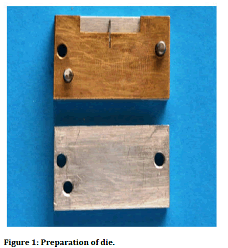
Figure 1: Preparation of die.
Preparation of specimen
The metallic die was first seated on to the metallic base properly. The inner surfaces of the die were coated with petroleum jelly for ease of removal of specimen from the die. All the core build up materials were manipulated according to manufacturer’s instructions and poured into mold. A glass slab was placed over the material to form a flush surface with the top of the mold. The light cured specimens were subjected to curing through open parts of die [7].
After complete setting, the plates were detached and the specimens were removed from the die by removing the walls of the die. The specimens were inspected for presence of any void or irregularities. The specimens with any void or surface irregularities were discarded. All excess flash were trimmed gently with Bard Parker knife (Figure 2) [8].
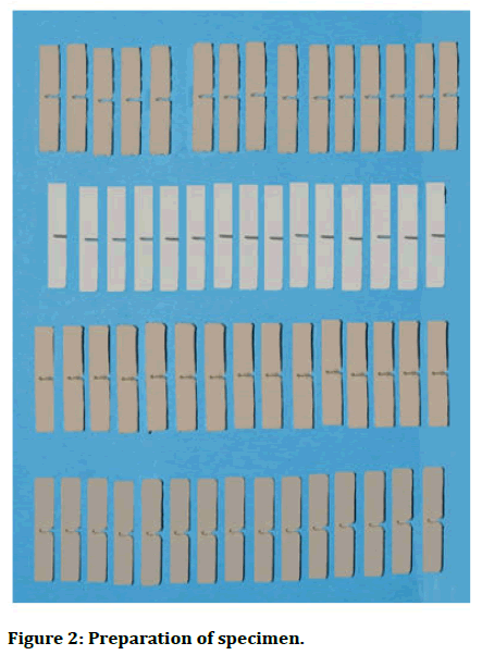
Figure 2: Preparation of specimen.
The samples were prepared at the relative humidity and temperature. The samples of each material were placed in tightly sealed plastic bags and tested 24 hours after preparation [9].
The fracture toughness and flexural strength was tested by using a universal testing machine (STAR testing system, India model no. STS 248, accuracy: ± 1%) (Figure 3) [10].
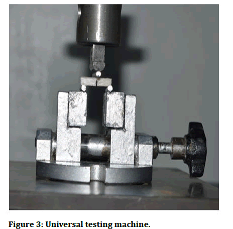
Figure 3: Universal testing machine.
Each of the test samples were loaded on the universal testing machine and subjected to load at constant speed and the results obtained on the computer screen along with plotted graph [11].
Results
Group A showed highest flexural strength (48.65 MPa) amongst all the groups while group D showed lowest flexural strength (17.90 MPa). The mean flexural strength for group B and group C were 40.18 MPa and 28.18 MPa respectively which were intermediate between group A and group D. When mean flexural strength values of all four groups were compared by using one way ANOVA, the data were statistically significant (Tables 1, 2 and Figure 4) [12].
| Variables | N | Mean | Std. deviation | F | Df | P | Inference | |
|---|---|---|---|---|---|---|---|---|
| Flexural strength | Group A | 15 | 48.65 | 13.11 | 30.14 | 3 | 0.0001 (<0.001) | Highly significant |
| Group B | 15 | 40.18 | 10.73 | |||||
| Group C | 15 | 28.18 | 8.17 | |||||
| Group D | 15 | 17.9 | 2.82 | |||||
| Total | 60 | 33.73 | 14.98 | |||||
Table 1: Comparison of flexural strength between groups A, B, C and D.
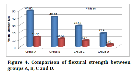
Figure 4: Comparison of flexural strength between groups A, B, C and D.
Post Hoc Tukey’s HSD test showed that no significant difference was found between group A and group B. It also showed that significant difference was found between group A and group C, group A and group D, group B and group C, group B and group D and group C and group D.
| Variables | Group B | Group C | Group D |
|---|---|---|---|
| Group A | 8.47 | 20.46* | 30.74* |
| Group B | - | 11.99* | 22.27* |
| Group C | - | - | 10.27* |
Note: *Indicates that the difference is significant at 0.05 level
Table 2: Post Hoc Tukey’s HSD test.
Group A showed highest fracture toughness (99.12 MPa) amongst all the groups while group D showed lowest fracture toughness (36.41 MPa.cm-0.5). The mean fracture toughness for group B and group C were 81.83 MPa.cm-0.5 and 57.59 MPa.cm-0.5 respectively which was intermediate between group A and group D. When mean fracture toughness values of all four groups were compared by using one way ANOVA, the compared data were statistically significant (Tables 3, 4 and Figure 5) [13].
| Variables | N | Mean (MPa.cm-0.5) | Std. deviation | F | df | P | Inference | |
|---|---|---|---|---|---|---|---|---|
| Fracture toughness | Group A | 15 | 99.12 | 26.71 | 30.15 | 3 | 0.0001 (<0.001) | Highly significant |
| Group B | 15 | 81.83 | 21.9 | |||||
| Group C | 15 | 57.59 | 16.6 | |||||
| Group D | 15 | 36.41 | 5.73 | |||||
| Total | 60 | 68.74 | 30.53 | |||||
Table 3: Comparison of fracture toughness between groups A, B, C and D.
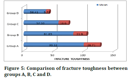
Figure 5: Comparison of fracture toughness between groups A, B, C and D.
Post Hoc Tukey’s HSD test showed that no significant difference was found between group A and group B. It also showed that significant difference was found between group A and group C, group A and group D, group B and group C, group B and group D, group C and group D.
| Variables | Group B | Group C | Group D |
|---|---|---|---|
| Group A | 17.28 | 41.53* | 62.71* |
| Group B | - | 24.24* | 45.42* |
| Group C | - | - | 21.18* |
Note: *Indicates that the difference is significant at 0.05 level
Table 4: Post Hoc Tukey’s HSD test.
Discussion
A core build-up is a restoration placed to provide the foundation for a restoration that will endure the masticatory stress that occurs in the oral cavity for prolonged periods and to provide satisfactory strength and resistance to fracture before and after crown preparation. It forms an integral part of the tooth. Cores are usually retained by pin, post and/or a bonding system to facilitate their retention and to restore the tooth to the extent necessary to support a crown or bridge abutment [14].
Flexural strength is used to evaluate the strength of the material and the amount of the distortion expected under bending stress. Flexural strength tests are considered to be sensitive to surface imperfections such as cracks, voids and related flaws which can influence the fracture strength of brittle materials. High flexural strength values reflect a limited tendency to crazing and high resistance to surface defects and erosion. Fracture toughness is an intrinsic property of a material and is a measure of the energy required to propagate a crack from an existing defect. Flexural strength and fracture toughness are considered to be the most important properties of core build up materials [15].
Several materials including amalgam, Glass Ionomer Cement (GIC), hybrid glass ionomer cement, composite resin and cast metal alloys have been used so far for core build up with various degrees of success. Initially amalgam was the best material for core build up with high mechanical properties, but the recent advances in the composite resins and glass ionomer cements have improved the mechanical properties. Composite resin core build-up materials have been widely used, owing to their high compressive strength, good adhesive properties, low modulus of elasticity and their economic affordability. According to the George, resin composite core build up materials showed better mechanical properties than silver amalgam core, which is similar to the results of study done by Bonilla et al. Choi et al have found that, some resin composites exhibited more compressive strength than that of amalgam and could be used as alternatives to amalgam. Capp and Warren also stated that, composite and acrylic resins can be used as alternatives to amalgam for core structures and that glass ionomer cement is the weakest core build up material. Cohen et al have shown that composite resin had statistically higher fracture resistance compared to GIC and amalgam.
Agrawal A et al showed that, on the basis of their mechanical properties, dual cure core build up resin composites with nano-fillers may be used as alternatives to amalgam core, which is similar to the results of study done by Jayanthi N et al. Mitra et al have found that, nano-composites to be superior to hybrid, micro-hybrid or micro-fill composite resins.
The flexural strength values for light cure composite resin containing titanium (Prisma APH) (171.7 MPa) were significantly higher than chemically cured composite resin (92.0 MPa) and glass ionomer cement (30.6 MPa). In the present study, the light cure composite resin(Lumiglass deepcure) was more superior than glass ionomer cement (Zirconomer improved) in term of flexural strength. This is in accordance to the study done by Kumar G et al,
The result of present study stated that dual cure composite resin (Luxacore Z) has more fracture toughness (99.12+26.71 MPa.cm-0.5) than light cure composite resin (Lumiglass deepcure) 81.83+21.90 MPa.cm-0.5. This is in accordance to study done by kumar L et al. It may be because of its composition. Type of fillers in dual cure composite resin (Luxacore Z) is aluminoborosilicate glass, fumed silica, and titanium oxide, which could be the reason for their high strength. The nanotechnology used in dual cure composite resin (Luxacore Z) eliminates particle agglomeration by incorporating a proprietary coating process during particle manufacture. Dual cure composite resin reinforced with zirconia particle (Luxacore Z DMG) possesses strength, flexibility, and insulation properties similar to that of dentin. The zirconia-silica nano fibers play the “bridge” role in the fracture regions. When a microcrack is initiated in a dental composite, zirconiasilica nanofibers remain intact across the crack planes and support the applied load. The stress-induced phase transformation of zirconia contributes to the toughening effect.
Steven G et al suggested that the fracture strength of composite resin (900 N) was at least 1.9 times that of glass ionomer based core build up materials (500 N), similar to the studies done by Lloyd and Butchart. The result of present study was in consistent with the results shown by Bonilla ED et al. They concluded that, the highest fracture toughness values were shown by dual cure composite resin (166.0+9.2 MPa.cm-0.5) while the lowest fracture toughness values were shown by chemically cure composite resin (72.32+7.2 MPa.cm-0.5).
Goldman concluded that fracture toughness values of glass ionomer materials were lower than the composite resins. In the present study, when fracture resistance of zirconia reinforced glass ionomer core build up was more superior than chemically cure composite resin. This was in accordance to the study done by Preetam shah. The main reason for the improved strength was incorporation of nano filler particle and zirconia filler particle which prevents crack propagation.
When choosing the core build up material, the amount and mode of stress must be considered because it affects the stress transmission to the core. As the firmness increases, the stress goes more directly to the root and less to the core. It is also important to choose a core that has similar physical properties with the tooth structure because of the favorable strong interface and lower risk of micro-leakage and failure. The results of our study indicate that, on the basis of fracture toughness and flexural strength, dual cure composites resin with zirconia filler particles may be used as a material of choice for core build up.
Conclusion
Within the limitations of the study, the following conclusions were drawn:
• Dual cure composite resin reinforced with zirconia filler particles (Luxacore Z) exhibited higher flexural strength followed by light cure composite resin (Lumiglass deepcure); however these findings were statistically insignificant. • Dual cure composite resin reinforced with zirconia filler particles (Luxacore Z) exhibited highest flexural strength than zirconia reinforced glass ionomer cement (Zirconomer improved) and chemically cure composite resin (Self comp) and the difference was statistically significant. • Dual cure composite resin reinforced with zirconia filler particles (Luxacore Z) exhibited higher fracture toughness followed by light cure composite resin (Lumiglass deepcure); however these findings were statistically insignificant. • Dual cure composite resin reinforced with zirconia filler particles (Luxacore Z) exhibits highest in fracture toughness than zirconia reinforced glass ionomer cement (Zirconomerimproved) and chemically cured composite resin (Self comp) and the difference was statistically significant. • Dual cure composite resin reinforced with zirconia filler particles (Luxacore Z) was the material of choice in terms of flexural strength and fracture toughness for core build up material.
References
- Piwowarczyk A, Ottl P, Lauer HC, et al. Laboratory strength of glass ionomer cement, compomers, and resin composites. J Prosthodont 2002; 11:86-91.
[Crossref] [Google Scholar] [PubMed]
- Combe EC, Shaglouf AM, Watts DC, et al. Mechanical properties of direct core build-up materials. Dent Mat 1999; 15:158-165.
[Crossref] [Google Scholar] [PubMed]
- O'Mahony A, Spencer P. Core build-up materials and techniques. J Irish Dent Assoc 1999; 45:84-90.
[Google Scholar] [PubMed]
- Fernandes A, Rodrigues S, Sardessai G, et al. Retention of endodontic post-a review. Endodontol 2001; 13:11-18.
- Maulana TI. Grindability analysis of dental core build-up materials under clinically relevant test procedure. J Med Mat Technol 2017; 1:1-21.
- Sidhu SK. Glass‐ionomer cement restorative materials: A sticky subject. Aus Dent J. 2011; 56:23-30.
[Crossref] [Google Scholar] [PubMed]
- Lynch CD, Shortall AC, Stewardson D, et al. Teaching posterior composite resin restorations in the United Kingdom and Ireland: Consensus views of teachers. Br Dent J. 2007; 203:183-7.
[Crossref] [Google Scholar] [PubMed]
- Wassell RW, Smart ER, St George G. Crowns and other extra-coronal restorations: Cores for teeth with vital pulps. Br Dent J 2002; 192:499-509.
[Crossref] [Google Scholar] [PubMed]
- Schwartz RS, Robbins JW. Post placement and restoration of endodontically treated teeth: A literature review. J Endodont 2004; 30:289-301.
[Crossref] [Google Scholar] [PubMed]
- Michael MC, Husein A, Bakar WZ, et al. Fracture resistance of endodontically treated teeth: An in vitro study. Arch Orofac Sci 2010; 5:36-41. [Google Scholar]
- Jotkowitz A, Samet N. Rethinking ferrule a new approach to an old dilemma. Br Dent J 2010; 209:25-33.
[Crossref] [Google Scholar] [PubMed]
- Jayanthi N, Vinod V. Comparative evaluation of compressive strength and flexural strength of conventional core materials with nanohybrid composite resin core material an in vitro study. J Ind Prosthodont Soc 2013; 13:281-289.
[Crossref] [Google Scholar] [PubMed]
- Huysmans MC, van der Varst PG. Mechanical longevity estimation model for post-and-core restorations. Dent Mat 1995; 11:252-257.
[Crossref] [Google Scholar] [PubMed]
- Saygili G, Sahmali S. Comparative study of the physical properties of core materials. Int J Periodont Restor Dent. 2002; 22:355-363.
[Google Scholar] [PubMed]
- Bonilla ED, Mardirossian G, Caputo AA. Fracture toughness of various core build‐up materials. J Prosthodont 2000; 9:14-18.
[Crossref] [Google Scholar] [PubMed]
Author Info
Prashant Nakade1, Bhushan R Bangar2, Sonam Thaore3*, Ajit S Jankar2, Madhuri Wadhwani2 and Varsha Joteppa4
1Department of Prosthodontics, M A Rangoonwala Dental College and Hospital, Maharashtra, India2Department of Prosthodontics, MIDSR Dental College and Hospital, Maharashtra, India
3Department of Conservative Dentistry and Endodontics, SMBT Dental College and Hospital, Maharashtra, India
4Department of Prosthodontics, S D Dental College and Hospital, Maharashtra, India
Citation: Prashant Nakade, Bhushan R Bangar, Sonam Thaore, Ajit S Jankar, Madhuri Wadhwani, Varsha Joteppa, Comparative Evaluation of Fracture Toughness and Flexural Strength of Four Different Core Build up Materials An In vitro Study, J Res Med Dent Sci, 2023, 11 (08): 099-104.
Received: 18-May-2020, Manuscript No. JRMDS-23-10952; , Pre QC No. JRMDS-23-10952 (PQ); Editor assigned: 21-May-2020, Pre QC No. JRMDS-23-10952 (PQ); Reviewed: 04-Jun-2020, QC No. JRMDS-23-10952; Revised: 21-Jun-2023, Manuscript No. JRMDS-23-10952 (R); Published: 22-Aug-2023
